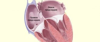
Cardiac aneurysm is a disease characterized by thinning and protrusion of the myocardial wall, lack of contractility in this area of the heart muscle. As a rule, an aneurysm forms in the wall of the left, sometimes in the area of the right ventricle. In newborns, protrusions of the interatrial and interventricular septa are more common. Acquired cardiac aneurysm is one of the common complications of myocardial infarction; the disease is diagnosed in 10-35% of patients in the post-infarction period. Men over forty are at risk.
Classification of cardiac aneurysms
Cardiac aneurysms are classified according to many criteria: the cause and course of the disease, cellular structure and mechanism of formation, shape and size. A detailed classification of cardiac aneurysms is given in the table.
Types of cardiac aneurysms
| Classification sign | Type of aneurysm | Decoding |
| Cause of the disease | congenital | are formed due to disorders of intrauterine development of the fetus |
| acquired | in the vast majority of cases occur after a myocardial infarction | |
| Flow | spicy | form within one to two weeks after a heart attack |
| subacute | are formed from the third to the eighth week after a heart attack | |
| chronic | occur later than the eighth week after myocardial infarction | |
| Cellular structure | muscular | made up of muscle fibers |
| fibrous | the walls of the aneurysm are formed by fibrous tissue | |
| fibromuscular | include muscle and fibrous tissue | |
| Education mechanism | true | have a three-layer structure, consist of dead scar tissue |
| false | are formed when the myocardium ruptures and the formation of a cavity limited by the pericardium (the outer lining of the heart) | |
| functional | areas of viable myocardium that protrude during ventricular contraction | |
| Form | diffuse | have a flat shape |
| saccular | characterized by the presence of a “neck”, which, when expanded, forms a sac-like cavity | |
| delaminating | are formed due to rupture of the endocardium (the inner lining of the heart) |
Cardiac aneurysms can come in different sizes. There are significant aneurysmal protrusions - more than eight centimeters, medium - from three to seven centimeters in size, and small (hidden) aneurysms - less than three centimeters.
What is the aorta
The aorta is the largest vessel in the human body, which carries blood from the heart to the organs and limbs. The upper section of the aorta runs inside the chest, this section is called the thoracic aorta . The lower part is in the abdominal cavity and is called the abdominal aorta . It delivers blood to the lower part of the body. In the lower abdomen, the abdominal aorta divides into two large vessels - the iliac arteries , which carry blood to the lower extremities.
The aortic wall consists of three layers: internal (intima), middle (media), external (adventitia).
Why is a cardiac aneurysm dangerous?
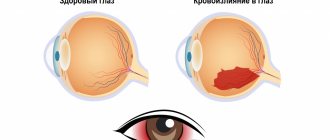
A cardiac aneurysm is dangerous due to the formation of blood clots. Echocardiography often reveals blood clots in a distended cavity. A blood clot can break off at any time and lead to serious consequences.
Common complications of cardiac aneurysm include:
- ischemic stroke - a violation of cerebral circulation with damage to brain tissue;
- thromboembolism (blockage) of the pulmonary artery;
- kidney infarction - an acute circulatory disorder leading to the death of organ tissue;
- gangrene – necrosis of body tissues, accompanied by their rotting;
- repeated myocardial infarction is an acute disturbance of blood circulation in the heart muscle.
In addition to the risk of blood clot rupture, a cardiac aneurysm contributes to the development of heart failure with characteristic symptoms: shortness of breath, interruptions in heart function, chest pain, and swelling.
Clinical manifestations of complications in subarachnoid hemorrhage
After suffering a subarachnoid hemorrhage due to a rupture of a cerebral artery aneurysm, the patient may experience complications. Such complications include:
- thrombophlebitis with pulmonary embolism
- stress-induced perforated duodenal ulcer
- ECG changes indicating myocardial infarction or ischemia
Subarachnoid hemorrhage in the cranial cavity triggers increased activity of the sympathetic nervous system. This causes myofibrillar degeneration of the heart muscle (myocardium). When an aneurysm ruptures with subarachnoid hemorrhage, cardiac arrhythmia may develop. For such patients, beta-blockers are recommended. Their use in patients should be carried out with caution, especially in the presence of atrioventricular block. A possible complication for the patient is a decrease in the level of sodium in the blood plasma (hyponatremia). Hyponatremia occurs when there is inappropriate secretion of antidiuretic hormone (ADH) or secretion of natriuretic hormone. Treatment of such patients is limited to free fluid restriction while maintaining sufficient intravascular volume.
How long do people live with a cardiac aneurysm?
Life expectancy with a cardiac aneurysm depends on several factors: the size of the protrusion, location, rate of increase, concomitant diseases and the age of the patient. People with small aneurysms live for years without surgical treatment, but under the constant supervision of a cardiologist. In cases where the size of the aneurysm is so large that the disease threatens a person’s life, surgery is necessary; without it, death is inevitable. An aneurysm is not a death sentence, but in no case should you let the situation take its course; only an experienced cardiologist can decide on the choice of treatment tactics.
Causes of cardiac aneurysm formation
The causes of congenital and acquired cardiac aneurysm differ. In adult patients, in 95-97% of cases, pathology develops after extensive myocardial infarction. Less commonly, the formation of a cardiac aneurysm is caused by trauma, injury and infection (rheumatism, tuberculosis, syphilis, infective endocarditis).
Aggravating factors are:
- high blood pressure;
- chronic heart failure;
- post-infarction cardiosclerosis is a pathology characterized by the growth of scar tissue in the myocardium.
Among the causes of congenital cardiac aneurysms, researchers identify:
- disruption of the formation of connective tissue during intrauterine development of the fetus;
- genetic predisposition;
- infections suffered by a pregnant woman;
- negative environmental factors.
Congenital cardiac aneurysm is quite rare and accounts for slightly more than 1% of all anomalies of intrauterine fetal development.
Symptoms of a cardiac aneurysm
Symptoms of a cardiac aneurysm depend on the course of the disease.
Characteristic signs of acute cardiac aneurysm are:
- severe pain in the chest that appears suddenly and lasts from several hours to several days;
- a feeling of lack of air, turning into suffocation;
- pronounced pallor of the skin;
- blue discoloration of mucous membranes;
- with the development of pulmonary edema, coughing, wheezing appear, breathing becomes bubbling, and copious sputum is produced in the form of foam.
With chronic cardiac aneurysm, the symptoms are different.
Most often, patients complain about:
- minor periodic pain in the heart area;
- heartbeat;
- weakness and dizziness;
- shortness of breath;
- swelling.
The listed symptoms of a cardiac aneurysm can also occur with other diseases of the cardiovascular system. Additional research methods help make an accurate diagnosis.
Initial clinical manifestations of aneurysm rupture, acute extensive subarachnoid hemorrhage
During the short moment of aneurysm rupture, when extensive subarachnoid hemorrhage acutely develops, intracranial pressure reaches the value of mean arterial pressure, with a decrease in cerebral blood supply (CPP, cerebral perfusion pressure).
In 45% of cases, after this, patients suddenly lose consciousness, which is then restored. Sometimes a patient's sudden loss of consciousness is preceded by a short moment of excruciating headache, but most patients, upon regaining consciousness, first of all complain of a headache.
In 10% of cases, bleeding from a ruptured aneurysm can be quite massive and cause loss of consciousness in the patient for several days.
Approximately 45% of patients after a rupture of an aneurysm complain of intense headache, usually worsening with tension, but they do not experience loss of consciousness. Patients often describe this headache as “the worst headache of their entire life.” Designations such as “tearing” and “breaking” headaches are often used. Typically, patients indicate that they have pain in “the whole head” or “the back of the head and neck.”
Whatever the onset of the disease, one of its symptoms of aneurysm rupture is vomiting. When vomiting and sudden headache are combined, the patient should always suspect acute subarachnoid hemorrhage.
Although the hallmark of a ruptured aneurysm is sudden headache in the absence of focal neurological symptoms, patients often also experience neurological deficits.
Paralysis of the muscles innervated by the third oculomotor nerve that occurs in the patient on the side of the hemorrhage makes one think about a ruptured aneurysm of the posterior communicating artery.
The often observed paralysis of the rectus externus muscle of the eye (innervated by the abducens VI nerve) is of little importance for the location of the lesion, but is often observed with rupture of a subtentorial aneurysm.
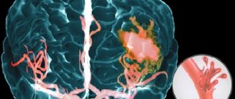
When an aneurysm of the anterior communicating artery or bifurcation of the middle cerebral artery ruptures, blood flows into the subdural space or into the basal cisterns of the subarachnoid space, and the resulting blood clot can be quite large and cause a local mass effect. As a result, a neurological deficit develops, including weakness of the muscles of one half of the body (hemiparesis), speech disorder (aphasia) with damage to the dominant hemisphere, impaired recognition (hemitype anosognosia), and with damage to the non-dominant hemisphere - memory loss and abulia.
When an aneurysm, located at the bifurcation of the middle cerebral artery, ruptures, blood can enter the Sylvian cistern, the temporal lobe, or upward into the frontal and parietal lobes. Such hemorrhages may be characterized by manifestations of a volumetric process in the patient and may be mistaken by the doctor for intracerebral hemorrhages. Often the accompanying cerebral edema leads to aggravation of the patient’s condition, which in some cases requires emergency neurosurgical intervention.
Sometimes patients immediately after aneurysm rupture develop acute unilateral edema of the cerebral hemisphere, combined with focal neurological symptoms and stupor. The causes of this cerebral edema have not been established. It is possible that there is a transient cessation of cerebral circulation in this arterial basin, possibly secondary to vascular spasm in the area of the artery trunk. It is often difficult to explain the cause of a patient's initial neurological deficit, and in most cases symptoms regress over time. To determine the tactics of patient management and the time of occurrence and development of residual neurological symptoms, careful documentation of the initial neurological deficit is of great importance. This is necessary to find out its cause, as well as carefully monitor its dynamics.
Subarachnoid hemorrhage severity rating scale:
Degree | Hunt-Hess scale | Glasgow Coma Scale (GCS) |
| 1 | Moderate headache, clear consciousness, cranial nerves and motor functions are not impaired | GCS score 15, without motor deficit |
| 2 | Severe headache, clear consciousness, possible dysfunction of cranial nerves | 13–14 GCS points, without motor deficit |
| 3 | Drowsiness, confusion, possible cranial nerve dysfunction or mild motor deficits | 13–14 GCS points, presence of motor deficit |
| 4 | Stupor (lethargy), moderate or severe motor deficit, possible intermittent reflex muscle hypertonicity | GCS score 7–12, with or without motor deficit |
| 5 | Coma, reflex hypertonicity or muscle flaccidity | GCS score 3–6, with or without motor deficit |
In case of subarachnoid hemorrhage, the initial clinical manifestations of aneurysm rupture can be assessed using the Hunt-Hess scale or the Glasgow Coma Scale (GCS) of the World Association of Societies of Neurosurgeons. When an aneurysm ruptures, the patient's prognosis worsens as the grade on this scale increases. For example, for patients scoring 1 on the Hunt-Hess scale, death is not typical if the ruptured aneurysm was treated in a timely manner. At the same time, the mortality rate of patients with grade 4 or 5 on the Hunt-Hess scale can be above 80%.
Diagnosis of the disease
Laboratory diagnostic methods are not very informative for cardiac aneurysm, therefore they are used only as auxiliary studies.
The main diagnostic methods for this disease are:
- electrocardiogram (ECG) - shows changes characteristic of an aneurysm (pathological O wave and persistent ST segment elevation in the precordial leads);
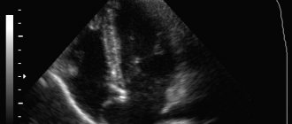
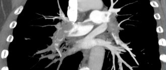
- echocardiography (ultrasound of the heart) – allows you to see the location, shape and size of the aneurysm;
- contrast ventriculography - a picture of the heart with the introduction of radiopaque substances;
- magnetic resonance imaging (MRI) – layer-by-layer images of the heart;
- coronary angiography – allows you to determine the nature, location and size of the aneurysm.
Sources
- S. Z. Amrakhov, A. S. Vischipanov. Surgical tactics and principles of performing operations in patients with coronary heart disease with post-infarction aneurysm of the left ventricle of posterobasal localization. Bulletin of the Scientific Center for Agricultural Sciences named after. A.N. Bakulev RAMS. 2013; 14 (1): 26-33.
- Koon K. Teo, MBBCh, PhD. Aneurysms of peripheral arteries. // MSD Disease Directory - 2021.
- Mark A. Farber, MD; Thaniyyah S. Ahmad, MD. Aneurysms of the thoracic aorta. // MSD Disease Directory - 2021.
- Brain aneurysms - Research Institute of Emergency Medicine named after. N.V. Sklifosovsky.
Treatment of cardiac aneurysm
The choice of treatment method is selected individually depending on the size of the protrusion, the rate of increase and the age of the patient.
In adults
Drug therapy includes taking medications aimed at treating heart rhythm disturbances and restoring blood circulation to the organ.
Adult patients are prescribed medications for:
- reducing blood viscosity and cholesterol levels;
- stabilization of blood pressure;
- preventing attacks of arrhythmia;
- reducing swelling.
Indications for surgical treatment are:
- large size of the aneurysm;
- increasing heart failure;
- thromboembolism;
- myocardial rupture;
- progressive mitral regurgitation (dysfunction of the mitral valve of the heart).
The main goal of surgery is removal of the aneurysm followed by reconstruction (restoration) of the heart.
In children
When diagnosing an aneurysm of the interatrial septum in a child, which is small in size and does not affect the quality of life, conservative treatment methods are used:
- taking medications that help improve the structure of connective tissue;
- taking medications to strengthen the heart muscle;
- antiarrhythmic drugs;
- physical therapy and massage.
Congenital left ventricular aneurysm is a direct indication for surgery. If the protrusion does not increase in size and does not pose a threat to life, surgery is performed after one year. From the moment of birth until the operation, the child is under constant supervision of a cardiologist. Surgical treatment of congenital aneurysm is carried out in different ways: complete removal (resection) of the protrusion and verticuloplasty - correction (restoration) of the structure of the heart.
In newborns
The choice of treatment tactics for cardiac aneurysm in newborns is determined by the severity of the pathology. If there is no threat to life, the question of surgery is postponed until one year. Small aneurysms do not require surgical treatment, however, children with such pathology are registered with a cardiologist and undergo regular examinations. Surgery to remove a cardiac aneurysm in newborns is performed only in emergency cases when there is a clear threat to the baby’s life. The surgical procedure and timing are determined individually by a medical council.
There are a number of contraindications for surgical treatment of cardiac aneurysm in children: severe heart and kidney failure, cancer, severe diabetes, intolerance to anesthesia.
Medicines
Photo: grud.guru
If a strange and sharp headache occurs, a person must immediately contact the nearest medical institution for qualified help. The disease is not treated with medication, but there is prevention and rehabilitation after surgery.
Surgical intervention is currently the only and most promising method of treating aneurysm. Treatment with special drugs is used only to stabilize the patient or in a situation where surgery is contraindicated or not possible at all.
Chemical drugs cannot eliminate an aneurysm; they only reduce the likelihood of vessel rupture by eliminating critical factors. Some medications are included in a complex of general therapy, which is aimed primarily at alleviating the symptoms of the initial pathology in patients. What vitamins and medications are taken for cerebral aneurysm?
Calcium channel blockers
The main representative of the nimodipine group. The chemical drug reliably blocks calcium channels in the muscle cells of the vascular walls. The vessels dilate. Blood circulation in the cerebral arteries is significantly improved. These drugs are simply irreplaceable in the prevention of dangerous arterial spasms.
Antacids
The principle of action is based on blocking H2 histamine receptors in the stomach. As a result, its acidity decreases and the secretion of gastric juice is significantly reduced. This group includes Ranitidine.
Anticonvulsants
Today, Fosphenytoin is the main representative of this group. The drugs cause reliable stabilization of membranes in nerve cells. Pathological nerve impulses noticeably slow down and do not spread.
Antiemetic drugs
Prochlorperazine is mainly used. The gag reflex is reduced by blocking postsynaptic dopamine receptors in the mesolimbic region of the brain.
Painkillers
Morphine is very effective for pain relief. The level of pain is reduced as a result of the effect on specific opioid receptors.
Antihypertensive drugs
Recently, three main drugs have been used: labetalol, captopril, hydralazine. Due to the effect on enzymes and receptors, the overall tone of the arteries is reduced and rupture is prevented.
How to live with a heart aneurysm
Cardiac aneurysm is a serious disease that requires a person to follow certain rules of life. However, if you follow your doctor’s recommendations and undergo regular monitoring with a cardiologist, the risk of blood clot formation and separation is significantly reduced.
For patients with cardiac aneurysm, it is recommended to:
- maintain a daily routine;
- eat a balanced diet;
- exclude strong physical activity;
- engage in physical therapy;
- undergo a heart examination at least twice a year;
- take maintenance medications as prescribed by your doctor.
Following simple rules and taking care of your health will help maintain quality and increase life expectancy.
Disease prognosis
The prognosis for cardiac aneurysm depends on many factors. First of all, it depends on the size and location of the protrusion, the state of the cardiovascular system, concomitant pathologies, and the person’s age. If the aneurysm is small in size and you see a doctor in a timely manner, the prognosis for the disease is favorable. An aggravating prognostic factor is the addition of heart failure. Lack of treatment for extensive post-infarction aneurysms also significantly worsens the prognosis. Uncomplicated flat cardiac aneurysms occur most easily; saccular protrusions are often complicated by intracardiac thrombosis.