What is cardiac radiography?
X-ray of the heart is a fairly old research method and is to a certain extent inferior to some modern diagnostic procedures. However, to this day, radiography is quite informative. It allows you to quickly and accurately find out the condition of the heart chambers and great vessels. You can also obtain data on the contractility of the organ. Heart disease can be detected without radiography; it is often prescribed as an adjunct. X-rays of the heart are possible in both public and private clinics. This diagnostic method is very common.
X-ray anatomy
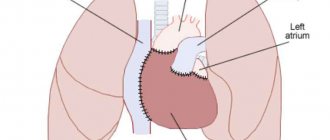
If you need to take a picture, it is worth learning the x-ray anatomy of large vessels and the heart. The X-ray anatomy of this organ consists of its shadow, the pulmonary artery, the aorta, and the vena cava. 2/3 of the listed structures are located to the left of the midline of the chest, only 1/3 protrudes to the right side. Chest X-ray anatomy includes the lungs, bronchi and trachea.
There are several types of location of the heart shadow, depending on its angle and the patient’s constitution. Namely:
- vertical – the angle is more than 45 degrees;
- oblique – the angle is 45 degrees;
- horizontal – angle less than 45 degrees.
As X-rays of the heart show, the horizontal position is usually characteristic of women, and the vertical position is usually characteristic of tall people.
Acute coronary syndrome in children.
G.E. Sukhareva, N.V. Lagunova, N.N. Rudenko, A.K. Kurkevich, I.G. Lebed, I.N. Imnadze, L.I. Ustenko, G.D. Levchenko, I.B. Zyukova, S.P. Bulatova.
- Crimean State Medical University named after. S.I. Georgievsky;
- Republican Children's Clinical Hospital;
- Republican Diagnostic Center, Simferopol
- Scientific and Practical Center for Pediatric Cardiology and Cardiac Surgery of the Ministry of Health of Ukraine, Kyiv.
Many pediatricians and cardiologists believe that acute coronary syndrome (ACS) in childhood is a casuistic case. If in adults the main cause of myocardial infarction (MI) is atherosclerotic lesion of the coronary arteries (CA), then the cause of the development of ACS in children is congenital or acquired VA pathology [1, 3, 6–8, 13, 14]. Considering the rare occurrence, the difficulty of diagnosis and the lack of alertness of practitioners towards the pathology of VA in children with the development of ACS, we want to present our own observations and draw the attention of practitioners to the problem under consideration.
Among the causes of ACS development in children, the most common are congenital anomalies and acquired pathology (coronaritis) of the VA.
The most common congenital pathology of VA is the Bland-White-Garland anomaly - an anomalous origin of the left coronary artery from the pulmonary artery (AOLVA from the PA), which accounts for 0.5% in the pediatric population [1, 3, 4, 8].
There are two main types of defect: infantile, or child, and adult [1, 6, 8].
The presence of an ischemic area supplied by an anomalous vessel (if there are no anastomoses between the left and right VAs) causes the development of extensive MI with rapidly progressing circulatory failure and death of the patient. However, collaterals are usually present. In the infantile type, such blood supply is clearly insufficient, and severe myocardial ischemia develops 6–8 weeks after birth.
The second type is an adult. It is characterized by well-developed intercoronary anastomoses and is detected in preschool or adulthood. In this type, the blood supply to the myocardium is determined by the degree of development of anastomoses. In typical cases, there is moderate left ventricular (LV) myocardial ischemia due to steal syndrome.
In most patients, symptoms of the disease appear in the first 3 months of life, less often in the second half of the year. The first signs are a violation of the general condition, lethargy, pale skin, increased sweating, vomiting, regurgitation, shortness of breath, tachycardia, fainting, cough. Half of the patients experience attacks of sudden anxiety with increased shortness of breath and pallor, often during or after feeding. At this time, the child’s facial expression is pained, grimaces appear, as if in severe pain, he screams piercingly, the pulse becomes threadlike. Sometimes sudden death occurs. Sometimes the first symptoms of this pathology may be fever, shortness of breath and diarrhea, possibly associated with the reflex nature of gastrointestinal disorders. Much less frequently, MI occurs without pain. Some children may develop cardiogenic shock (cold gray-pale skin covered with sticky sweat, oligoanuria, thready pulse, decreased pulse blood pressure (BP) less than 20–30 mm Hg, decreased systolic blood pressure). Many children are lagging behind in physical development. They develop a left-sided heart hump early. The apical impulse is diffuse, weakened, displaced to the 6th, 7th intercostal space. The borders of the heart are shifted to the left. The sounds are often muffled; the cause of the systolic murmur of mitral valve insufficiency (MV) may be chronic ischemia or infarction of the papillary muscles, dilatation of the LV cavity. Sometimes a three-part rhythm is heard. X-ray examination reveals cardiomegaly. It can reach such a degree that the X-ray shows darkening of the entire left half of the chest [1, 8, 13].
The ECG of the children we observed revealed a characteristic pattern of the defect described in the literature [1, 8]:
- pathological Q wave in leads I, AVL (max in AVL);
- failure of the R waves in leads V3-V4 (the morphology of the complex becomes rS,QS,Qr), indicating a previous MI;
- T wave inversion in leads I, AVL and left chest. Pathological deviation of the electrical axis of the heart (EOS) to the left due to blockade of the anterior left branch of the His bundle.
Under our supervision in the cardiorheumatology department of the Republican Children's Clinical Hospital (RCCH) in Simferopol there were two children with Bland-White-Garland syndrome. One of the first descriptions of MI in a child with this anomaly in Ukraine was made by us in 1999 [13]. In this message we would like to provide another personal observation of the development of ACS in a child with AOLVA from LA.
Child M., 12 months old, has been treated by a local pediatrician for acute obstructive bronchitis for the last three months. At the age of 3 months, cough, shortness of breath, and tachycardia appeared, for which the parents turned to a pediatric cardiorheumatologist in Yalta. They were recommended for an in-depth inpatient examination, which the parents refused. The condition worsened on March 27, 2003 – shortness of breath, cough, and tachycardia intensified. The child was hospitalized at the Yalta Children's Hospital, where a chest x-ray revealed cardiomegaly. With suspicion of congenital heart disease, the patient was transferred to the intensive care unit of the Russian Children's Clinical Hospital.
From the anamnesis of life and illness it is known that the child is from the 7th pregnancy, 2 term births. Pregnancy and childbirth proceeded without pathology. He was born with a weight of 3400 g, height – 51 cm, 8 points on the Apgar scale. He immediately screamed and was put to the chest in the maternity hospital. Vaccinated according to age. The neonatal period was uneventful. Allergy history is not burdened. Mother is 37 years old, healthy. Father is 50 years old, healthy. Heredity is not burdened.
Upon admission to the hospital, the child’s condition is serious due to heart failure. Meningeal symptoms are negative. The child is restless. The skin is pale with an earthy tint, cyanosis of the nasolabial triangle. Moaning breath. Swelling of the face, eyelids. Mixed shortness of breath with retraction of the pliable parts of the chest. Respiratory rate (RR) – 60 per minute. Above the lungs, percussion - clear pulmonary sound, auscultation - hard breathing, moist rales on both sides. The heart area is not changed. The apical impulse is palpated in the 4th intercostal space and is weakened. The boundaries of relative cardiac dullness are expanded in diameter: right - 1.5 cm outward from the right edge of the sternum; upper – upper edge of the 2nd rib; left - along the anterior axillary line. Heart sounds are weakened, tachycardia. Heart rate (HR) – 170 per 1 min with anxiety. Blood pressure – 90/45 mm Hg. Art. The abdomen is moderately distended, painless. Liver +4.5 cm from under the edge of the costal arch, the edge is elastic, painless. The spleen is not palpable. Diuresis is reduced. The stool is regular and mushy. General clinical examination upon admission was unremarkable; according to a general blood test over time - anemia of the 1st degree (Hb - 100 g/l).
Echocardiography: expansion of all cavities of the heart with pronounced dilatation of the left chambers of the heart. Moderate relative mitral and tricuspid insufficiency. Defects of septa are not located. Overall contractility is reduced (ejection fraction (EF) – 45%). General hypokinesia. The blood flow in the abdominal aorta is pulsating. Moderate pulmonary hypertension.
The chest x-ray shows cardiomegaly (cardiothoracic index (CTI) – 75%). Consultation with a neurologist: hypoxic encephalopathy, hyperexcitability syndrome.
He received therapy: antibacterial, inotropic (dobutamine), cardiometabolic (Neoton), diuretics (furosemide), painkillers (analgin, promedol), nitroglycerin (0.5 mcgChkg-1Chmin-1). During the therapy, the child’s condition stabilized, and he was sent to determine further management tactics at the Scientific Research Center for Pediatric Cardiology and Cardiac Surgery with a diagnosis of Bland-White-Garland anomaly. Endomyocardial fibroelastosis. Circulatory failure (CI) IIB. The child underwent probing of the heart cavities and coronary angiography, which confirmed the diagnosis of AOLVA from PA. A coronary artery bypass surgery was performed.
In the observations of L.A. Bockeria et al. [4], who presented the results of surgical treatment of 91 patients with AOLVA from pulmonary arterial valve, the range of surgical interventions for this pathology was diverse: direct implantation of an abnormally draining VA into the aorta was carried out in 53.8% of patients, in 10.9% a aortopulmonary tunnel (Takeuchi operation). The operation of simple ligation of an abnormally extending left VA was performed in 15.4% of cases, transpulmonary suturing of the mouth of the left VA - in 5.5% of cases, coronary artery bypass grafting - in 11%. Yu.M. Belozerov and co-authors believe that surgical treatment is not effective enough in children under 2 years of age due to an inadequate number of intercoronary anastomoses. If patients survive a critical period of life (1–2 years) with drug treatment, then their condition stabilizes. Children over 2 years of age tolerate ligation of the left VA well. The prognosis of the defect without surgery is in most cases unfavorable. Almost 2/3 of patients without treatment die in the 1st year of life, 12–15% survive to an older age, of which later half die suddenly after physical exertion or mental trauma [1, 8].
Acquired lesions of the VA include acute and chronic coronaritis. The true prevalence of coronaritis due to the complexity of diagnosis is unknown; in most cases, this diagnosis is made at autopsy. Coronaritis occurs in various infectious diseases, rheumatic and non-rheumatic carditis, infective endocarditis, systemic lupus erythematosus, systemic (primary) vasculitis (giant cell (temporal arteritis), Takayasu arteritis; polyarteritis nodosa (classic), Kawasaki disease; Wegener's granulomatosis, Churg-Strauss syndrome , microscopic polyangiitis, Henoch-Schönlein purpura, etc.) [15], heart tumors (secondary vasculitis).
The clinical picture of coronaritis is the same regardless of the etiological factor and is manifested by the development of ACS up to MI.
Takayasu's disease - nonspecific aortoarteritis (NAA), pulseless disease - belongs to the group of systemic vasculitis and is a chronic granulomatous arteritis with predominant damage to the aorta and its main branches [9, 11, 12, 15]. VA lesions occur in 10–25% of patients with NAA and are considered a potentially fatal complication. As a rule, coronary artery disease is combined with damage to other arteries, but isolated damage to the VA is also possible. Involvement of VA in the process is characterized by clinical signs of ACS.
Considering the rare occurrence and difficulty of diagnosing Takayasu's disease and Kawasaki disease, we want to demonstrate our own observations of the development of ACS in this pathology. We present our observation of the development of ACS in Takayasu disease in a 12-year-old girl.
Patient V., 12 years old, was admitted to the cardiorheumatology department of the Russian Children's Clinical Hospital in Simferopol with complaints of weakness, fatigue, dizziness, shortness of breath at rest, worsening with physical activity, palpitations, a feeling of interruptions in heart rhythm, severe cardialgia. From the medical history, it is known that she considers herself sick for 1.5 years, when, after suffering from the flu, the above complaints appeared, but she was not examined or treated in the hospital. The condition worsened in the last 3 months, the parents turned to a cardio-rheumatologist, after whose consultation the patient with suspected non-rheumatic carditis was hospitalized in the Central District Hospital and received therapy for myocarditis, but there was no effect. She was transferred to the cardiorheumatology department of the Russian Children's Clinical Hospital for further examination and treatment. Upon admission, the child's condition was serious. Lethargic, pale. There is pastiness of the face and upper body, a pronounced vascular network on the anterior surface of the chest, and visible pulsation of the vessels of the neck. Pulse 140 per minute, weak filling, asymmetrical. There is no pulse in the left radial artery. In the remaining vessels, pulsation is detected. The boundaries of relative cardiac dullness are expanded to the left: the left border is along the anterior axillary line. Auscultation: heart sounds are arrhythmic, single extrasystoles are recorded. A rough systolic murmur is heard with a maximum in the second intercostal space to the right of the sternum and at Botkin's point. A systolic murmur is heard in the vessels of the neck. The asymmetry of blood pressure in the extremities is determined: blood pressure in the left arm is 90/50 mm Hg. Art.; Blood pressure on the right arm is 110/60 mm Hg. Art. Pulsation in the femoral artery is preserved. Blood pressure in the legs – 130/80 mm Hg. Art. Above the lungs, percussion - clear pulmonary sound, auscultation - vesicular breathing, no wheezing. Shortness of breath, respiratory rate 28–30 per minute. The abdomen is soft, palpable, the liver is +2 cm from under the edge of the costal arch.
General clinical tests: grade I anemia, moderate leukocytosis with a band shift, persistently increased ESR (up to 60 mm/h), an increase in acute-phase indicators is noted. The ECG shows sinus tachycardia, single supraventricular extrasystole, ST-T changes in V1-V4. A chest x-ray showed a change in the configuration of the cardiac shadow, LV enlargement, hypervolemia, and expansion of the shadow of the vascular bundle.
Echocardiography reveals pronounced mitral regurgitation. Expansion of the ascending aortic arch up to 4.7 cm, in the thoracic region - up to 3.7 cm. The aortic valve is compacted, the diameter of the aortic valve ring is 2.2; sunrise. – 4.0. Pulmonary hypertension – 50 mm Hg. Art. EF – 56%. Computed tomography of the chest cavity - aneurysm of the ascending aorta.
Doppler examination of the vessels of the aortic arch revealed: sonographic changes are characteristic of nonspecific aortoarteritis (Takayasu's disease) with predominant damage to the common carotid arteries (CCA) on both sides, the brachiocephalic trunk, the first segment of the subclavian artery on the left, vertebral-subclavian steal syndrome on the right. Stenoses are most pronounced: in the subclavian artery on the left (stenosis of about 50% in diameter). At the mouth and throughout the CCA on the left, the diameter is reduced by 3 times. The intima-media of the vessel is unevenly thickened, differentiation into layers is impaired due to stenoses, hemodynamically significant asymmetry is observed in the CCA and subclavian arteries with a reduction in blood flow on the left. Compensatory dilatation of the vertebral artery on the left. The blood flow in the renal artery on the right is changed, the diameter of the artery is up to 2 mm (acquired hypoplasia). Conclusion: Takayasu's disease with predominant involvement of the CCA on both sides and the subclavian on the left.
Based on the above, a clinical diagnosis was substantiated: Takayasu's disease (nonspecific aortoarteritis), complicated by an aneurysm of the ascending aorta, development of mitral-aortic insufficiency. Acute coronary syndrome. NK IIA.
Due to deterioration of her condition, the patient was sent to the Research Institute of Agricultural Medicine named after. N.M. Amosov Academy of Medical Sciences of Ukraine, where she developed a papillary muscle infarction with rupture of the MV leaflet, which required MV replacement (MIKS-25). The MK specimen revealed myxomatous degeneration with superficial proliferation. The aortic valve was inspected and a band of the ascending aorta was applied. During pathological examination, the specimen obtained from the aortic wall showed changes characteristic of NAA (Takayasu's disease). Currently, the girl is under the supervision of a cardiorheumatologist, and her health is satisfactory. Receives glucocorticoids in a maintenance dose in combination with methotrexate, antiplatelet therapy.
Kawasaki disease (mucocutaneous lymph node syndrome) is a systemic vasculitis primarily affecting the VA. It is more common in early childhood [2, 5, 8, 10, 16, 17]. Heart damage can be of the type of myocarditis (in the acute stage), coronaritis with the development of multiple, sometimes giant aneurysms (at 6–8 weeks), stenosis and occlusion of the VA, which can lead to the development of ACS even in the long term from the onset of the disease, which illustrates our observation.
Child M., 3 years 7 months, was admitted to the cardiorheumatology department of the Russian Children's Clinical Hospital on May 29, 2006, with complaints of weakness and shortness of breath on exertion. From the anamnesis of life and illness, it is known that the child has been ill for a year, when in June 2005, episodes of fever to febrile levels, difficult to respond to antipyretic therapy and resistant to antibiotics, rashes on the body, regarded as an allergic reaction, and arthralgia were first noted. He was hospitalized in the Central District Hospital with a diagnosis of acute respiratory viral infection, severe course, hyperthermic syndrome. Atopic dermatitis. Acute pyelonephritis. Exudative pericarditis. According to the general blood test, an accelerated ESR was noted up to 64 mm/h, leukocytosis up to 14.4-1012/l with a band shift up to 17%; high serological activity - SRP +++, ASL-O - negative, according to a general urine test - leukocyturia. According to echocardiography, there are signs of pericarditis.
Antibacterial therapy and glucocorticosteroid therapy were carried out, against the background of which some positive dynamics were noted, and the child was discharged at the place of residence with a recommendation for re-examination in a month and consultation at the Russian Children's Clinical Hospital. However, the child’s parents did not contact doctors throughout the year.
In the department, a general blood test was performed: pronounced leukocytosis up to 30.2ґ1012/l, band shift up to 17%, CRP++++, ESR acceleration up to 61 mm/h, CEC 283 U, ASL-O - neg. twice, increased transaminase levels. A SIPS puncture was performed: according to the myelogram, there was no evidence of acute leukemia. A computed tomography scan was performed: no organic pathology was detected. X-ray examination of the chest organs: the heart is enlarged in diameter, CTI - 78%. When analyzing a series of electrocardiograms in dynamics, starting from June 2005, attention is drawn to the obvious underestimation by doctors of electrocardiographic changes and negative dynamics, expressed in an increase in the failure of the R wave in V1-V4, ST elevation above the isoelectric line with a negative T wave in the chest leads.
The ECG on August 1, 2005 recorded sinus rhythm with a heart rate of 94–120 per minute. EOS was not rejected. Changes are recorded in the form of an arcuate rise above the isoline of the RS-T segment in leads V2-V3 (sharply expressed elevation +9–15 mm); in I, AVL, V4-V5 – moderately pronounced elevation. The QRS complex is presented in leads I–qR; AVL–qr; V4-V5–Rs; V2-V3–rs.
On subsequent ECGs, starting from August 12, 2005 to June 2006, there was no dynamics of graphic changes in the form of QS V1-V5 with deep inverted T wave in V1-V6, moderately pronounced ST elevation; in leads I, AVL the qR complex is recorded. These changes are characteristic of post-transmural MI of the anterior wall of the left ventricle and the anteroseptal region. A “frozen” ECG waveform may indicate an aneurysm.
Negative dynamics of echocardiographic examination were also noted: LV dilatation increased, myocardial contractility progressively decreased (end-diastolic volume (EDV) - 83 ml, end-diastolic index (EDI) - 126 ml/m2, EF (Simpson) - 28%, contractility was sharply reduced, dyskinesia of the anterior apical zone with the formation of an aneurysm. The pathology of the left VA is visualized: an enlarged trunk with dilatation up to 7 mm at the bifurcation site.
The child was sent to the Research and Medical Center for Pediatric Cardiology and Cardiac Surgery of the Ministry of Health of Ukraine, where, after an examination, a diagnosis was made: Kawasaki disease. Multiple VA aneurysms. Post-infarction (2005) cardiosclerosis. Aneurysm of the anterior apical zone of the left ventricle.
The child underwent surgical intervention: the LV aneurysm (along the anterior, lateral and posterior wall) was opened with a 7 cm long incision, and a 6-1.5 cm aneurysm was resected. Endoventriculoplasty was performed. Mammary coronary bypass surgery was performed (a shunt was placed: LIMA - LAD). In the long-term postoperative period (one year after the operation), the child feels well and has no complaints. With echocardiography: the LV cavity remains dilated (EDV - 73 ml, ESV - 43 ml, EDI - 96 ml/m2, EF - 59%). Right VA – up to 6 mm. Akinesia of the apical (anterior, lateral) segments persists. Hypokinesia of the septal segments.
Thus, ACS in children occurs more often than is diagnosed. The vigilance of practitioners and timely diagnosis of this pathology will improve the prognosis of the disease, reduce the risk of death and reduce disability.
Literature
- Belozerov Yu.M. Myocardial infarction in children // Ros. Bulletin of Perinatology and Pediatrics. – 1996. – No. 3. – P. 36-40.
- Belozerov Yu.M. Kawasaki disease // Ros. Bulletin of Perinatology and Pediatrics. – 1995. – No. 3. – P. 41-47.
- Berishvili I.I., Vakhromeeva M.N., Katsitadze Z.D. Anomalous origin of the left coronary artery from the pulmonary artery // Pathology Archives. – 1998. – No. 2. – P. 35-39.
- Bockeria L.A., Shatalov K.V., Podzolkov V.P., Arnautova I.V. Surgical treatment of the syndrome of anomalous origin of the left coronary artery from the pulmonary artery // Materials of the second All-Russian conference “Topical issues of cardiology of early childhood”. – M., 2006. – P. 48.
- Bregel L.V., Subbotin V.M., Belozerov Yu.M., Tolstikova T.V. Features of myocarditis in Kawasaki disease // Abstracts of the All-Russian Congress “Pediatric Cardiology 2002”. – M.: Medpraktika, 2002. – P. 88.
- Volosovets A.P., Krivopustov S.P. Cerebral stroke and myocardial infarction in children: a modern view of the problem // Child’s Health. – 2006. – No. 2. – P. 63-69.
- Kubyshkin V.F., Filin P.I., Ushakov A.V. and others. Acute coronary syndrome. Guide for doctors and interns. – K.: SPD Kolyada O.P., 2004. – 40 p.
- Leontyeva I.V., Tsaregorodtseva L.V., Belozerov Yu.M. et al. Myocardial infarction in children: possible causes, modern approaches to diagnosis // Pediatrics. – 2001. – No. 1. – P. 32-37.
- Lectures on cardiology / Ed. L.A. Boqueria, E.Z. Golukhova. – M.: Publishing house NTsSSKh im. A.N. Bakuleva RAMS, 2001. – P. 167-171.
- Lyskina G.A. Clinical picture, treatment and prognosis of mucocutaneous lymphonodular syndrome (Kawasaki) // Ros. Bulletin of Perinatology and Pediatrics. – 2007. – No. 2. – P. 31-35.
- Marushko T.V., Todurov B.M. Features of the course and diagnosis of Takayasu’s disease in children // Modern Pediatrics. – 2003. – No. 1. – P. 89-92.
- Murzina O.Yu., Kovalev I.A., Varvarenko V.I. and others. A case of nonspecific aortoarteritis in a 3-year-old child // Materials of the fifth Russian Congress “Modern technologies in pediatrics and pediatric surgery.” – M.: Overley, 2006. – P. 176-177.
- Sukhareva G.E., Trofimishin V.V., Levchenko G.D. Myocardial infarction in children // Tauride Medical and Biological Bulletin. – 1999. – No. 1-2. – pp. 58-61.
- Sharykin A.S. Congenital heart defects. Guide for pediatricians, cardiologists, neonatologists. – M.: Teremok, 2005. – P. 289-294.
- Shuba N.M. Systemic vasculitis // Doctor. – 2002. – No. 1. – P.42-47.
- Benseler SM, McCrindle BW, Silverman ED et al. Infections and Kawasaki disease: implications for coronary artery outcome // Pediatrics. – 2005. – 116:6:760-766.
- Tanaka H., Narisawa T., Hirano J. et al. Coronary artery bypass grafting for coronary aneurysms due to Kawasaki disease // Ann. Thorac Cardiovasc Surg. – 2001. – Vol. 7. – P. 307-310.
Ukrkardio
What can an x-ray reveal?
An X-ray of the heart will provide a large amount of information regarding the position, size and condition of the organ and its structures. Based on these data, the doctor will be able to draw a conclusion about the presence of pathologies and identify the cause of the patient’s deterioration in well-being.
Heart sizes
An X-ray of the heart must be taken to find out whether the size of the organ is normal. They are very important in determining pathologies. The size is determined not by the width and length that are visible on an x-ray, but by the cardiothoracic index. To do this, you need to measure the width of the chest at the level of the fourth rib, and also find out the width of the heart in the same area. If the cell is twice as wide as the organ, the index will be 50%. If it is higher, then the heart muscle is enlarged. Myocardial hypertrophy may indicate ischemia or hypertension. Dilation of the chambers may be associated with cardiomyopathy or heart failure.
Condition of the great vessels
When thinking about what an X-ray of the heart shows, it is worth knowing that the condition of the great vessels will become known. The doctor will be able to find out if there are plaques, calcium deposits, or any lumps in the aorta. It will also be possible to identify a disease that is characterized by the accumulation of fluid in the pericardial sac. It is otherwise called “pericarditis”. If the fluid accumulates slowly, the organ becomes like a bag, but if it accumulates quickly, it becomes like a ball. If the condition is aggravated by calcium salt deposits, the doctor will diagnose constrictive pericarditis.
What symptoms should not be ignored
The following symptoms may indicate an enlarged heart in a child:
- increased heart rate;
- pallor or bluishness of the skin;
- rapid breathing;
- blueness of the nasolabial triangle;
- weak appetite.
Since the heart rate in children is always higher than in adults, it is impossible to say for sure whether your baby’s heart beats often or not. Only a doctor can determine this. But if the pulse is more than 160 beats per minute, then this is clearly not a good signal.
Breathing with cardiomegaly is not just rapid, but also shallow, uneven in inhalation and exhalation.
Pale skin occurs as a result of poor circulation due to poor functioning of the heart muscle and the organ itself.
Features of the procedure in children
Sometimes heart x-rays may be performed in children. This type of study is very rarely prescribed to children, since the procedure is contraindicated under 15 years of age. However, it may be required to identify heart defects, as well as when planning surgical interventions or to evaluate the effectiveness of the chosen treatment method. X-rays may also be needed to identify tumor processes.
If we are talking about a very young child, an X-ray of the heart will be taken using a special device. It is a stand on which the child is placed and his torso, head and limbs are secured with belts. This will ensure that the patient remains completely still while the doctor takes the image. The procedure will take no more than a couple of seconds, and discomfort is possible only from the belts with which the fixation is carried out. If we are talking about older children, they can undergo the procedure even without adults.
Causes
The causes of an enlarged heart in a child depend on the type of cardiomegaly. It can be primary or secondary. The secondary course of the disease occurs due to:
- severe toxic injuries;
- past infectious diseases that provoked various heart complications;
- respiratory failure of the lungs;
- acute viral infections.
If we are talking about the primary type of enlarged heart in a child, then the reasons for its occurrence are still not fully understood.
Preparation and carrying out diagnostics
You will only need to prepare if you need to undergo a cardiac X-ray with contrast. It involves taking tests to determine if you have an allergy to contrast, as well as visiting the office on an empty stomach, which will help avoid nausea.
To begin the procedure, you need to remove all jewelry or metal accessories that may affect the quality of the photo. The procedure is performed in 4 different projections:
- front;
- lateral left;
- oblique left (at an angle of 45 degrees);
- right.
Thanks to this, the description and conclusion of the heart x-ray will be as accurate as possible. The doctor will examine all planes of the organ and note the presence of pathological processes.
Contraindications
Such a study cannot be called completely safe. In this regard, there are a number of situations in which such diagnostics are not performed, but a more gentle method is selected. Often, an ultrasound
or MRI, as well as other studies.
Pregnant women
Before finding out what a heart x-ray is and what this procedure shows, a woman should be absolutely sure that there is no pregnancy. This situation is an absolute contraindication to the procedure, since there is a risk that X-ray radiation will have an adverse effect on the body of both the expectant mother and the baby. It is also not recommended for breastfeeding mothers to undergo diagnostics. However, in some situations, a doctor may prescribe such a procedure if its benefits are significantly higher than the likely risks. Therefore, it is best to trust the doctor.


