Dopplerography is a modern diagnostic method that makes it possible to visualize large vessels in real time. The method is based on the “Doppler effect” - the ability of an ultrasound beam to pass through motionless tissue. In most cases, Doppler is used to diagnose cardiovascular pathologies. Using this study, you can evaluate the speed and turbulence of blood flow, and also makes it possible to determine the condition of the walls of blood vessels. Based on the results of Doppler sonography, the doctor will be able to identify abnormalities, make a diagnosis and decide on treatment tactics.
General characteristics of the examination
A procedure such as Doppler ultrasound can be performed both to identify violations when indicated, and for preventive purposes. The whole point is the absence of radiation exposure to the patient’s body, as well as the fact that it will be possible to visualize areas that are inaccessible to other research methods.
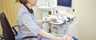
Ultrasound scanning of the heart vessels is done using a special sensor that generates ultrasound signals and receives the echo reflected by the tissues. Special software is responsible for converting such signals into images.
Preparation
- Duplex and triplex ultrasound is performed on an empty stomach, the last time to eat is better than 3 hours before the examination.
- On the day of ultrasound examination, smoking, drinking alcohol and caffeine-containing drinks is prohibited.
- The day before and on the day of the examination, you should avoid physical activity and not be nervous.
- If the patient’s blood pressure is elevated to 160 mmHg or higher or the pulse is increased to 90 beats per minute or more, they must be normalized before the procedure with the help of medications prescribed by the cardiologist who refers the patient to ultrasound.
You can check the condition of the heart vessels and get advice from an experienced cardiologist at a medical clinic.
Doppler ultrasound is performed by our doctors in the clinic and at home. We accept patients by appointment, which can be arranged by calling the phone number listed on the website. Using the same number, you can invite a specialist to your place and find out the cost of the service. Come to us at the first alarming symptoms and for a preventive examination, and be healthy!
Diseases that can be detected using Doppler ultrasound
Doppler sonography plays an important role in identifying diseases of the cardiovascular system. These diseases are today considered one of the most dangerous and widespread in the world; they can lead not only to human disability, but also to death. Cardiac Dopplerography is prescribed to narrow the range of diagnostic alternatives; it allows one to identify abnormalities and make a decision on treatment, significantly increasing the chance of a positive outcome.
Congenital heart defect in a child
Heart defects can manifest themselves in both acute and latent forms. The cause of their occurrence is heredity, influenced by the age of the spouses, as well as external influences in the form of viral or infectious diseases. In this case, ultrasound examination of the heart vessels is extremely necessary, since the pathology can lead to tragic consequences.
Myocardial infarction (or pre-infarction condition)
Narrowing of the lumen of a blood vessel is a pre-infarction condition. Complete closure of the vessel and lack of oxygen supply to the heart muscle is a myocardial infarction. The cause may be a plaque or blood clot in the arteries. Such conditions are usually preceded by angina pectoris in an advanced form. Ultrasound of the heart and echocardiography
allow you to examine the organ and identify any abnormalities, make a conclusion about the condition of the blood vessels and determine whether conservative or surgical treatment is needed.
Pressure disorders - hypotension and hypertension
First of all, it is necessary to examine the heart in case of pressure disorders. It is not enough to select drugs that normalize blood pressure and intensity of blood flow; it is important to identify the cause of this phenomenon, since it may lie in heart pathology.
Acquired vices
Ultrasound scanning of the heart is often prescribed for suspected valve defects, otherwise known as “acquired” defects. They lie in the fact that due to infections, inflammatory processes, autoimmune reactions, dilatation and overload of the chambers of the heart, certain changes occur. They can be morphological or functional or combined.
Indications and contraindications
Doppler ultrasound is not performed for serious damage to the skin in the epigastric and chest areas (trauma, exacerbation of eczema or psoriasis, herpes zoster).
Otherwise, the method is absolutely safe and painless, but at the same time very informative. Medical cardiologists recommend it for determining the fetal heartbeat during pregnancy, identifying heart defects in the youngest patients, elderly people, anyone who has had a heart attack, and those who have been suffering from chronic diseases for a long time.
Research is also required if:
- changes in electrocardiogram readings;
- shortness of breath, difficulty breathing, worsening when lying down, dry cough;
- chest pain that appears during physical activity;
- tachycardia, bradycardia;
- swelling.
For children, Doppler ultrasound is prescribed for increased fatigue, a sharp decrease in physical and mental activity, increasing weakness, pallor and bruises under the eyes, and decreased appetite. All these are signs of circulatory disorders that can be caused by cardiovascular pathologies.
How is research done?
The procedure does not involve surgical intervention, that is, it is non-invasive. There is also no radiation exposure, so the study can be carried out both for certain indications and for preventive purposes.
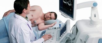
After the patient comes to see the doctor, he is asked to remove clothes that interfere with the examination and lie down on a special couch. After this, a special gel is applied to the sternum, which will help the sensor glide over the skin. Using a sensor, the doctor examines the organ and takes pictures, and also notes the condition and size of all structures. The data obtained will be compared with the norm of Doppler ultrasound of the heart, after which the doctor will draw up a conclusion and give it to the patient.
Ultrasound of the heart: when is it prescribed and what will it show?
To clarify a cardiac diagnosis, one of the most frequently prescribed studies is an ECG. However, often the doctor refers the patient to an ultrasound of the heart. What does this diagnosis show and is it worth doing it at all?
Today we are having an unusual conversation, as our consultants will be two doctors.
Olga Nikolaevna Vitkova, and her colleague, an ultrasound diagnostics doctor in pediatric practice, Inna Vyacheslavovna Popova, tell us about heart ultrasound and its capabilities.
— Olga Nikolaevna, Inna Vyacheslavovna, please tell us what ultrasound of the heart is and when is it prescribed?
This is a research method based on the use of ultrasound with certain characteristics for diagnostic purposes. Ultrasound is beyond the audibility of the human ear. During the examination, the ultrasound machine generates a stream of ultrasonic waves. The doctor directs them using a sensor to one or another part of the body. Ultrasound, having reached anatomical formations, is reflected from them and returns. These signals are recorded by an ultrasound machine, which, based on their characteristics, “constructs” an image of the organ being examined on the monitor screen.
Ultrasound examination allows us to judge the size of the heart, its layers, valves, and the characteristics of blood circulation.
It is prescribed for pain in the heart, high blood pressure, edema, shortness of breath, wheezing in the lungs; for dizziness, loss of consciousness, general weakness, prolonged increase in body temperature of unknown origin, patients with diabetes mellitus, and those with a history of myocardial infarction; people whose relatives suffered from certain diseases.
It's off scale! We are looking for the causes of high blood pressure. Read here
In children, ultrasound is performed on the direction of a cardiologist or pediatrician - most often if a heart defect is suspected. More than half of young patients come for examination precisely for this reason.
Ultrasound is prescribed for children with serious infections, in particular bacterial ones. Occasionally - if a heart tumor is suspected, it is extremely rare, but still a common diagnosis in children.
Preventive examinations are also performed for babies up to one year old.
— Are cardiac ultrasound and echocardiography the same study or are there differences between them? And if so, what is the difference?
Ultrasound of the heart and echocardiography are the same thing.
— Can ultrasound of the heart replace an ECG? What is the difference between these studies?
No, these studies are not interchangeable. It is correct to say that they complement each other. These methods characterize different aspects of the heart’s work. An ECG indicates the function of the conduction system of the heart and its possible disorders. With its help, you can indirectly judge the size of the cavities of the heart.
However, the ECG is not informative regarding the structural features of the organ - for example, the valve apparatus, the presence of defects, blood clots or inflammatory changes in the heart. In these cases, compared with ECG, ultrasound is the method of choice. It will allow you to directly evaluate the chambers of the heart, its layers, valves and their defects, aneurysms after a heart attack, the amount of blood ejected by the chambers of the heart, etc.
Specific example. The patient's ultrasound revealed tachycardia (rapid heartbeat), but we cannot say anything about its origin or type. This is the power of an ECG.
What causes tachycardia? Elena Viktorovna Novikova, a cardiologist at the Expert Kursk Clinic, says:
— What diseases can ultrasound of the heart diagnose?
Taking into account what has already been listed above, ultrasound also reveals signs of congestive heart failure (for example, fluid in the pericardial cavity), neoplasms and aneurysms of the heart, dilation of the heart chambers and blood clots in them, dissecting aortic aneurysm.
— In order to identify heart pathology, is it enough for a patient to undergo only an ultrasound scan or should the diagnosis be comprehensive?
No, ultrasound alone is definitely not enough. The examination must be comprehensive and must include a survey and examination of a doctor, an ECG and/or - as indicated - Holter monitoring (24-hour ECG), laboratory tests (depending on the intended diagnosis), ultrasound of the vessels of the head and neck, gastrointestinal tract, as indicated - coronary angiography (x-ray examination of the heart vessels using a contrast agent).
Why are x-rays dangerous? Yulia Aleksandrovna Rutskaya, head of the radiation diagnostics department at Clinic Expert Kursk, says:
— Is there an alternative to cardiac ultrasound?
It depends on what you mean by alternative. If the method is safe, non-invasive and accessible, then there is no alternative. If it refers to diagnostic capabilities, to a certain extent, transesophageal echocardiography, as well as spiral computed tomography with a contrast agent, can be an alternative. These methods can be used in cases where traditional echocardiography is difficult for some reason or does not answer some of the doctor’s questions.
— How safe is heart ultrasound and how often can this study be done?
Completely safe. It can be performed as often as required.
— How is heart ultrasound performed for adults and children?
There are no fundamental differences between adults and children. It is performed with the patient lying down, more often on the left side. Using a sensor, the doctor examines all heart formations and evaluates a number of functional parameters.
The peculiarity of children is that it is often difficult to convince them to lie on their left side, and therefore studies are often performed on their back. Another point is the location of the sensor. In childhood, research is often carried out through the so-called subcostal access (when the sensor is located in the epigastrium). In adults, this approach is rarely used due to their anatomical features.
— Is preparation necessary for an ultrasound examination of the heart?
There is no special preparation for adults.
It is important to calm children before the test. Due to their inherent mobility and restlessness, they may not lie as they should. Therefore, in this regard, parents play a big role, who must explain to the child what is happening and calm him down. Before the planned examination, you can play ultrasound with your child at home.
It is recommended that before the study the child is well-rested and well-fed (but preferably not immediately after eating, i.e. not with a full stomach). Or, if conditions permit, a small child can first be euthanized and only then examined.
Occasionally, examinations are performed under anesthesia - usually in a hospital setting and for certain indications.
— Can ultrasound examination of the heart be recommended as a preventive method?
Yes. In addition, in children under 1 year of age, echocardiography for preventive purposes is mandatory, not to mention examination after infectious diseases.
— In order to undergo a heart ultrasound at the Expert Clinic, do you need a doctor’s referral?
You can undergo the examination with or without a referral. The advantage of a referral is that the doctor can provide some additional information about the patient (for example, about a heart murmur detected during an examination). Algori makes more sense.
You can sign up for a cardiac ultrasound here
Please note: diagnostics are not available in all cities
For reference:
Vitkova Olga Nikolaevna
Graduate of the Faculty of Medicine of the Smolensk State Medical Academy in 2012.
From 2012 to 2014 she completed clinical residency in the specialty “Therapy”.
In 2014, she underwent professional retraining in the specialty “Cardiology”, in 2015 - in the specialty “Ultrasound Diagnostics”.
Since 2021, he has been working at Clinic Expert Smolensk LLC as an Ultrasound Doctor.
Popova Inna Vyacheslavovna
Graduate of the pediatric faculty of the Smolensk State Medical Academy in 2007.
In 2008 she completed an internship in the specialty “Pediatrics”
In 2009, she underwent professional retraining in the specialty “Functional Diagnostics”, and in 2010 - in the specialty “Ultrasound Diagnostics”.
Since 2021, he has been working at Clinic Expert Smolensk LLC as an Ultrasound Doctor.