X-ray (x-ray) is a non-invasive medical test that helps doctors diagnose and treat various diseases. The images are obtained by exposing a specific part of the body to a small dose of ionizing radiation. This radiation is able to penetrate or be retained in the tissues of organs, thereby making it possible to obtain photographs or screen images. X-rays are the oldest and most commonly used form of medical imaging.
A chest x-ray is a very common test. This method is used to diagnose heart diseases separately or together with other chest organs. It is an X-ray examination that is among the first procedures that a patient will undergo if the doctor suspects a heart or lung disease. It is also used to test the effectiveness of treatment.
What is cardiac radiography?
X-ray of the heart is a fairly old research method and is to a certain extent inferior to some modern diagnostic procedures. However, to this day, radiography is quite informative. It allows you to quickly and accurately find out the condition of the heart chambers and great vessels. You can also obtain data on the contractility of the organ. Heart disease can be detected without radiography; it is often prescribed as an adjunct. X-rays of the heart are possible in both public and private clinics. This diagnostic method is very common.
X-ray anatomy
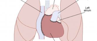
If you need to take a picture, it is worth learning the x-ray anatomy of large vessels and the heart. The X-ray anatomy of this organ consists of its shadow, the pulmonary artery, the aorta, and the vena cava. 2/3 of the listed structures are located to the left of the midline of the chest, only 1/3 protrudes to the right side. Chest X-ray anatomy includes the lungs, bronchi and trachea.
There are several types of location of the heart shadow, depending on its angle and the patient’s constitution. Namely:
- vertical – the angle is more than 45 degrees;
- oblique – the angle is 45 degrees;
- horizontal – angle less than 45 degrees.
As X-rays of the heart show, the horizontal position is usually characteristic of women, and the vertical position is usually characteristic of tall people.
What can an x-ray reveal?
An X-ray of the heart will provide a large amount of information regarding the position, size and condition of the organ and its structures. Based on these data, the doctor will be able to draw a conclusion about the presence of pathologies and identify the cause of the patient’s deterioration in well-being.
Heart sizes
An X-ray of the heart must be taken to find out whether the size of the organ is normal. They are very important in determining pathologies. The size is determined not by the width and length that are visible on an x-ray, but by the cardiothoracic index. To do this, you need to measure the width of the chest at the level of the fourth rib, and also find out the width of the heart in the same area. If the cell is twice as wide as the organ, the index will be 50%. If it is higher, then the heart muscle is enlarged. Myocardial hypertrophy may indicate ischemia or hypertension. Dilation of the chambers may be associated with cardiomyopathy or heart failure.
Condition of the great vessels
When thinking about what an X-ray of the heart shows, it is worth knowing that the condition of the great vessels will become known. The doctor will be able to find out if there are plaques, calcium deposits, or any lumps in the aorta. It will also be possible to identify a disease that is characterized by the accumulation of fluid in the pericardial sac. It is otherwise called “pericarditis”. If the fluid accumulates slowly, the organ becomes like a bag, but if it accumulates quickly, it becomes like a ball. If the condition is aggravated by calcium salt deposits, the doctor will diagnose constrictive pericarditis.
Indications for cardiac x-ray
A heart x-ray is performed for the following indications:
- planned treatment of patients suffering from coronary heart disease;
- suspicion of existing heart defects;
- asymptomatic, unstable angina;
- tracking conditions in the pulmonary circulation;
- asymptomatic hidden diseases of the cardiovascular system;
- to identify calcifications of the aortic valves, mitral valves, pericardial sac, myocardium after acute cardiac ischemia.
Cardiologists and surgeons refer their patients who complain of:
- having symptoms of angina pectoris - this is compressive pain in the chest, a feeling of lack of air, interruptions in heart function, rhythm disturbances;
- shortness of breath at rest and during physical activity, increased fatigue and severe weakness;
- tachycardia, arrhythmia - disturbances in the rhythm of heart contractions;
- swelling of the lower extremities and pronounced pallor of the skin;
- enlarged liver;
- persistent cough and fever.
X-ray of the heart makes it possible to suspect the following pathologies in a patient:
- exudative pericarditis - infectious inflammatory lesions of the pericardium (pericardium);
- abnormal increases in heart size called myocardial hypertrophy, which is found in coronary heart disease and hypertension;
- aneurysms of the walls after myocardial infarction, manifest themselves in the form of protrusions;
- dilated cardiomyopathy - damage to the heart muscles in the form of stretching of the heart chambers;
- a pronounced anatomical defect in the membranes of the heart muscle, most often these are various valve defects;
- calcium deposits on the walls of the coronary artery, the presence of seals, detection of atherosclerotic and thrombotic plaques;
- cloudiness, expansion of the root of the lungs, which means the possible presence of cardiac pathology.
Features of the procedure in children
Sometimes heart x-rays may be performed in children. This type of study is very rarely prescribed to children, since the procedure is contraindicated under 15 years of age. However, it may be required to identify heart defects, as well as when planning surgical interventions or to evaluate the effectiveness of the chosen treatment method. X-rays may also be needed to identify tumor processes.
If we are talking about a very young child, an X-ray of the heart will be taken using a special device. It is a stand on which the child is placed and his torso, head and limbs are secured with belts. This will ensure that the patient remains completely still while the doctor takes the image. The procedure will take no more than a couple of seconds, and discomfort is possible only from the belts with which the fixation is carried out. If we are talking about older children, they can undergo the procedure even without adults.
Who can benefit from CT coronary angiography?
To examine in detail the condition of the heart vessels, assess blood circulation in them and determine the location of blockages, CT coronary angiography is used.
Vladislav Vasilievich Babenko, a radiologist at the advisory and diagnostic department of the Expert Institute, told us in detail about this study.
– Vladislav Vasilievich, what is CT coronary angiography?
– This is a non-invasive and effective screening method for examining the heart vessels using computed tomography (CT). A more medically correct name for coronary angiography is coronary angiography or coronary angiography.
Read more about the computed tomography method in our article
During CT coronary angiography, the coronary calcium index and the risk of acute coronary events are determined. This technique is carried out using an iodine-containing contrast agent and is a radiation research method.
The procedure is recommended for patients with a low and intermediate risk of pre-test probability of stable coronary artery disease.
– When is coronary angiography prescribed?
– The patient is referred for coronary angiography in the following cases:
- for differential diagnosis in the presence of unclear pain in the chest (with a questionable or negative stress test);
— for uninformative stress tests or as an alternative to stress tests if there are contraindications to them;
— to decide whether further in-depth examination and treatment is necessary (for example, performing invasive coronary angiography, stenting or coronary artery bypass grafting);
— for preventive examinations (screening) of people without symptoms of coronary heart disease in order to identify patients with atherosclerosis of the coronary arteries;
- to control the progression of atherosclerosis of the heart vessels;
- to determine the effectiveness of the treatment.
Read materials on the topic:
Coronary heart disease: diagnosis and treatment
Serious question: what happens to the heart during an angina attack?
How to prevent myocardial infarction?
– What does CT coronary angiography show?
– This method allows you to visualize the vessels of the heart, assess the condition of atherosclerotic plaques, determine the variant anatomy (structural features) of the vascular bed and exclude developmental anomalies.
“We must remember that atherosclerosis, which develops against the background of increased cholesterol concentrations, is a chronic pathology that requires long-term, many-year cooperation between the doctor and the patient.” Quote from the material “How to distinguish good and bad cholesterol?”
– Do you need special preparation for the examination?
- Yes. The patient must bring the results of a blood creatinine test from a week ago and ECG results with interpretation. In order to achieve stabilization of the heart rate (HR) before the examination, special medications are prescribed. Heart rate should be in the range of 50-65 beats per minute. If the heart rate is higher than the permissible value, CT coronary angiography is not advisable, since the study will be uninformative.
Before the procedure, all patients undergo a consultation with a therapist or cardiologist.
– How is CT coronary angiography performed?
– At the first stage (before the administration of a contrast agent), the coronary calcium index is calculated and the patient is familiarized with the upcoming examination, and instructions are given. Next, preliminary medicinal preparation of the patient and stabilization of heart rate are performed. The third stage is the introduction of an intravenous catheter into the right ulnar vein. The fourth is the research itself. The patient is placed in the machine and a contrast scan is performed. And the last, fifth stage is monitoring the patient to exclude side effects from the administration of a contrast agent and providing assistance if they occur.
– What are the contraindications to CT coronary angiography?
– Relative contraindications include:
- allergic reactions to the administration of iodine-containing contrast agent;
- the patient’s inability to lie still for a long time;
— the patient’s inability to follow the operator’s and doctor’s instructions;
- uncorrectable heart rate (HR);
- decreased kidney function (glomerular filtration rate);
- thyrotoxicosis (increased levels of thyroid hormones) and thyrotoxic crisis;
— the patient’s body weight is more than 120 kg;
— pregnancy regardless of term (including potential pregnancy);
- children's age, a clear justification (vital indications) for conducting the study is necessary.
“There are two concepts: hyperthyroidism and thyrotoxicosis. Sometimes they are combined, in particular in foreign sources. But there is a difference." Quote from the material “If the thyroid gland is “raging”: what is hyperthyroidism?”
It should be added that if this study is vitally necessary, it is performed in the presence of contraindications in a hospital setting.
– Tell us about other types of coronary angiography. What is the difference between them?
– In addition to CT coronary angiography, invasive coronary angiography (sometimes called traditional coronary angiography) is used. This procedure is performed in a hospital setting. Surgical access is through the femoral artery, in contrast to CT coronary angiography, which uses intravenous access. The contrast agent is administered in a larger volume than during CT coronary angiography.
– Which is better: CT coronary angiography or traditional coronary angiography?
Each method has its own advantages and disadvantages. Traditional invasive coronary angiography has higher resolution and a higher percentage of specificity in assessing stenosis (narrowing) of blood vessels, but is more likely to cause adverse reactions. With invasive coronary angiography, it is possible to measure the fraction of reserve blood flow (this is the “gold standard” in the assessment of cardiac stenosis), and also immediately install a stent if necessary. But this procedure is usually carried out within the framework of compulsory medical insurance only for health reasons. If the patient does not have vital indications, then this study will be very expensive: in comparison, CT coronary angiography is approximately 10 times cheaper. The advantage of CT coronary angiography is its speed and the ability to perform the study on an outpatient basis.
– Vladislav Vasilievich, is CT coronary angiography different from CT of the heart, and if so, how?
– It differs in its methodology, as well as in the fact that cardiac CT has other research purposes and patients with different pathologies.
The patient may be referred for a cardiac CT scan, for example, to evaluate the left atrium and left atrial appendage. For what? In order to confirm or exclude the presence of a blood clot, assess the condition of the mouths of the pulmonary veins, their diameters, the presence or absence of stenoses. That is, these are patients who have arrhythmia and need to find out the cause. Depending on what was revealed on a CT scan of the left atrium, further surgical or conservative treatment will be performed.
The editors recommend:
What will a heart ultrasound tell you?
Holter (24-hour) ECG monitoring – complete instructions for the patient
When is daily blood pressure monitoring prescribed?
Interested in other articles on CT scanning? Read more materials in our section
For reference:
Babenko Vladislav Vasilievich
In 2015 he graduated from the Faculty of Medicine of Voronezh State Medical University. N.N. Burdenko.
2015 – 2021 – residency in the specialty “Radiology”.
Currently, he is a radiologist at the advisory and diagnostic department of the Expert Institute.
Preparation and carrying out diagnostics
You will only need to prepare if you need to undergo a cardiac X-ray with contrast. It involves taking tests to determine if you have an allergy to contrast, as well as visiting the office on an empty stomach, which will help avoid nausea.
To begin the procedure, you need to remove all jewelry or metal accessories that may affect the quality of the photo. The procedure is performed in 4 different projections:
- front;
- lateral left;
- oblique left (at an angle of 45 degrees);
- right.
Thanks to this, the description and conclusion of the heart x-ray will be as accurate as possible. The doctor will examine all planes of the organ and note the presence of pathological processes.
Disadvantages and advantages of the method
Advantages of the method:
- widespread availability of X-ray rooms, accessibility of the method for patients;
- low cost of the procedure;
- high detail of images thanks to high-resolution film, which allows you to identify and assess the condition of surrounding tissues, neighboring organs and blood vessels;
- ease of comparison of two images taken at different times in order to assess the dynamics of the development of pathology or taken routinely during preventive examinations.
The disadvantages of the method include:
- insufficient information content;
- difficulties in diagnosing organs that are in constant motion, which includes the heart due to its regular contractions (the image is blurred);
- impossibility of frequent implementation due to radiation exposure;
- the need to develop and process the film, which takes a lot of time.
An annual examination, as a rule, reveals pathologies before the first symptoms appear and the patient independently seeks medical attention, becoming the basis for prescribing more detailed studies and subsequent diagnosis.
Contraindications
Such a study cannot be called completely safe. In this regard, there are a number of situations in which such diagnostics are not performed, but a more gentle method is selected. Often, an ultrasound
or MRI, as well as other studies.
Pregnant women
Before finding out what a heart x-ray is and what this procedure shows, a woman should be absolutely sure that there is no pregnancy. This situation is an absolute contraindication to the procedure, since there is a risk that X-ray radiation will have an adverse effect on the body of both the expectant mother and the baby. It is also not recommended for breastfeeding mothers to undergo diagnostics. However, in some situations, a doctor may prescribe such a procedure if its benefits are significantly higher than the likely risks. Therefore, it is best to trust the doctor.
Decoding the results
The initial description of the image (protocol) is performed by the radiologist after receiving the result (film or image on the screen). The doctor (surgeon, cardiologist, therapist) additionally examines the image and protocol, makes a transcript and gives a conclusion. The specialist evaluates the shape of the heart, the presence of seals, the shape of the edge of the shadow and other parameters. The procedure can only be performed by a doctor with extensive experience in interpreting images due to the difficulty of determining deviations from shadows superimposed on each other.


