Chronic hypertension is accompanied by constant changes in blood pressure, which negatively affects the condition of the vascular bed. Against the background of prolonged stenosis, microcirculation disorder occurs in all organs and tissues of the body. Among the complications of arterial hypertension, hypertensive angiopathy occupies the first position in terms of prevalence. The pathological condition is characterized by expansion of the vascular lumen of the fundus veins, disproportionality of capillary tortuosity, as well as pinpoint hemorrhages. When the disease is diagnosed, immediate treatment is required as the patient may remain blind. What are the causes of the development of pathology and what treatment methods are there to eliminate it?
General characteristics of the condition
Hypertensive retinal angiopathy is not an independent disease, but is a symptom of various pathologies associated with impaired functional state of the fundus vessels and changes in their structure. Signs of damage to the vascular system are changes in their elasticity, tone and temporary reversible spasms.
The effect of chronic hypertension on the vessels of the fundus
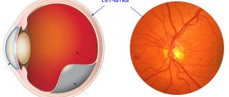
Angiopathy leads to irreversible complications when the necrotic area of the retina, which was supplied with nutrients through a deformed vessel, undergoes thinning, rupture or detachment. These negative phenomena are combined into the group of retinopathy.
The functional state of the retina depends on the quality of hemodynamic processes, and when degenerative changes form, visual impairment occurs that is not restored. If the area in the macula area is affected, central visual function is impaired. Complete retinal detachment subsequently results in blindness.
No ads 1
Symptoms
Symptoms usually do not appear until late in the disease and include1:
- Decreased vision: You see blurry or just worse than before
- Significant redness of the eyes: as a rule, it is associated with rupture of the blood vessels of the conjunctiva (atherosclerotically damaged), which could not withstand the increased pressure
- Double vision: You see double
- Headaches: occur due to impaired blood flow
Seek medical help immediately if you have hypertension and suddenly have decreased vision!
Causes
Hypertensive angiopathy occurs as a result of exposure to high blood pressure levels over a long period of time. Prolonged spasm of the arteries contributes to the development of structural disorders in the vascular wall, which leads to circulatory disorders in the body, disruption of the functioning of the main organs, which can aggravate the course of the disease and increase the number of complications. Pathology can occur at any age, but most often it is observed in people after 30 years of age.
Pathological changes in the vessels of the fundus characterize the severity of damage to the vascular system of the whole body
The most common causes of the disease are:
- hypertension of various etiologies;
- diabetes;
- atherosclerosis;
- intoxication of the body;
- congenital defects in the formation of the vascular system;
- old age.
Provoking factors:
- genetic predisposition;
- excess body weight;
- alcohol abuse, smoking;
- physical inactivity;
- abnormal consumption of table salt;
- deficiency of microelements in the body (potassium, magnesium).
The disease can be complicated by various injuries, osteochondrosis, metabolic disorders, and pathologies of the hematopoietic system.
Complications
People with hypertensive retinopathy are at risk of developing retinal complications. These include the following2,3:
- Ischemic optic neuropathy occurs when high blood pressure interferes with normal blood flow in the eyes, causing damage to the optic nerve. As a result, the brain cannot form an image normally.
- Retinal artery occlusion occurs when the arteries that carry blood to the retina become blocked by blood clots. When this happens, the retina does not have enough oxygen or blood. This leads to loss of vision.
- Malignant hypertension is a rare disease that causes a sudden increase in blood pressure, affecting vision and causing sudden loss of vision. This is a potentially life-threatening condition.
Patients with GAD are also at increased risk of stroke or heart attack. One study found that people with GAD are more likely to have a stroke than people without the condition.
Stages
According to statistics, at the first stage of hypertension, 26% of patients have a normal condition of the fundus of the eye, at the second stage, degenerative changes are recorded in 4%, and at the third stage, all hypertensive patients are susceptible to disorders.
There are several main stages of pathology development, each of which is characterized by some changes in the fundus:
- Functional disorders. They manifest themselves as uneven expansion of venules and narrowing of arterioles. The vessels acquire a more tortuous shape, as a result of which microcirculation is disrupted. There are no symptoms; defects can be identified during a special diagnostic examination of the fundus.
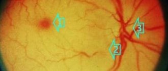
- Organic changes. They are characterized by pronounced changes in the structure of blood vessels; they become denser, subsequently being replaced by connective tissue. Thickening of the lumen causes a lack of blood circulation in the retina, which is why the manifestations of the disease become intense. On examination, retinal edema is revealed, and in some cases, pinpoint hemorrhages are formed. Arteries and veins take on a branched shape.
- Angioretinopathy. Hemodynamic disorder becomes critical, which is manifested by the formation of exudate in the fundus of the eye, soft or hard in consistency. The formation is filled with a liquid consisting of plasma, proteins, blood cells, minerals and pathogenic microflora, which provokes the inflammatory process. There is a significant decrease in visual function, and there is the possibility of complete loss of vision.
No ads 2
Course of the disease
Retinal angiopathy of the hypertensive type has some features in its development:
- This type of pathology is a consequence of prolonged exposure to high blood pressure. This condition leads to dilation of the veins of the fundus, minor hemorrhages in the area of the eyeball, and a neuroregulatory disorder of mechanisms.
- In the absence of adequate treatment, the disease progresses, contributing to an increase in the number of pathological changes. Over time, the retina becomes cloudy, which can be corrected with timely anti-hypertensive therapy.
- The disease can be diagnosed at the initial stage during a thorough examination of the fundus, when the patient has no signs of developing pathology.
Angiopathy against the background of arterial hypertension can be complicated by the addition of pathological vascular changes in the excretory system, myocardium, and nervous regulation.
Treatment
Effective treatment includes reducing high blood pressure with medications and lifestyle changes.
To keep your blood pressure under control, you need4:
- Exercise regularly
- Avoid coffee and alcohol
- Reduce the amount of salt in your diet
- Lose weight if you are overweight
Your doctor may also prescribe blood pressure medications: diuretics, beta blockers, or ACE inhibitors.
Bibliography:
- Kiseleva T. N., Ezhov M. V. et al. Retinal circulation disturbances in arterial hypertension. Pharmateka. 2014; 20: 14-18.
- Moshetova L., Vorobyova I. et al. Hypertensive angioretinopathy: clinical and morphological manifestations, diagnosis. Doctor. 2018; 3: 82-84.
- Zadionchenko V. S., Adasheva T. V. et al. The eye is a mirror of cardiovascular pathology. Evolution of ideas about hypertensive retinopathy. Rational pharmacotherapy in cardiology. 2010; 6 (6): 853-858.
- Arterial hypertension in adults. Clinical recommendations. Approval year 2021.
THE INFORMATION PROVIDED ON THE SITE DOES NOT CONSTITUTE AND DOES NOT REPLACE PROFESSIONAL ADVICE
Manifestations of the disease depending on the stage
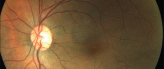
As a rule, the initial stage of the disease does not cause discomfort in patients. Complaints appear at a later stage, when the patient experiences a decrease in visual acuity and blurred vision. Upon examination, the ophthalmologist reveals a narrowing of the lumen of the retinal arterioles, their excessive tortuosity, and corkscrew-shaped tortuosity of the venules. In severe cases, vascular obstruction, the formation of hemorrhages, and the appearance of extravasation from blood clots are recorded.
Corkscrew tortuosity of venules near the macula
Characteristic signs of the disease:
- Decreased visual acuity (appearance of “cloudiness” before the eyes).
- Addition of myopia (inability to focus vision on distant objects).
- The appearance of bright flashes, “lightning” before the eyes (disorder of the blood supply to light receptors).
- Narrowing of visual fields (decreased peripheral vision).
- Headache (provoked by hypoxia).
- A feeling of pulsation occurs in the eyeballs (increased blood circulation through the narrowed arterial lumen).
- Visual dysfunction (as the disease progresses, blindness is possible).
- Bleeding from the nose (the area located on the surface of the mucous membrane of the nose and eye may bleed).
Minor hemorrhage in the eyeball
Since hypertension affects the vascular system throughout the body, hypertensive angiopathy of both eyes is observed. In medical documentation, this condition is recorded as a lesion of the retina OU, that is, of both eyes. The disease is aggravated by obstruction of the central retinal artery, the lumen of which is subsequently blocked. Malnutrition of the optic nerve leads to its atrophy.
No ads 3
Diagnosis of hypertensive retinal angiopathy
As a rule, the first signs of hypertensive damage to the vessels of the retina are noted during ophthalmoscopy. An ophthalmologist can determine the following negative changes:
- narrowing of arterioles in combination with dilated venules;
- thickening of the walls of blood vessels (angiosclerosis);
- retinopathy (due to hemorrhages, the retina becomes saturated with blood fractions);
- damage to the optic nerve head, that is, neuroretinopathy.
To confirm the diagnosis, the following examination techniques can be used:
- Doppler ultrasound;
- Ultrasound scanning of the vitreous body;
- fluorescein angiography.
These diagnostic methods allow ophthalmologists to thoroughly study the condition of the blood vessels of the eye.
Diagnostic approaches
At the first manifestations of pathology, you must contact an ophthalmologist, who will prescribe a series of special studies to confirm the diagnosis.
If you suspect the development of the disease, the patient should contact a qualified specialist
Special diagnostic methods:
- Ophthalmochromoscopy (the technique includes studying the functional state of the fundus arteries using red and non-red light).
- Fluorescent angiorrhaphy (the method allows you to establish a clear picture of the condition of the capillaries).
- Ultrasound of blood vessels (allows us to establish circulatory processes).
- Doppler scanning (reveals dystrophic changes in arterioles and venules).
- Contrast radiography (assessment of the patency of the venous lumen).
Important
All treatment methods are important in the fight against a detected disease. Only a variety of medications that change blood pressure and reduce the likelihood of blood clots will help you cope with hypertensive macroangiopathy, the signs of which we listed above.
Only if the whole body is treated can the disease be overcome. An important condition is an integrated approach. If high blood pressure is observed along with an altered blood chemical composition, then the entire treatment plan is adjusted and complex treatment regimens are prescribed.
To identify the disease at an early stage of its development, you need to undergo a complete examination of the body at least twice a year by all specialists. Early treatment gives a good chance of a complete recovery for the patient.


