Angioma is a benign vascular formation. As a rule, the neoplasm does not cause discomfort to its owner. Unless it causes deformity if it is located on the face, neck, or other open parts of the body. But sometimes small red spots are dangerous. Pathology can degenerate and spread. Therefore, vascular anomalies always require observation, and in some cases, treatment.
Let's figure out what angiomas are, why they appear, and how to remove vascular pathology. Frequently asked questions from patients are answered by doctors at the Lasersvit Center for the Diagnosis of Moles and Vascular Pathologies. We conduct consultations in Kharkov, offering children and adults ultra-precise diagnosis, treatment and prevention of skin and cutaneous-vascular abnormalities.
If you have an angioma on your skin, we suggest you make an appointment with a dermatologist. We will not only conduct a thorough diagnosis of the skin, but also tell you which skin tumors pose a hidden threat. A comprehensive study allows you to accurately determine the need for treatment of skin abnormalities.
Make an appointment with a dermatologist
Important : Angioma is often compared to a red mole. However, the vascular formation is not a mole. Red moles are formed by melanocyte cells, and skin angioma is a bundle of dilated lymphatic or blood vessels. In the first case it is a lymphangioma, and in the second it is a hemangioma.
Vascular angiomas appear in various parts of the body. They can be seen on the face, torso, and scalp. Medical statistics show that 80% of people over 50 have these formations. Sometimes the tumors disappear on their own. This happens with infantile hemangiomas. But often their number only increases with age. There is even such a thing as multiple senile angiomas. They appear due to the fragility of capillaries and their loss of elasticity. The vessels dilate and are unable to regain their previous diameter.
Types of angiomas
According to the etiology and pathogenesis, formations caused by the dilation of small vessels are divided into hemangiomas, formed from a cluster of capillaries, and lymphangiomas, consisting of small lymphatic vessels. Formations of the lymphatic system are less common. Their peculiarity is that the skin with this pathology does not change color. Flesh-colored nodules simply appear on the skin. Angiomas on the body are another matter. This disease always causes red spots to appear. And according to the type of structure, such formations come in several types.
Simple angiomas
Vascular formations of this type are, as a rule, congenital pathologies. They can be smooth or protruding above the surface of the skin. The color of the new growths can be scarlet, burgundy, sometimes with a bluish tint. Vascular angioma grows on any part of the body, but mainly on the upper part of the body. The formation of dilated capillaries reaches a size of 10 cm in diameter. The anomaly is not dangerous, but its presence on the skin can lead to emotional imbalance. After all, the stain greatly spoils the appearance and becomes the reason for the close attention of others and their ridicule. Capillary hemangioma can especially ruin life when it is located on the face, neck, or arms.
Cavernous angiomas
A benign formation of this type looks like a pulsating purple spot. The structure of the pathology is characteristic. Blood enters the cavernous chambers through narrow arteries and is drained from the formation through wide venous channels. The spot is soft on palpation, and after squeezing it quickly restores its shape and appearance.
Branched angioma
The pathology often affects the limbs, less often appears on the scalp. The structure of the formation looks like intertwined dilated vessels. The skin over such a pathology is often affected by simple angioma. This is why capillary hemangioma must be deeply investigated. After all, a branched angioma may be hidden under it.
Intraosseous angiomas
These formations develop on the bones and are detected when they spread to nearby tissues. Bleeding becomes a symptom of the pathology. Accurate diagnosis is possible after obtaining radiographic images. The photo clearly shows the boundaries of the pathology.
Of all the listed types of angiomas, it is simple capillary formations that affect the skin.
Classification
In world medical practice, there are several varieties of this tumor:
- cavernous hemangioma of the liver, which is several free cavities connected into a single one;
- capillary hemangioma of the liver forms a large number of small cavities, each of which houses a blood or venous vessel.
Along with the most common ones, venous, cluster-shaped hemangioma, and hemangioendothelioma can also occur.
Angiomas: causes of appearance
Until now, scientists cannot name the exact reasons for the appearance of vascular formations. Doctors agree that vascular pathology begins during embryonic development. Infectious diseases suffered by the mother can affect this. Toxicosis, anemia of a woman during pregnancy, and serious hormonal fluctuations also contribute to the appearance of abnormalities. Acquired angiomas occur due to sunburn, liver disease, skin injuries, and increased fragility of blood vessels.
Angiomas in children
Angioma in children is usually diagnosed at birth or during the first weeks of life. Girls get sick twice as often as boys. Angioma in a child goes through several stages. In the first few months, the formation actively grows, the size of the spot increases. At the next stage, growth stops. By the age of 9, in 90% of cases, self-healing occurs - spontaneous involution of the pathology. The stain disappears, leaving no traces in its place.
Unfortunately, in some cases, benign formation is accompanied by the following complications:
- ulceration and bleeding of the skin as a result of trauma, for example, due to rubbing with a diaper when the formation is located on the leg or buttock;
- formation of scars at the site of healing ulcers after ulceration of the skin at the site of formation;
- dysfunction of internal organs due to the germination of the formation or compression of them by its boundaries;
- When the formation is located on the forehead, ophthalmological diseases develop.
Important: Very often, angioma in a child is located on the face, neck, or chest. This leads to a pronounced cosmetic defect.
It is recommended to observe uncomplicated disease in children. To do this, during the appointment, photographs of the formation are taken using a dermatoscope. At subsequent appointments, images can be compared to analyze the behavior of the formation.
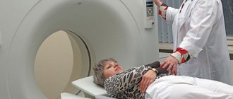
How to diagnose
Hemangioma of the 7th cervical vertebra and other cases of the disease are often determined by chance during examination. X-rays and MRIs are used to examine the body. Sometimes contrast enhancement is used. It helps to “highlight” the layers of capillaries or arteries in the tissue of such a neoplasm. The patient is sent for examination if he exhibits a group of symptoms:
- The occurrence of local pain in the thoracic or cervical spine. Most often they are aching in nature and are especially intensified after physical activity and a night's rest.
- Spinal cord irritation. This symptom of a cervical hemangioma indicates that the tumor is actively developing and is already beginning to exert strong pressure on the spinal cord.
- Pathological fractures. Such symptoms are very rare. Fractures are observed against the background of developing osteoporosis or when the tumor begins to put strong pressure on the bones. The need for treatment is also indicated by the occurrence of swelling, loss of sensitivity and paralysis of the limbs.
Angiomas: treatment
Angioma in adults and children is easily diagnosed. The doctor performs a dermatoscopy to examine the formation. A digital dermatoscope repeatedly magnifies the skin, allowing you to determine the depth, nature, and boundaries of the pathology. In some cases, angiography, blood flow analysis, and histology may be prescribed.
Important: Many patients are interested in why angiomas are removed if the formation is benign and non-aggressive. The main reasons have already been mentioned above. A large angioma on the face disfigures the appearance and there is a risk of complications. In this case, it is enough to observe congenital small spots, since there is a high probability of their disappearance without a trace.
When an angioma on the body or face is perceived as a serious aesthetic defect, if the risk of complications is high, in case of bleeding the formation must be removed. Today, several methods of treating vascular formations are practiced: radio wave therapy, cold treatment (cryotherapy), electrocoagulation, surgery and laser treatment. Each method has its own advantages. But there are also disadvantages. For example, removing part of the skin with a scalpel leaves scars and does not allow you to work with pathology accurately. Cold treatment can negatively affect adjacent tissues. And electrocoagulation is not suitable for neoplasms of significant depth and large area.
The most effective treatment for pathology of any complexity is laser removal of angioma. The laser beam removes abnormally dilated capillaries without affecting adjacent tissue. The doctor controls the intensity and depth of the impact, so he works with high precision. It is important that the treatment is bloodless, painless, and safe. The beam also cauterizes blood vessels, preventing infection. There are no scars left on the skin.
Important: Many people try to get rid of senile moles and red spots on their legs, arms, and face with dubious means at home. Aggressive agents and various ointments, including those containing hormones, are used. Doctors strongly do not recommend doing this, since you most likely will not be able to get rid of the pathology. But serious harm to health can be caused. Moreover, it is not difficult to treat skin lesions professionally today.
In each case, the dermatologist practices an individual approach to the patient. This allows you to get optimal results.
Important: If the formation does not bother you and is located in an inconspicuous place, you can replace treatment with a wait-and-see approach. Because there is a high chance that it will disappear.
Advantages of treating angiomas at the Lazersvit clinic
Our specialists have extensive experience in treating skin pathologies and vascular formations of the skin. Highly qualified dermatologists, the use of international protocols, and the use of the latest laser equipment in treatment ensure high cosmetic and therapeutic results. In the vast majority of cases, we completely remove vascular anomalies without consequences or complications. In the case of large and deep pathology, several sessions may be required at intervals of 2-3 weeks.
Important: Laser removal of angiomas does not require complex preparation and does not require a long rehabilitation period. The procedure is performed on an outpatient basis. In one session you can completely get rid of a small formation. Immediately after the intervention, the patient returns to his usual lifestyle.
You can learn more about the work of the Mole Diagnostic Center and directly about the treatment of angiomas during a face-to-face consultation with a doctor. You can make an appointment by phone.
The main types of the disease and its symptoms
Depending on the morphological characteristics, the following types of hemangiomas are distinguished:
- simple (capillary). It has clear boundaries, red or purple-blue color. It turns pale when pressed, and then restores the shade;
- cavernous (cavernous). Lumpy, nodular formation located subcutaneously. It is characterized by a soft elastic consistency, which is explained by the structure - inside there are cavities filled with blood;
- combined. It has a cutaneous and subcutaneous part. Clinical manifestations are characteristic of the capillary and cavernous type;
- mixed. A tumor with a complex structure, containing elements of vascular, nervous, connective and lymphoid tissues. Color, consistency and appearance vary depending on the fabrics included.
Tumors are single and multiple in nature, and can be small or large. In 95% of cases in pediatrics, simple forms are diagnosed. Symptoms of hemangioma appear only externally; rarely the patient complains of itching and discomfort in the affected area.
Treatment and examination of a child with hemangioma provides:
- pediatrician;
- surgeon;
- dermatologist.
Additionally, consultation with an ophthalmologist, otolaryngologist, urologist, dentist and pediatric gynecologist is indicated. In order to determine the depth of spread and evaluate the structure, an ultrasound scan of the affected area is performed. The complex evaluates the speed of blood flow in the tumor itself and the vessels supplying it.
The prognosis for patients is favorable. The tumor may regress on its own. A soft tissue tumor that does not have indications for surgical removal does not need to be eliminated. The decision on the treatment scheme or dynamic control method is always made by the doctor.
Are you experiencing symptoms of hemangioma?
Only a doctor can accurately diagnose the disease. Don't delay your consultation - call
Prevention of angioma
It is impossible to prevent the occurrence of congenital vascular pathologies, so effective prevention does not exist. There are several ways to reduce the risk of abnormalities such as red spots. Women are advised to carefully plan their pregnancy, and while carrying a child, follow all the recommendations of the obstetrician-gynecologist.
As for senile vascular anomalies, their occurrence can be prevented by avoiding prolonged exposure to the sun and cold, and by building a healthy diet rich in minerals and vitamins. It is also recommended that older people lead a healthy lifestyle and do not ignore physical activity agreed upon with their family doctor.