Historical reference
In the past, when scientists did not yet have informative instruments at hand that could study physiological processes in a living organism, the greatest scientists were forced to search for anatomical features in corpses. Naturally, the heart of a deceased person does not contract, so some nuances had to be figured out on their own, and sometimes simply fantasized. Thus, back in the second century AD, Claudius Galen, studying from the works of Hippocrates himself, assumed that the arteries contained air instead of blood in their lumen. Over the next centuries, many attempts were made to combine and link together the existing anatomical data from the standpoint of physiology. All scientists knew and understood how the circulatory system works, but how does it work?
Miguel Servetus and William Harvey made a tremendous contribution to the systematization of data on the work of the heart in the 16th century. Harvey, the scientist who first described the systemic and pulmonary circulation , in 1616 determined the presence of two circles, but he could not explain in his works how the arterial and venous beds were connected. And only later, in the 17th century, Marcello Malpighi, one of the first to use a microscope in his practice, discovered and described the presence of tiny capillaries, invisible to the naked eye, which serve as a connecting link in the blood circulation.
Phylogeny, or the evolution of blood circulation
Due to the fact that, as animals of the vertebrate class evolved, they became more and more progressive in anatomical and physiological terms, they required a complex structure of the cardiovascular system. Thus, for faster movement of the liquid internal environment in the body of a vertebrate animal, the need for a closed blood circulation system arose. Compared to other classes of the animal kingdom (for example, arthropods or worms), the rudiments of a closed vascular system appear in chordates. And if the lancelet, for example, does not have a heart, but there is an abdominal and dorsal aorta, then in fish, amphibians (amphibians), reptiles (reptiles) a two- and three-chambered heart appears, respectively, and in birds and mammals a four-chambered heart appears, the peculiarity of which is is the focus in it of two circles of blood circulation that do not mix with each other.
Thus, the presence of two separated circulatory circles in birds, mammals and humans, in particular, is nothing more than the evolution of the circulatory system, necessary for better adaptation to environmental conditions.
Circulatory systems of vertebrates (complex)
Fish
The heart of fish has 4 cavities connected in series: sinus venosus, atrium, ventricle and conus arteriosus/bulb.
- The venous sinus (sinus venosus) is a simple extension of a vein that receives blood.
- In sharks, ganoids and lungfishes, the conus arteriosus contains muscle tissue, several valves and is capable of contraction.
- In bony fishes, the conus arteriosus is reduced (has no muscle tissue and valves), therefore it is called the “arterial bulb”.
The blood in the heart of fish is venous, from the bulb/cone it flows to the gills, there it becomes arterial, flows to the organs of the body, becomes venous, returns to the venous sinus.
Lungfish
In lungfishes, a “pulmonary circulation” appears: from the last (fourth) branchial artery, blood flows through the pulmonary artery (PA) into the respiratory sac, where it is additionally enriched with oxygen and returns through the pulmonary vein (PV) to the heart, to the left
part of the atrium. Venous blood from the body flows, as it should, into the venous sinus. To limit the mixing of arterial blood from the “pulmonary circle” with venous blood from the body, there is an incomplete septum in the atrium and partially in the ventricle.
Thus, arterial blood in the ventricle appears before
venous, therefore it enters the anterior branchial arteries, from which a direct road leads to the head. The smart fish brain receives blood that has passed through the gas exchange organs three times in a row! Bathing in oxygen, the rogue.
Amphibians
The circulatory system of tadpoles is similar to that of bony fish.
In an adult amphibian, the atrium is divided by a septum into left and right, resulting in a total of 5 chambers:
- venous sinus (sinus venosus), in which, like in lungfishes, blood flows from the body
- the left atrium (left atrium), into which, like in lungfishes, blood flows from the lung
- right atrium
- ventricle
- arterial cone (conus arteriosus).
1) The left atrium of amphibians receives arterial blood from the lungs, and the right atrium receives venous blood from organs and arterial blood from the skin, so in the right atrium of frogs the blood is mixed.
2) As can be seen in the figure, the mouth of the conus arteriosus is shifted towards the right atrium, so blood from the right atrium enters there first, and from the left - last.
3) Inside the conus arteriosus there is a spiral valve that distributes three portions of blood:
- the first portion of blood (from the right atrium, the most venous of all) goes to the pulmonary cutaneous artery (pulmocutaneous artery), to be oxygenated
- the second portion of blood (a mixture of mixed blood from the right atrium and arterial blood from the left atrium) goes to the body organs through the systemic artery
- the third portion of blood (from the left atrium, the most arterial of all) goes to the carotid artery (carotid artery) to the brain.
4) In lower amphibians (tailed and legless) amphibians
- the septum between the atria is incomplete, so mixing of arterial and mixed blood occurs more strongly;
- the skin is supplied with blood not from the cutaneous pulmonary arteries (where the most venous blood is possible), but from the dorsal aorta (where the blood is average) - this is not very beneficial.
5) When a frog sits under water, venous blood flows from the lungs into the left atrium, which, in theory, should go to the head. There is an optimistic version that the heart begins to work in a different mode (the ratio of the pulsation phases of the ventricle and the arterial cone changes), complete mixing of the blood occurs, due to which not completely venous blood from the lungs enters the head, but mixed blood consisting of venous blood of the left atrium and mixed blood of the right. There is another (pessimistic) version, according to which the brain of an underwater frog receives the most venous blood and becomes dull.
Reptiles
In reptiles, the pulmonary artery (“to the lung”) and two aortic arches emerge from a ventricle partially divided by a septum. The division of blood between these three vessels occurs in the same way as in lungfish and frogs:
- The most arterial blood (from the lungs) enters the right aortic arch. To make it easier for children to learn, the right arch of the aorta begins from the very left part of the ventricle, and it is called the “right arch” because, having gone around the heart to the right
, it is included in the spinal artery (you can see how it looks in the next and subsequent figures). The carotid arteries depart from the right arch - the most arterial blood enters the head; - mixed blood enters the left aortic arch, which goes around the heart on the left and connects with the right aortic arch - the spinal artery is obtained, carrying blood to the organs;
- The most venous blood (from the body organs) enters the pulmonary arteries.
Crocodiles
Crocodiles have a four-chambered heart, but they still mix blood through a special foramen of Panizza between the left and right aortic arches.
It is believed, however, that mixing does not normally occur: due to the fact that there is higher pressure in the left ventricle, blood from there flows not only into the right aortic arch (Right aorta), but also - through the foramen of Panicia - into the left aortic arch (Left aorta), thus the crocodile’s organs receive almost entirely arterial blood.
When a crocodile dives, the blood flow through its lungs decreases, the pressure in the right ventricle increases, and the flow of blood through the foramen of panicia stops: the left aortic arch of an underwater crocodile flows blood from the right ventricle. I don’t know what the point is in this: all the blood in the circulatory system at this moment is venous, why should it be redistributed where? In any case, blood enters the head of the underwater crocodile from the right aortic arch - when the lungs are not working, it is completely venous. (Something tells me that the pessimistic version is also true for underwater frogs.)
Birds and mammals
The circulatory systems of animals and birds in school textbooks are presented very close to the truth (all other vertebrates, as we have seen, are not so lucky with this). The only little thing that you are not supposed to talk about in school is that in mammals (B) only the left aortic arch is preserved, and in birds (B) only the right one is preserved (under the letter A is the circulatory system of reptiles, in which both arches are developed) - There is nothing else interesting in the circulatory system of either chickens or people. Except for the fruits...
Fruit
Arterial blood received by the fetus from the mother comes from the placenta through the umbilical vein. Part of this blood enters the portal system of the liver, part bypasses the liver, both of these portions ultimately flow into the inferior vena cava (interior vena cava), where they mix with venous blood flowing from the fetal organs. Entering the right atrium (RA), this blood is once again diluted with venous blood from the superior vena cava (superior vena cava), thus resulting in hopelessly mixed blood in the right atrium. At the same time, some venous blood from the non-functioning lungs enters the left atrium of the fetus - just like a crocodile sitting under water. What shall we do, colleagues?
The good old incomplete septum, which the authors of school textbooks on zoology laugh at so loudly, comes to the rescue - in the human fetus, right in the septum between the left and right atria, there is an oval hole (Foramen ovale), through which mixed blood from the right atrium enters the left atrium. In addition, there is a ductus arteriosus (Dictus arteriosus), through which mixed blood from the right ventricle enters the aortic arch. Thus, mixed blood flows through the fetal aorta to all its organs. And to the brain too! And you and I pestered frogs and crocodiles!! And themselves.
Tests
1. Cartilaginous fish lack:
a) swim bladder; b) spiral valve; c) conus arteriosus; d) chord.
2. The circulatory system in mammals contains:
a) two aortic arches, which then merge into the dorsal aorta; b) only the right aortic arch c) only the left aortic arch d) only the abdominal aorta, and there are no aortic arches.
3. The circulatory system of birds contains:
A) two aortic arches, which then merge into the dorsal aorta; B) only the right aortic arch; B) only the left aortic arch; D) only the abdominal aorta, and there are no aortic arches.
4. The arterial cone is present in
A) cyclostomes; B) cartilaginous fish; B) cartilaginous fish; D) bony ganoid fish; D) bony fish.
5. Classes of vertebrates in which blood moves directly from the respiratory organs to the tissues of the body, without first passing through the heart (select all correct options):
A) Bony fish; B) adult amphibians; B) Reptiles; D) Birds; D) Mammals.
6. The heart of a turtle in its structure:
A) three-chamber with an incomplete septum in the ventricle; B) three-chamber; B) four-chamber; D) four-chamber with a hole in the septum between the ventricles.
7. Number of blood circulation in frogs:
A) one in tadpoles, two in adult frogs; B) one in adult frogs, tadpoles have no blood circulation; C) two in tadpoles, three in adult frogs; D) two in tadpoles and adult frogs.
8. In order for a carbon dioxide molecule that has passed into the blood from the tissues of your left foot to be released into the environment through the nose, it must pass through all of the following structures of your body except:
A) right atrium; B) pulmonary vein; B) alveoli of the lungs; D) pulmonary artery.
9. There are two circles of blood circulation (choose all the correct options):
A) cartilaginous fish; B) ray-finned fish; B) lungfishes; D) amphibians; D) reptiles.
10. A four-chambered heart has:
A) lizards; B) turtles; B) crocodiles; D) birds; D) mammals.
11. Here is a schematic drawing of a mammalian heart. Oxygenated blood enters the heart through the following vessels:
A) 1; B) 2; AT 3; D) 10.
12. The figure shows arterial arches:
A) lungfish; B) tailless amphibian; B) tailed amphibian; D) reptile.
You can also read
Circulatory systems of vertebrates (school level)
Evolution of the circulatory system (even more complex than here)
© D.V. Pozdnyakov, 2009-2020
Anatomical features of the blood circulation
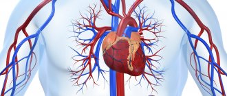
The circulatory system is a set of blood vessels, which is a closed system for the supply of oxygen and nutrients to the internal organs through gas exchange and nutrient exchange, as well as for the removal of carbon dioxide and other metabolic products from cells. The human body is characterized by two circles - the systemic, or large circle, and the pulmonary, also called the small circle.
Video: blood circulation circles, mini-lecture and animation
Vessels
Vessels differ in structure and function:
- arteries;
- capillaries;
- veins
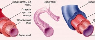
Arteries are vessels with the most elastic and thick walls. They carry blood away from the heart and withstand the highest pressure during heart contraction.
The largest vessel in diameter is the aorta, which extends in an arc from the left ventricle. The other major artery is the pulmonary artery, which arises from the right ventricle.
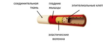
The thinnest, most numerous vessels that penetrate the entire body and all organs are called capillaries .
The capillary wall consists of a single layer of cells. The speed of blood flow in the capillaries is minimal. Capillaries supply all organs and tissues with oxygen and nutrients.
Veins are vessels that collect venous blood, saturated with carbon dioxide, from throughout the body. Blood pressure in veins is lower than in arteries and capillaries.
From the back of the body, blood collects in the caudal and iliac veins, which drain into the posterior vena cava.
From the head, blood collects in the jugular veins. Merging with the subclavian, they form the anterior vena cava. Both vena cava flow into the right atrium.
Systemic circulation
The main function of the large circle is to ensure gas exchange in all internal organs except the lungs. It begins in the cavity of the left ventricle; represented by the aorta and its branches, the arterial bed of the liver, kidneys, brain, skeletal muscles and other organs. Further, this circle continues with the capillary network and venous bed of the listed organs; and through the entry of the vena cava into the cavity of the right atrium it ends in the latter.
So, as already said, the beginning of the great circle is the cavity of the left ventricle. Arterial blood flow, which contains more oxygen than carbon dioxide, is sent here. This flow enters the left ventricle directly from the circulatory system of the lungs, that is, from the small circle. The arterial flow from the left ventricle is pushed through the aortic valve into the largest great vessel - the aorta. The aorta can be figuratively compared to a kind of tree that has many branches, because arteries extend from it to the internal organs (to the liver, kidneys, gastrointestinal tract, to the brain - through the system of carotid arteries, to skeletal muscles, to the subcutaneous fat fiber, etc.) Organ arteries, which also have numerous branches and bear names corresponding to their anatomy, carry oxygen to each organ.
In the tissues of internal organs, arterial vessels are divided into vessels of smaller and smaller diameter, and as a result, a capillary network is formed. Capillaries are the smallest vessels, practically without a middle muscular layer, and are represented by an inner membrane - intima, lined with endothelial cells. The gaps between these cells at the microscopic level are so large compared to other vessels that they allow proteins, gases and even formed elements to easily penetrate into the intercellular fluid of the surrounding tissues. Thus, intense gas exchange and exchange of other substances occurs between the capillary with arterial blood and the liquid intercellular medium in a particular organ. Oxygen penetrates from the capillary, and carbon dioxide, as a product of cell metabolism, enters the capillary. The cellular stage of respiration occurs.
After more oxygen has passed into the tissues and all carbon dioxide has been removed from the tissues, the blood becomes venous. All gas exchange occurs with each new influx of blood, and during the period of time while it moves along the capillary towards the venule - a vessel that collects venous blood. That is, with each cardiac cycle, in one or another part of the body, oxygen enters the tissues and carbon dioxide is removed from them.
These venules unite into larger veins, and a venous bed is formed. Veins, similar to arteries, are named according to the organ in which they are located (renal, cerebral, etc.). From large venous trunks, tributaries of the superior and inferior vena cava are formed, and the latter then flow into the right atrium.
Features of blood flow in the organs of the systemic circle
Some of the internal organs have their own characteristics. So, for example, in the liver there is not only a hepatic vein, which “carries” the venous flow away from it, but also a portal vein, which, on the contrary, brings blood to the liver tissue, where blood purification is performed, and only then the blood collects in the tributaries of the hepatic vein to enter to a big circle. The portal vein brings blood from the stomach and intestines, so everything that a person eats or drinks must undergo a kind of “purification” in the liver.
In addition to the liver, certain nuances exist in other organs, for example, in the tissues of the pituitary gland and kidneys. Thus, in the pituitary gland the presence of a so-called “wonderful” capillary network is noted, because the arteries that bring blood to the pituitary gland from the hypothalamus are divided into capillaries, which then collect into venules. The venules, after the blood with the molecules of releasing hormones are collected, are again divided into capillaries, and then veins are formed that carry the blood from the pituitary gland. In the kidneys, the arterial network is divided twice into capillaries, which is associated with the processes of excretion and reabsorption in the kidney cells - in the nephrons.
Pulmonary circulation
Its function is to carry out gas exchange processes in the lung tissue in order to saturate the “waste” venous blood with oxygen molecules. It begins in the cavity of the right ventricle, where venous blood flow with an extremely small amount of oxygen and a large content of carbon dioxide enters from the right atrial chamber (from the “end point” of the great circle). This blood moves through the pulmonary valve into one of the large vessels called the pulmonary trunk. Next, the venous flow moves along the arterial bed in the lung tissue, which also breaks up into a network of capillaries. By analogy with capillaries in other tissues, gas exchange occurs in them, only oxygen molecules enter the lumen of the capillary, and carbon dioxide penetrates into the alveolocytes (cells of the alveoli). With each act of breathing, air enters the alveoli from the environment, from which oxygen penetrates through the cell membranes into the blood plasma. When exhaling, the carbon dioxide that enters the alveoli is expelled with the exhaled air.
After being saturated with O2 molecules, the blood acquires the properties of arterial blood, flows through the venules and ultimately reaches the pulmonary veins. The latter, consisting of four or five pieces, open into the cavity of the left atrium. As a result, venous blood flows through the right half of the heart, and arterial blood flows through the left half; and normally these flows should not mix.
Lung tissue has a double network of capillaries. With the help of the first, gas exchange processes are carried out in order to enrich the venous flow with oxygen molecules (relationship directly with the small circle), and in the second, the lung tissue itself is supplied with oxygen and nutrients (relationship with the large circle).
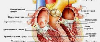
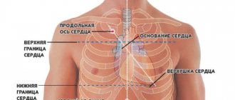
Heart
The heart is a muscular organ located in the chest. It works rhythmically throughout life, ensuring the movement of blood through the vessels.
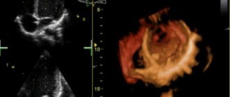
Rice. 1. Diagram of the mammalian heart.
The heart consists of 4 chambers:
- left atrium;
- left ventricle;
- right atrium;
- right ventricle.
Between the left and right halves of the heart there is a complete septum. Blood does not pass through it. That is why, unlike amphibians and reptiles, arterial and venous blood does not mix in the heart. The blood flow always goes from the atrium to the ventricle.
The heart has valves that open in one direction and prevent blood from returning back through the bloodstream.
The size of the heart and pulse rate correspond to the intensity of metabolism. The higher the heart rate and the smaller the heart, the more intense the metabolism.
TOP 3 articles
who are reading along with this
Circulatory organs
Arthropod circulatory system
Mammal heart
Rice. 2. Whale heart model.