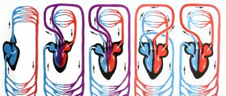
The heart is a muscular pump that ensures continuous movement of blood through the vessels. Together, the heart and blood vessels make up the cardiovascular system. This system consists of the systemic and pulmonary circulation. From the left side of the heart, blood first moves through the aorta, then through large and small arteries, arterioles, and capillaries. In the capillaries, oxygen and other substances necessary for the body enter the organs and tissues, and from there carbon dioxide, metabolic products, are removed. After this, the blood turns from arterial to venous and again begins to move towards the heart. First along the venules, then through smaller and larger veins. Through the inferior and superior vena cava, blood again enters the heart, only this time into the right atrium. A large circle of blood circulation is formed.
Venous blood from the right side of the heart is sent through the pulmonary arteries to the lungs, where it is enriched with oxygen, and returns to the heart again - this is the pulmonary circulation.
Inside, the heart is divided by partitions into four chambers. The two atria are divided by the interatrial septum into the left and right atria. The left and right ventricles of the heart are separated by the interventricular septum. Normally, the left and right parts of the heart are completely separate. The atria and ventricles have different functions. The atria store blood that flows into the heart. When the volume of this blood is sufficient, it is pushed into the ventricles. And the ventricles push blood into the arteries, through which it moves throughout the body. The ventricles have to do more hard work, so the muscle layer in the ventricles is much thicker than in the atria. The atria and ventricles on each side of the heart are connected by the atrioventricular orifice. Blood moves through the heart in only one direction. In the systemic circle of blood circulation from the left side of the heart (left atrium and left ventricle) to the right, and in the small circle from the right to the left.
The correct direction of blood flow is ensured by the valve apparatus of the heart:
Valves:
- tricuspid
- pulmonary
- mitral
- aortic
They open at the right time and close, preventing blood flow in the opposite direction.
Aortic valve
Closes the entrance to the aorta. It also consists of three valves, which look like crescents. Opens when the left ventricle contracts. In this case, blood enters the aorta. When the left ventricle relaxes, it closes. Thus, venous blood (poor in oxygen) from the superior and inferior vena cava enters the right atrium. When the right atrium contracts, it moves through the tricuspid valve into the right ventricle. Contracting, the right ventricle ejects blood through the pulmonary valve into the pulmonary arteries (pulmonary circulation). Enriched with oxygen in the lungs, the blood turns into arterial blood and moves through the pulmonary veins to the left atrium, then to the left ventricle. When the left ventricle contracts, arterial blood enters the aorta through the aortic valve under high pressure and spreads throughout the body (systemic circulation).
The heart muscle is called the myocardium
There are contractile and conductive myocardium. The contractile myocardium is the actual muscle that contracts and produces the work of the heart. In order for the heart to contract in a certain rhythm, it has a unique conduction system. The electrical impulse to contract the heart muscle occurs in the sinoatrial node, which is located in the upper part of the right atrium and spreads through the conduction system of the heart, reaching every muscle fiber
First, both atria contract, then both ventricles, thereby ensuring the flow of blood to all organs and tissues of the body. The heart muscle has two membranes (external and internal). The inner lining of the heart is called the endocardium. The outer lining of the heart is called the pericardium.
Circulation circles
From previous articles you already know the composition of blood and the structure of the heart. It is obvious that the blood performs all functions only thanks to its constant circulation, which is carried out thanks to the work of the heart. The work of the heart resembles a pump that pumps blood into the vessels through which blood flows to the internal organs and tissues.
The circulatory system consists of a large and small (pulmonary) circulation, which we will discuss in detail. They were described by William Harvey, an English physician, in 1628.
Systemic circulation (BCC)
This circulatory system serves to deliver oxygen and nutrients to all organs. It begins with the aorta, the largest vessel, emerging from the left ventricle, which sequentially branches into arteries, arterioles and capillaries. The famous English scientist, physician William Harvey discovered the BCC and understood the meaning of the blood circulation.
The capillary wall is single-layered, so gas exchange occurs through it with surrounding tissues, which also receive nutrients through it. Respiration occurs in the tissues, during which proteins, fats, and carbohydrates are oxidized. As a result, carbon dioxide and metabolic products (urea) are formed in the cells, which are also released into the capillaries.
Venous blood collects through venules into veins, returning to the heart through the largest - the superior and inferior vena cava, which flow into the right atrium. Thus, BCC begins in the left ventricle and ends in the right atrium.
Blood passes through the BCC in 23-27 seconds. Arterial blood flows through the arteries of the BCC, and venous blood flows through the veins. The main function of this circulation is to provide oxygen and nutrients to all organs and tissues of the body. In the vessels of the BCC there is high blood pressure (relative to the pulmonary circulation).
Pulmonary circulation (pulmonary)
Let me remind you that the BCC ends in the right atrium, which contains venous blood. The pulmonary circulation (PCC) begins in the next chamber of the heart - the right ventricle. From here, venous blood enters the pulmonary trunk, which divides into two pulmonary arteries.
The right and left pulmonary arteries with venous blood are directed to the corresponding lungs, where they branch to capillaries intertwining the alveoli. Gas exchange occurs in the capillaries, as a result of which oxygen enters the blood and combines with hemoglobin, and carbon dioxide diffuses into the alveolar air.
Oxygenated arterial blood collects in venules, which then drain into the pulmonary veins. The pulmonary veins with arterial blood flow into the left atrium, where the ICC ends. From the left atrium, blood flows into the left ventricle, the site of origin of BCC. Thus, two circles of blood circulation are closed.
ICC blood passes through in 4-5 seconds. Its main function is to saturate venous blood with oxygen, as a result of which it becomes arterial blood rich in oxygen. As you noticed, venous blood flows through the arteries in the ICC, and arterial blood flows through the veins. Blood pressure here is lower than BKK.
Interesting Facts
On average, a person’s heart pumps about 5 liters every minute, and over 70 years of life - 220 million liters of blood. In one day, the human heart makes approximately 100 thousand beats, over a lifetime - 2.5 billion beats.
© Bellevich Yuri Sergeevich 2018-2021
This article was written by Yuri Sergeevich Bellevich and is his intellectual property. Copying, distribution (including by copying to other sites and resources on the Internet) or any other use of information and objects without the prior consent of the copyright holder is punishable by law. To obtain article materials and permission to use them, please contact Yuri Bellevich
.
Conduction system of the heart
The heart, like any organ, has its own nervous system. The nervous system of the heart has several levels. The first and main pacemaker of the heart is the sinus node, located in the right atrium. It is subject to the atrioventricular node, which is located on the border between the atria and ventricles and quite often slows down the heart rate set by the sinus node. Then the nerve impulse goes to the ventricles of the heart along the branches of the Hiss bundle, which are divided into the smallest nerve endings - Purkinje fibers.
Blood vessels
Blood vessels have different shapes, structures and volumes, depending on their role in the body.
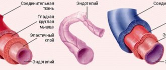
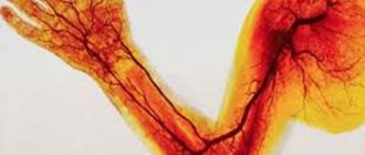
1. Arteries are the strongest vessels in the human body. Their walls are dense and elastic, consisting of three layers - endothelium, smooth muscle fibers and fibrous tissue. The task of the arteries is to saturate all organs and tissues with blood enriched with oxygen and nutrients. An exception is the arteries of the pulmonary circulation, through which venous blood flows from the heart to the lungs. The largest arterial vessel is the aorta.
2. Veins perform the function of transporting waste blood, saturated with carbon dioxide, back to the heart. This vein fluid is obtained from capillaries. Like an artery, a vein consists of several layers - endothelial, soft connective, dense connective and muscle. Venous walls are several times thinner and more vulnerable than arterial walls. For this reason, as you move away from the heart, the movement of venous blood may be disrupted - the pressure in the capillaries is almost equal to atmospheric pressure, and a normal flow is not created. Therefore, in hemodynamics, the vessels are assisted by the venous valves and the venous pulse.
3. Capillaries are the thinnest vessels, similar in volume to human hair. They are branches of large peripheral arteries. It is through them that tissues and organs are supplied with oxygen and nutrients. They also communicate with the veins to carry cellular waste. Consequently, these tiny vessels are at the same time the breadwinners and caretakers of our body.
Normal blood circulation within the vascular system is ensured by blood pressure.
Arteries
Not a single cell can survive without nutrients and oxygen. They are delivered by arteries. They carry oxygen-rich blood throughout the body. When you breathe, oxygen enters your lungs. where the delivery of oxygen throughout the body begins. First to the heart, then through the systemic circulation to all parts of the body. There, the blood exchanges oxygen for carbon dioxide and then returns to the heart. The heart pumps it back to the lungs, which take in carbon dioxide and give out oxygen, and so on endlessly. There are also pulmonary arteries of the pulmonary circulation, they are located in the lungs and through them blood, poor in oxygen and rich in carbon dioxide, enters the lungs, where gas exchange occurs. This blood then returns to the heart through the pulmonary veins.
Heart and blood vessels
The basis of the circulatory system is the heart, which, like a pump, pumps blood through the arteries, ensuring the delivery of oxygen and nutrients to all organs and tissues. The heart is located in the chest along the projection of the sternum, slightly to the left, and is a hollow muscular organ the size of a fist. The heart has 4 chambers separated by septa. Between the left atrium and the left ventricle, as well as the right atrium and the right ventricle, there are openings with valves that regulate the direction of blood flow from the atria to the ventricles. Blood circulation occurs through the systemic and pulmonary circulation. Blood collects from all organs and tissues and enters the right atrium through the veins. From the right atrium, through the corresponding opening, venous blood flows into the right ventricle. Both of these right reservoirs (atrium and ventricle) are also called the right heart (“venous”). The pulmonary circulation begins in the right ventricle. From the right ventricle, due to the contraction of its muscular wall, dark, oxygen-poor and carbon dioxide-rich blood is pushed into the pulmonary artery and flows through it into the lungs. There it enters small arteries and capillaries, is cleared of carbon dioxide by diffusion and enriched with oxygen, acquires a bright red color and is already called arterial blood. Through four pulmonary veins, arterial blood enters the left atrium, and this completes the pulmonary circulation. Considering that the left cavities of the heart (atrium and ventricle) or the left heart are already filled with arterial blood, the left heart is called “arterial”. The systemic circulation begins in the left atrium. From the left atrium, arterial blood flows into the left ventricle, which is an even more powerful pump than the right ventricle. Contracting, the left ventricle pushes blood into the aorta and its branches, through which it enters all organs and tissues, down to the smallest capillaries. Having given oxygen to the tissues and taken away carbon dioxide from them, the blood again becomes venous. Venous capillaries gradually connect with each other into larger veins, which, in turn, form two wide ones: the superior and inferior vena cava. The superior vena cava collects blood from the head, neck, upper limbs and torso walls, and the inferior vena cava collects blood from the lower limbs, abdominal organs and pelvic region. Both vena cavae carry blood to the right atrium, where the systemic circulation ends.
In this way, 2 closed circles of blood circulation are obtained, which are connected by the motor of the human body - the heart. So, the systemic circulation begins in the left ventricle of the heart and ends in the right atrium. Its function is to supply all organs and tissues with nutrients and oxygen. The pulmonary circulation begins from the right ventricle and ends in the left atrium. Its function is to enrich the blood with oxygen in the lungs. The heart muscle, constantly performing enormous work, itself needs nutrition and oxygen. Blood flows to the heart through vessels that extend directly from the aorta and surround the heart like a crown or crown, which is why they are called coronary or coronary arteries.
Movement of blood through vessels.
The heart does a lot of work. So, in one minute it pumps 4.5–5 liters of blood in only one direction. Blood movement is ensured by valves located between the atria and ventricles, between the left ventricle and the aorta, pulmonary vessels and the right atrium. The speed of blood movement through the vessels depends on their diameter: if in the aorta the blood moves at high speed, then in the capillaries this speed is minimal. When the cardiovascular system is damaged by an atherosclerotic, inflammatory or degenerative process, both general and local circulatory disorders can be observed. An example of general circulatory disorders is heart failure with shortness of breath, palpitations, cough, bluish skin and swelling. An example of local circulatory disorders, when the blood supply to any organ is affected, is a heart attack (of the heart, lung or kidneys), or gangrene of a limb. But since the circulatory system functions as a single whole, even local circulatory disorders in any organ eventually affect the entire system. The activity of the heart is regulated by the central nervous system. In addition, the heart also has its own intracardiac regulatory mechanisms that promote rhythmic contraction (systole phase) and relaxation (diastole phase) of the heart. In an adult, the number of heartbeats per minute normally ranges from 60 to 80 beats; in athletes, the heart works more economically. Their heart rate is 40–50 beats per minute.
Arteries of our body
Before talking about atherosclerosis, let us recall the anatomical features of the arteries. Arteries are cylindrical elastic tubes of various diameters. The wall of arteries is much thicker than that of veins, the vessels that carry blood back to the heart. This difference in the thickness of the vessels is not accidental and is due to the fact that the blood pressure in the arteries is much greater than in the veins. The arterial wall consists of three layers: outer, middle and inner. The outer layer or serosa is a framework of connective tissue; the middle (muscle) layer consists of smooth muscle fibers; the inner layer (intima) is lined with a single layer of cells and is called endothelium. It is the endothelium, or rather its damage or dysfunction, that plays a major role in the development of atherosclerosis. However, we will talk about this later. The lumen of the arteries can change as a result of contraction or relaxation of the smooth muscle fibers of the middle layer. The dilation of blood vessels (for example, in hot conditions) helps to increase blood flow and more intense metabolism, and conversely, their narrowing (in low temperatures) slows down these processes in the body. If the vessels are constantly narrowed for some reason, then the organs and tissues receive little blood, and therefore oxygen. Over time, this leads to disruption of the functioning of those organs and tissues that are nourished by narrowed arteries.
Vascular changes in atherosclerosis
Atherosclerosis (from the Greek words “ather” - gruel and “sclerosis” - hardening), exactly as the name suggests, is the process of accumulation of soft deposits of lipids (fats, fat-like substances, primarily cholesterol) on the walls of arteries.
It has been established that atherosclerosis develops in response to damage to the endothelium (the inner lining of blood vessels).
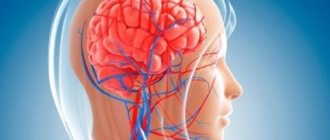
Damage or dysfunction of the endothelium can be caused by a number of reasons, in particular, smoking, significant increases in blood lipid levels, high blood pressure, acute or chronic psycho-emotional stress, viral or bacterial infection. Following damage to the endothelium, fats, fat-like substances, and cholesterol begin to penetrate into the arterial wall. Leukocytes, or rather their special varieties, monocytes and macrophages, rush here from the blood. This is the beginning of the formation of an atherosclerotic plaque. The “mush” formed on the wall of the artery is covered with a thin connective tissue capsule consisting of fibrin threads. Such an atherosclerotic plaque is called young. Over time, as the atherosclerotic process progresses, calcium begins to accumulate in the plaques, and fibrous and connective tissue grows. The plaque becomes covered with a thick capsule (this is a formed plaque), increases in size and significantly narrows the lumen of the artery. Often several plaques form and merge with each other, further narrowing the lumen of the vessel. Due to the narrowing of the lumen of the vessel, the organ supplied with blood does not receive the required amount of oxygen and chronic ischemia occurs (from the Greek words “ishe” - I retain and “hemo” - blood). Thus, when one or more coronary arteries narrows, chronic myocardial ischemia occurs (chronic coronary heart disease).
Acute ischemia (acute vascular insufficiency) develops differently. The fact is that the body produces special enzymes that “eat away” the connective tissue membrane of the atherosclerotic plaque from the edges, reaching its soft, mushy core. When the plaque capsule is opened, this mass enters the blood. An open wound on the capsule of an ulcerated plaque becomes covered with sticky platelets. Gradually, a blood clot forms - a thrombus, which bulges into the lumen of the vessel, sharply narrowing it. A thrombus can break away from the vessel wall and, moving with the blood flow, clog a smaller vessel, creating acute local vascular insufficiency and stopping tissue nutrition, which leads to tissue necrosis (death). For example, when one of the coronary arteries of the heart is blocked, myocardial infarction occurs; when thrombosis of the femoral artery or leg artery occurs, foot necrosis (gangrene) occurs. Atherosclerosis can completely affect the aorta and its branches, but more often arteries of various sizes are affected, and not throughout, but in certain areas. Atherosclerotic plaques “love” to form in places where arteries bend and branch, which are especially numerous in vital organs. Thus, the coronary arteries of the heart, the vessels of the brain, the abdominal aorta and its branches that supply blood to the abdominal organs, kidneys, pelvic organs, and lower extremities are most susceptible to atherosclerosis. Often, atherosclerosis initially favors one organ. For example, with damage to the coronary arteries, coronary heart disease develops; with atherosclerosis of the cerebral arteries, coronary artery disease develops, including stroke. When the iliac or femoral vessels are narrowed by atherosclerotic plaques, obliterating atherosclerosis of the vessels of the lower extremities occurs. If the renal vessels are affected by atherosclerosis, then hypertension may develop with a predominant increase in the “lower” numbers of blood pressure. Depending on the location of the affected vessels, clinical manifestations of atherosclerosis of a particular vital organ are formed. Atherosclerosis in its development is not similar to any other disease, since its first signs appear in early childhood. Thus, at six months of age, some babies begin to develop fatty spots and stripes on the inner wall of blood vessels. During the growth process, a significant part of these spots resolves and only a few remain. If you do not engage in prevention, then by the age of 35-50 in men and somewhat later in women, clinical signs of atherosclerosis appear in the form of coronary heart disease, coronary disease of the brain and other organs. The resulting disease requires many years, and in fact lifelong treatment. Meanwhile, in medicine it has long been proven that serious diseases, including atherosclerosis, are better and easier to prevent than to treat later for many, many years. In order to prevent atherosclerosis, you need to know everything about the substrate of atherosclerotic plaques - cholesterol and its assistants - lipoproteins.