Blood circulation is a continuous flow of blood that moves through the vessels and cavities of the heart. This system is responsible for metabolic processes in the organs and tissues of the human body. Circulating blood transports oxygen and nutrients to the cells, taking carbon dioxide and metabolites from there. That is why any circulatory disorders threaten with dangerous consequences.
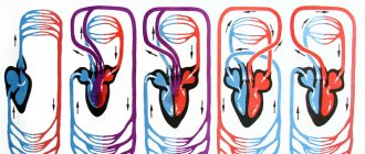
The blood circulation consists of a large (systemic) and a small (pulmonary) circle. Each turn has a complex structure and functions. The systemic circle originates from the left ventricle and ends in the right atrium, and the pulmonary circle originates from the right ventricle and ends in the left atrium.
Types of Blood Vessels
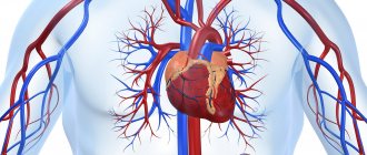
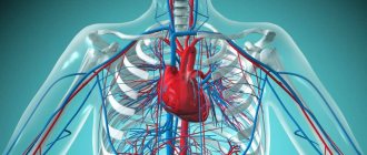
Blood circulation is a complex system that consists of the heart and blood vessels. The heart constantly contracts, pushing blood through the vessels to all organs and tissues. The circulatory system consists of arteries, veins, and capillaries.
The circulatory system is formed by arteries, veins and capillaries
The arteries of the systemic circulation are the largest vessels; they have a cylindrical shape and transport blood from the heart to the organs.
Structure of the walls of arterial vessels:
- outer connective tissue membrane;
- middle layer of smooth muscle fibers with elastic veins;
- strong elastic inner endothelial membrane.
Arteries have elastic walls that constantly contract, allowing blood to move evenly.
With the help of the veins of the systemic circulation, blood moves from the capillaries to the heart. Veins have the same structure as arteries, but they are less strong, since their middle layer contains less smooth muscle and elastic fibers. That is why the speed of blood movement in the venous vessels is largely influenced by nearby tissues, especially skeletal muscles. All veins, except the vena cava, are equipped with valves that prevent the backflow of blood.
Capillaries are small vessels that consist of endothelium (a single layer of flat cells). They are quite thin (about 1 micron) and short (from 0.2 to 0.7 mm). Due to their structure, microvessels saturate tissues with oxygen and useful substances, carrying away carbon dioxide, as well as metabolic products. Blood moves slowly through them; in the arterial part of the capillaries, water is removed into the intercellular space. In the venous part, blood pressure decreases and water flows back into the capillaries.
Anatomy of the cardiovascular system
In order to talk about diseases of the cardiovascular system, it is necessary to understand its structure. The circulatory system is divided into arterial and venous. Through the arterial system, blood flows from the heart, through the venous system it flows to the heart. There are large and small circles of blood circulation.The great circle includes the aorta (ascending and descending, aortic arch, thoracic and abdominal sections), through which blood flows from the left side of the heart. From the aorta, blood enters the carotid arteries that supply blood to the brain, subclavian arteries, blood supply to the arms, renal arteries, arteries of the stomach, intestines, liver, spleen, pancreas, pelvic organs, iliac and femoral arteries, and blood supply to the legs. Blood flows from the internal organs through veins that drain into the superior vena cava (collects blood from the upper half of the body) and the inferior vena cava (collects blood from the lower half of the body). The vena cava drains into the right heart.
The pulmonary circulation includes the pulmonary artery (through which, however, venous blood flows). Through the pulmonary artery, blood enters the lungs, where it is enriched with oxygen and becomes arterial. Through the pulmonary veins (four), arterial blood enters the left heart.
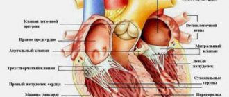
The heart pumps blood - a hollow muscular organ consisting of four sections. These are the right atrium and right ventricle, which make up the right heart, and the left atrium and left ventricle, which make up the left heart. Oxygen-rich blood coming from the lungs through the pulmonary veins enters the left atrium, from there into the left ventricle and then into the aorta. Venous blood enters the right atrium through the superior and inferior vena cava, from there into the right ventricle and further through the pulmonary artery into the lungs, where it is enriched with oxygen and again enters the left atrium.
There are pericardium, myocardium and endocardium. The heart is located in the cardiac sac - the pericardium. Cardiac muscle - the myocardium consists of several layers of muscle fibers; there are more of them in the ventricles than in the atria. These fibers, contracting, push blood from the atria into the ventricles and from the ventricles into the vessels. The internal cavities of the heart and valves are lined by the endocardium.
- Right coronary artery
- Anterior descending artery
- Ear
- Superior vena cava
- Inferior vena cava
- Aorta
- Pulmonary artery
- Branches of the aorta
- Right atrium
- Right ventricle
- Left atrium
- Left ventricle
- Trabeculae
- Chords
- Tricuspid valve
- Mitral valve
- Pulmonary valve
Valvular apparatus of the heart.
Between the left atrium and the left ventricle there is a mitral (bicuspid) valve, and between the right atrium and the right ventricle there is a tricuspid (three-leaf) valve. The aortic valve is located between the left ventricle and the aorta, the pulmonary valve is between the pulmonary artery and the right ventricle.
Work of the heart.
From the left and right atria, blood flows into the left and right ventricles, while the mitral and tricuspid valves are open, the aortic and pulmonary valves are closed. This phase in the work of the heart is called diastole. Then the mitral and tricuspid valves close, the ventricles contract and through the opened aortic and pulmonary valves, blood rushes into the aorta and pulmonary artery, respectively. This phase is called systole, systole is shorter than diastole.
Conduction system of the heart.
We can say that the heart works autonomously - it itself generates an electrical impulse that spreads through the heart muscle, causing it to contract. The pulse must be generated at a certain frequency - normally about 50-80 pulses per minute. In the conduction system of the heart, the sinus node is distinguished (located in the right atrium), from which nerve fibers go to the atrioventricular (atrioventricular) node (located in the interventricular septum - the wall between the right and left ventricles). From the atrioventricular node, nerve fibers run in large bundles (right and left bundle branches), dividing into smaller bundles in the walls of the ventricles (Purkinje fibers). An electrical impulse is generated in the sinus node and spreads through the conduction system throughout the myocardium (heart muscle).
Blood supply to the heart.
Like all organs, the heart must receive oxygen. Oxygen is delivered through arteries called coronary arteries. The coronary arteries (right and left) arise from the very beginning of the ascending aorta (at the origin of the aorta from the left ventricle). The trunk of the left coronary artery is divided into the descending artery (also known as the anterior interventricular) and the circumflex artery. These arteries give off branches - the artery of the obtuse edge, diagonal, etc. Sometimes the so-called median artery branches off from the trunk. The branches of the left coronary artery supply blood to the anterior wall of the left ventricle, most of the interventricular septum, the lateral wall of the left ventricle, and the left atrium. The right coronary artery supplies part of the right ventricle and the posterior wall of the left ventricle.
Now that you have become an expert in the anatomy of the cardiovascular system, let's move on to its diseases.
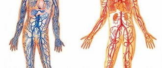
Structure of the systemic circulation
The aorta is the largest vessel of the great circle, with a diameter of 2.5 cm. This is a kind of source from which all other arteries emerge. The vessels branch, their size decreases, they go to the periphery, where they give oxygen to organs and tissues.
The largest vessel in the systemic circulation is the aorta
The aorta is divided into the following sections:
- ascending;
- descending;
- the arc that connects them.
The ascending section is the shortest, its length is no more than 6 cm. The coronary arteries emanate from it, which supply oxygen-rich blood to the myocardial tissues. Sometimes the term “cardiac circulation” is used to name the ascending section. From the most convex surface of the aortic arch, arterial branches depart that supply blood to the arms, neck, and head: on the right side there is the brachiocephalic trunk, divided in two, and on the left there is the common carotid, subclavian artery.
The descending aorta is divided into 2 groups of branches:
- Parietal arteries that supply blood to the chest, spinal column, and spinal cord.
- Visceral (splanchnic) arteries that transport blood and nutrients to the bronchi, lungs, esophagus, etc.
Under the diaphragm is the abdominal aorta, the parietal branches of which supply the abdominal cavity, the lower surface of the diaphragm, and the spine.
The internal branches of the abdominal aorta are divided into paired and unpaired. The vessels that extend from the unpaired trunks transport oxygen to the liver, spleen, stomach, intestines, and pancreas. The unpaired branches include the celiac trunk, as well as the superior and inferior mesenteric arteries.
There are only two paired trunks: renal, ovarian or testicular. These arterial vessels are adjacent to the organs of the same name.
The aorta ends with the left and right iliac arteries. Their branches extend to the pelvic organs and legs.
Many people are interested in the question of how the systemic circulatory system works. In the lungs, the blood is saturated with oxygen, after which it is transported to the left atrium, and then to the left ventricle. The iliac arteries supply blood to the legs, and the remaining branches supply blood to the chest, arms, and organs of the upper half of the body.
The veins of the systemic circulation carry oxygen-poor blood. The systemic circle ends with the superior and inferior vena cava.
The diagram of the veins of the systemic circle is quite clear. The femoral veins in the legs unite to form the iliac vein, which becomes the inferior vena cava. In the head, venous blood collects in the jugular veins, and in the arms - in the subclavian veins. The jugular as well as the subclavian vessels unite to form the innominate vein, which gives rise to the superior vena cava.
Topic 9.3 Veins of the systemic circulation Lymphatic system
Topic 9.3 Veins of the systemic circulation
Lymphatic system
Venous vessels of the systemic circulation are represented by: the superior vena cava system, the inferior vena cava system and the portal vein system
To the superior vena cava system
These include all venous vessels that collect blood from the head, brain, neck, upper limbs, from the walls of the chest cavity, organs of the chest cavity, and also partially from the abdominal cavity. They are represented by the brachiocephalic veins, internal jugular, subclavian, veins of the upper extremities, azygos and semi-gypsy veins.
Inferior vena cava
collects blood from the lower extremities, walls and organs of the pelvis and abdominal cavity.
Portal vein
collects blood from unpaired abdominal organs; spleen, stomach, gall bladder, pancreas, small and large intestines. This is a short thick trunk that runs deep into the hepatoduodenal ligament.
| Vein | Main branches | Area, organ from which blood is collected |
| I. Superior vena cava system | Head, neck, upper limbs, upper body | |
| 1. Veins head and neck | Superficial veins: external jugular vein | Temporal, parietal and occipital areas of the head, ears, anterior and lateral areas of the neck |
| Deep veins: internal jugular vein | The brain and its membranes; anterior and lateral areas of the face, tongue, pharynx, larynx, thyroid gland | |
| 2. Veins of the upper limb | Superficial veins | |
| Lateral saphenous vein | Skin, subcutaneous tissue of the lateral parts of the upper limb | |
| Medial saphenous vein | Skin, subcutaneous tissue of the medial parts of the upper limb | |
| Deep veins | ||
| Radial vein - steam room | Muscles, ligaments, bones of the lateral sides of the hand and forearm | |
| Ulnar vein - steam room | Muscles, ligaments, bones of the medial sides of the hand and forearm | |
| The brachial vein is initially a steam vein, then the two veins merge into one trunk | Free part of the upper limb (skin, ligaments, muscles, hand bones, forearm, shoulder) | |
| Axillary vein | Free part of the upper limb, skin, subcutaneous tissue of the lateral sections of the chest wall | |
| Subclavian vein | Upper limb, upper anterior and lateral chest wall | |
| 3. Veins of the chest | Azygos vein | Posterior wall of the abdomen and chest cavity, mediastinal organs |
| Hemizygos vein | Posterior wall of the abdomen and left half of the chest cavity, mediastinal organs | |
| Brachiocephalic vein | The anterior wall of the abdomen and chest cavity, mediastinal organs, thyroid gland, thymus, larynx, cervical spinal cord and its membranes, deep muscles of the neck, head, neck, upper limbs | |
| II. Inferior vena cava system | Lower limbs, walls and organs of the pelvis, diaphragm (partially), posterior, lateral and part of the anterior wall of the abdominal cavity, paired abdominal organs, unpaired abdominal organs (through the venous vascular system of the liver) | |
| Vein | Main branches | Area, organ from which blood is collected |
| 1. Veins of the lower limb | Superficial veins | |
| Great saphenous vein legs | Skin and subcutaneous tissue of the anteromedial parts of the foot, lower leg and thigh, external genitalia, anterior abdominal wall | |
| Small subcutaneous leg vein | Skin and subcutaneous tissue posterolateral sections of the bone and lower leg | |
| Deep veins | ||
| Anterior tibial vein—paired | Muscles, ligaments, bones of the dorsum of the foot and the front of the leg | |
| Posterior tibial vein - steamed | Muscles, ligaments, bones of the sole of the foot and the back of the leg | |
| Popliteal vein | Skin, ligaments, muscles, bones of the foot, leg and knee | |
| Femoral vein | Skin, ligaments, muscles, bones of the foot, lower leg, thigh, skin and subcutaneous tissue of the external genitalia, anterior abdominal wall | |
| 2. Veins of the pelvis | External iliac vein | Free part of the lower limb, anterior abdominal wall, external genitalia |
| Internal iliac vein | Walls and organs of the pelvis, external and internal genitalia | |
| Common iliac vein | Walls and organs of the pelvis, external and internal genitalia, lower limb | |
| III. Portal vein system | Unpaired organs of the abdominal cavity (stomach, small and large intestine, pancreas, spleen) | |
| Splenic vein | Spleen, area of the bottom and posterior wall of the body of the stomach, body and tail of the pancreas, left half of the greater omentum | |
| Superior mesenteric vein | Small intestine and its mesentery, cecum, ascending and right half of the transverse colon, appendix, head and part of the body of the pancreas, right half of the body of the stomach and greater omentum | |
| Inferior mesenteric vein | Upper rectum, sigmoid colon, descending colon and left transverse colon | |
| Portal vein | Receives venous blood from the splenic, superior mesenteric and inferior mesenteric veins | |
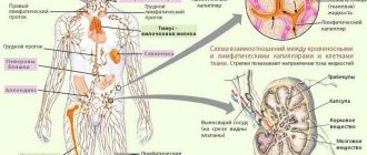
The lymphatic system is a closed part of the vascular system; it complements the venous bed, with which it jointly drains organs through the formation of lymph.
Along with this, the lymphatic system performs specific functions: transport - it transfers metabolic products from tissues into the blood, and into tissues - nutrients and hormones; hematopoietic and lymph-forming - forms lymphocytes; barrier - retains some particles (foreign) and microbial bodies, tumor cells; immune - produces immune bodies responsible for immunity.
The lymphatic system includes: lymph, lymphatic bed (capillaries, intra- and extraorgan vessels, lymphatic trunks and ducts), as well as lymphoid organs (lymph nodes).
Lymphatic system (general diagram)
1 - parotid lymph nodes; 2 - submandibular lymph nodes; 3 - cervical lymph nodes; 4 - superior vena cava; 5 - axillary lymph nodes; 6 - thoracic duct; 7 - cistern of the thoracic duct;
8 - inferior vena cava; 9 - iliac lymph nodes; 10 - superficial lymphatic vessels of the upper limb; 11 - inguinal lymph nodes;
12 - superficial lymphatic vessels of the lower limb; 13 - right lymphatic duct
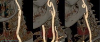
Blood supply to the head
The circulatory system of the head is the most complex structure of the body. The carotid artery, which is divided into 2 branches, is responsible for the blood supply to the parts of the head. The external carotid arterial vessel saturates the face, temporal region, oral cavity, nose, thyroid gland, etc. with oxygen and useful substances.
The main vessel supplying blood to the head is the carotid artery
The internal branch of the carotid artery goes deeper, forming the Circle of Wallisian, which transports blood to the brain. In the cranium, the internal carotid artery branches into the ophthalmic, anterior, middle cerebral, and communicating arteries.
This is how only ⅔ of the systemic circle is formed, which ends with the posterior cerebral arterial vessel. It has a different origin, the scheme of its formation is as follows: subclavian artery - vertebral - basilar - posterior cerebral. In this case, the brain is supplied with blood by the carotid and subclavian arteries, which are connected to each other. Thanks to anastomoses (vascular anastomoses), the brain survives minor disturbances in blood flow.
Principle of placement of arteries
The circulatory system of each body structure is approximately similar to that described above. Arterial vessels always approach organs along the shortest path. The vessels in the limbs pass precisely along the flexion side, since the extensor part is longer. Each artery originates at the embryonic site of the organ, and not at its actual location. For example, the arterial vessel of the testicle emerges from the abdominal aorta. Thus, all vessels are connected to their organs from the inside.
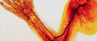
The arrangement of vessels resembles the structure of the skeleton
The placement of arteries is also related to the structure of the skeleton. For example, the brachial branch runs along the upper limb, which corresponds to the humerus; the ulnar and radial arteries also pass next to the bones of the same name. And in the skull there are openings through which arterial vessels transport blood to the brain.
Arterial vessels of the systemic circulation form networks in the joint area using anastomoses. Thanks to this scheme, the joints are continuously supplied with blood during movement. The size of the vessels and their number depend not on the size of the organ, but on its functional activity. Organs that work more intensively are saturated with a large number of arteries. Their placement around the organ depends on its structure. For example, the diagram of the vessels of parenchymal organs (liver, kidneys, lungs, spleen) corresponds to their shape.