In medicine, blood is usually divided into arterial and venous. It would be logical to think that the first flows in the arteries, and the second in the veins, but this is not entirely true. The fact is that in the systemic circulation, arterial blood (a.k.) actually flows through the arteries, and venous blood (v.k.) through the veins, but in the small circle the opposite happens: c. It enters from the heart into the lungs through the pulmonary arteries, releases carbon dioxide to the outside, is enriched with oxygen, becomes arterial, and returns from the lungs through the pulmonary veins.
How does venous blood differ from arterial blood? A.K. is saturated with O2 and nutrients, it flows from the heart to organs and tissues. V. k. - “spent”, it gives O2 and nutrition to the cells, takes CO2 and metabolic products from them and returns from the periphery back to the heart.
Human venous blood differs from arterial blood in color, composition and functions.
By movement
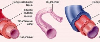
Blood circulation in the arterial and venous systems is significantly different. A. k. moves from the heart to the periphery, and v. k. - in the opposite direction. When the heart contracts, blood is ejected from it under a pressure of approximately 120 mmHg. pillar As it passes through the capillary system, its pressure decreases significantly and is approximately 10 mmHg. pillar Thus, a. k. moves under pressure at high speed, and c. It flows slowly under low pressure, overcoming the force of gravity, and its reverse flow is prevented by valves.
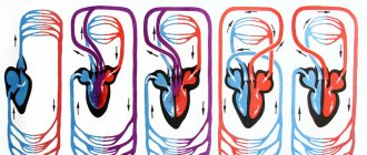
How the transformation of venous blood into arterial blood and vice versa occurs can be understood if we consider the movement in the pulmonary and systemic circulation.
Blood saturated with CO2 enters the lungs through the pulmonary artery, from where CO2 is excreted. Then O2 saturation occurs, and the blood already enriched with it enters the heart through the pulmonary veins. This is how movement occurs in the pulmonary circulation. After this, the blood makes a large circle: a. It carries oxygen and nutrition through the arteries to the cells of the body. Giving up O2 and nutrients, it is saturated with carbon dioxide and metabolic products, becomes venous and returns through the veins to the heart. This completes the large circle of blood circulation.
Principle of placement of arteries
The circulatory system of each body structure is approximately similar to that described above. Arterial vessels always approach organs along the shortest path. The vessels in the limbs pass precisely along the flexion side, since the extensor part is longer. Each artery originates at the embryonic site of the organ, and not at its actual location. For example, the arterial vessel of the testicle emerges from the abdominal aorta. Thus, all vessels are connected to their organs from the inside.
The arrangement of vessels resembles the structure of the skeleton
The placement of arteries is also related to the structure of the skeleton. For example, the brachial branch runs along the upper limb, which corresponds to the humerus; the ulnar and radial arteries also pass next to the bones of the same name. And in the skull there are openings through which arterial vessels transport blood to the brain.
Arterial vessels of the systemic circulation form networks in the joint area using anastomoses. Thanks to this scheme, the joints are continuously supplied with blood during movement. The size of the vessels and their number depend not on the size of the organ, but on its functional activity. Organs that work more intensively are saturated with a large number of arteries. Their placement around the organ depends on its structure. For example, the diagram of the vessels of parenchymal organs (liver, kidneys, lungs, spleen) corresponds to their shape.
By bleeding
Due to the characteristics of movement, bleeding will also differ. With arterial bleeding, the blood flows in full swing; such bleeding is dangerous and requires prompt first aid and medical attention. With venous flow, it calmly flows out in a stream and can stop on its own.
Cellular structure of blood
Blood consists of two components: plasma (50-60%) and suspended formed elements (40-50%).
The second category includes:
· Erythrocytes (red blood cells) are the most numerous of the formed elements. According to official studies, one drop of blood contains about 5 million red blood cells. Red blood cells are responsible for transporting gases - oxygen and carbon dioxide. They contain the protein hemoglobin, which binds oxygen molecules in the lungs. Red blood cells deliver oxygen to all tissues and organs, after which they absorb carbon dioxide and carry it to the lungs. It is removed from the body during respiration.
· Leukocytes (white blood cells) - elements that protect our body from foreign bodies and compounds, are part of the immune system. White blood cells recognize and attack pathogens through the production of antibodies and macrophages. When an infection enters the body, the production of leukocytes increases significantly. Normally, their quantity is inferior to the concentration in the blood of other formed elements.
· Platelets (blood platelets) are cells that provide coagulation (clotting) of blood flowing from a damaged vessel and protect the body from heavy blood loss. They stick to the hole in the damaged vessel, forming a “sealing” plug to stop bleeding. It is the platelets that can stick together and form pathological blood clots inside the vessels, called thrombi.
All formed elements are synthesized by the bone marrow and distributed through plasma, the liquid part of the blood.
Other differences
- A.K. is located on the left side of the heart, in. k. – in the right, blood mixing does not occur.
- Venous blood, unlike arterial blood, is warmer.
- V. k. flows closer to the surface of the skin.
- A.K. in some places comes close to the surface and here the pulse can be measured.
- The veins through which the v. flows. to., much more than arteries, and their walls are thinner.
- Movement a.k. is ensured by a sharp release during contraction of the heart, outflow into the. the valve system helps.
- The use of veins and arteries in medicine also differs - medications are injected into the vein, and it is from it that biological fluid is taken for analysis.
Blood supply to the head
The circulatory system of the head is the most complex structure of the body. The carotid artery, which is divided into 2 branches, is responsible for the blood supply to the parts of the head. The external carotid arterial vessel saturates the face, temporal region, oral cavity, nose, thyroid gland, etc. with oxygen and useful substances.
The main vessel supplying blood to the head is the carotid artery
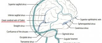
The internal branch of the carotid artery goes deeper, forming the Circle of Wallisian, which transports blood to the brain. In the cranium, the internal carotid artery branches into the ophthalmic, anterior, middle cerebral, and communicating arteries.
This is how only ⅔ of the systemic circle is formed, which ends with the posterior cerebral arterial vessel. It has a different origin, the scheme of its formation is as follows: subclavian artery - vertebral - basilar - posterior cerebral. In this case, the brain is supplied with blood by the carotid and subclavian arteries, which are connected to each other. Thanks to anastomoses (vascular anastomoses), the brain survives minor disturbances in blood flow.
Process description
Based on the above, you can understand where gas exchange occurs. But how exactly? It is worth trying to answer this question.
First you need to understand that the key task of the lungs is to carry out the process of gas exchange, and not just to distill air. And the inhaled composition in them changes. This is where the circulatory system comes into play. Namely, capillaries. They permeate all the alveoli.
Once in them, oxygen is sent to the walls of the capillaries. Why? Because there is different pressure in the blood and in the air contained in the alveoli. In venous blood it is much lower. That is why oxygen rushes from the alveoli into the capillaries. But the pressure of carbon dioxide is greater in the blood than in the alveoli. What, in turn, does this mean? The fact that carbon dioxide is transported from venous blood directly into the lumen of the alveoli.
Oxygen then joins the hemoglobin contained in red blood cells and moves throughout the body in this form. As a result, arterial blood enriched with oxygen is formed.
Process intensity
When talking about in which vessels gas exchange occurs in the lungs, it is worth noting an interesting nuance. The fact is that this process (and, accordingly, subsequent energy consumption) becomes less intense if body temperature decreases. This was first identified in cold-blooded creatures, and then a similar dependence was proven in warm-blooded mammals. Man, naturally, applies to them too.
The same thing is observed under conditions of artificial or natural hypothermia. But when body temperature rises, when a person gets sick or overheats, gas exchange, on the contrary, increases.
Features of the structure of blood vessels
The network of alveolar capillaries cannot be compared with anything. And when talking about in which vessels gas exchange occurs, it is simply impossible not to touch upon the specifics of their structure. Features can be identified in the following list:
- The capillary segments are very small.
- All of them are abundantly interconnected, as a result of which a looping network is formed.
- Individual capillary segments very densely fill all units of alveolar surface area.
- The blood flow rate is very low. Why? Because the walls of the alveoli have such a dense capillary network that some physiologists consider it to be a continuous layer of moving blood.
So, where gas exchange occurs is clear. What about sizes? The surface area of the studied capillary network is close to the alveoli (approximately 80 m2). And it contains about 200 ml of blood.
If we talk about alveolar blood capillaries, their diameter varies from 8.3 to 9.9 microns. In red blood cells it is smaller - 7.4 microns. What does it mean? The fact that red blood cells adhere very tightly to the capillary walls. This feature of the blood supply creates excellent conditions for effective and rapid gas exchange, which results in the normalization of the gas composition of arterial blood and alveolar air.
Oxygen value
In which blood vessels gas exchange occurs and how this process is carried out in general is clear. Now it’s worth talking about the importance of oxygen for the human body.
This is an organogenic element. The body contains up to 65%. And this is about 40 kilograms, if we take into account the average person.
The key function of oxygen is to participate in all redox reactions taking place in the body. It is thanks to it that the body can utilize proteins, fats and carbohydrates to extract energy for its needs.
According to research, 1.8 to 2.4 grams of oxygen are consumed per minute.
Pathologies[edit | edit code]
Pathology of the cardiovascular system includes, first of all, primary heart diseases: some forms of myocarditis, cardiomyopathy, heart tumors. This also includes heart damage in infectious, infectious-allergic, dysmetabolic and systemic diseases and diseases of other organs.
In the International Classification of Diseases, diseases of the heart and blood vessels are combined into a single class called “Diseases of the circulatory system” and divided into the following points [2]:
- Rheumatism in the active phase, including active rheumatism without heart damage, as well as active rheumatic pericarditis, endocarditis, myocarditis
- Chronic rheumatic heart diseases, including acquired heart defects
- Hypertonic disease
- Coronary heart disease, as well as acute myocardial infarction and various forms of angina, atherosclerotic cardiosclerosis and cardiac aneurysm
- Other heart diseases
- Vascular lesions of the brain, combining subarachnoid hemorrhages, cerebral hemorrhages, cerebral vascular thrombosis and cerebral embolism, transient cerebrovascular accidents, as well as generalized cerebral vascular lesions
- Diseases of arteries, arterioles, and capillaries
Heart[edit | edit code]
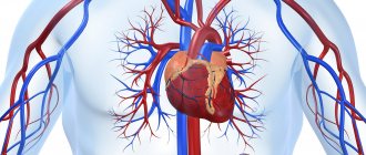
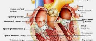
The heart (Latin cor, Greek καρδιά) is a hollow muscular organ that, through a sequence of contractions and relaxations, rhythmically pumps blood through the vessels. The function of the heart is carried out thanks to the alternating contractions and relaxations of the muscle fibers that form the walls of the atria and ventricles. Depending on the biological species, the inside can be divided by partitions into 2, 3 or 4 chambers. Mammals and birds have a 4-chambered heart. In this case, they distinguish (according to blood flow): the right atrium, the right ventricle, the left atrium and the left ventricle.
Briefly about the process
Before we talk about in which vessels gas exchange occurs, it is necessary to discuss the specifics of the process itself. Based on the name, one can understand that this is how it is customary to designate the exchange of gases between the body and the external environment.
You may be interested in:Senior is... Meaning of the word and spelling
The process is simple. Oxygen enters the body from the environment (without interruptions), which is consumed by all tissues, organs and cells. And it, in turn, releases back the carbon dioxide formed in the process, as well as some other metabolic products.
This process is necessary for almost all organisms. Because without it, normal energy metabolism is not possible.
Features of blood flow in the organs of the systemic circle
Some of the internal organs have their own characteristics. So, for example, in the liver there is not only a hepatic vein, which “carries” the venous flow away from it, but also a portal vein, which, on the contrary, brings blood to the liver tissue, where blood purification is performed, and only then the blood collects in the tributaries of the hepatic vein to enter to a big circle. The portal vein brings blood from the stomach and intestines, so everything that a person eats or drinks must undergo a kind of “purification” in the liver.
In addition to the liver, certain nuances exist in other organs, for example, in the tissues of the pituitary gland and kidneys. Thus, in the pituitary gland the presence of a so-called “wonderful” capillary network is noted, because the arteries that bring blood to the pituitary gland from the hypothalamus are divided into capillaries, which then collect into venules. The venules, after the blood with the molecules of releasing hormones are collected, are again divided into capillaries, and then veins are formed that carry the blood from the pituitary gland. In the kidneys, the arterial network is divided twice into capillaries, which is associated with the processes of excretion and reabsorption in the kidney cells - in the nephrons.
What happens in the lungs?
It is worth returning to this issue. It is clear how gas exchange occurs in the vessels and what path the oxygen-enriched blood then takes. But what exactly happens in the lungs?
They perform an excretory function. It manifests itself in the removal of more than 200 volatile substances formed in the body or entering it from the outside. Carbon dioxide, exogenous substances (ethyl ether and alcohol), acetone, methane, nitrous oxide and fluorotane - all of the above are removed from the blood to one degree or another through the lungs.
In addition to conditioning, this organ also performs a protective function. Microorganisms that enter during inhalation and then settle on the walls of the alveoli capture and destroy alveolar macrophages.
It is also worth recalling that immunoglobulins, interferon, specific leukocyte antibodies and lysozyme are formed in the lungs - those elements that play an important role in protecting the body from various infectious agents.
Efficiency. Professionalism. Mercy
At yesterday's first aid courses, conducted by the Tyumen Emergency Medical Service, there were at least ten people. The courses are free. Everyone listened with interest to emergency doctor Andrei Rogotnev.
Useful tips based on practical experience
It is immediately worth noting that there is a lot of information on first aid on the Internet. The courses conducted by emergency doctors in Tyumen are valuable because the training is based on practical experience. Let's give a simple example. How long do you think it is possible to clamp an artery on the thigh or shoulder? Many will answer that no more than 1.5 hours. In fact, this recommendation has long been outdated, because after an hour and a half of compression, the blood below the tourniquet will clot in all vessels, and the limb will then have to be amputated. We are not at war, and we not only need to save a person’s life, but also try to save an injured arm or leg. However, first things first.
We find out where the blood is coming from - from a vein or from an artery
Before giving useful tips and recommendations for stopping bleeding, Andrei Rogotnev reminded that bleeding can be venous and arterial. We did not consider ordinary cuts, since they are not life-threatening. In venous bleeding, the blood comes from a vein; in arterial bleeding, the blood comes from an artery. It's generally simple. It is much more difficult for people far from anatomy and medicine to distinguish one bleeding from another. Meanwhile, it is important to understand this before you start saving a person. In fact, everything is simple here: blood from the artery flows like a fountain, but not in a continuous flow, but pulsates in time with the heartbeat, since blood from the heart enters all organs through the arteries. The blood flows back through the veins, so during venous bleeding there is no such pressure, the blood flows in a continuous stream, like water from a tap.
In case of arterial bleeding, you have no more than 3-8 minutes left
With arterial bleeding, the probability of death is greatest; with venous bleeding, death is unlikely; damage to large veins in the neck is dangerous. In case of bleeding from large arteries, those around you have no more than 3-8 minutes left. In such a short period of time, the ambulance will not have time to get there. Therefore, we drive away all fears, put plastic bags or rubber gloves on our hands if they happen to be nearby (so that in case something happens we don’t catch dangerous viruses), and look for points on the body to press the arteries with fingers. There are more than 10 such points on the body at which pulsation is felt on each side of the vertical axis of the body. We need to remember only three main ones: the carotid, brachial and inguinal arteries.
How to clamp the carotid artery
The carotid artery in the neck is to the right and left of the trachea. The pulse is clearly felt in this place. We feel and pinch this artery below the wound with our fingers. We compress the artery until the blood stops gushing out. If this happens, you did everything right. Do not under any circumstances reduce the pressure or remove your hand; the artery will be more difficult to find later.
And only after you have managed to stop the bleeding, Andrei Rogotnev advises you to do everything else: scream, call for help, ask passers-by to call an ambulance. If it is not possible to squeeze the artery with your fingers, you can secure a bandage (tourniquet, belt or piece of fabric) through the arm opposite to the wound. Thus, the artery will be compressed only on the side where the wound is. The most correct option is to compress the carotid artery until the ambulance arrives.
How to compress the brachial artery
We do the same with the brachial artery. The shoulder is the part of the arm from the elbow to the shoulder joint. Press your thumb against the inside of your shoulder to the bone and you will feel a pulsation. This is where you need to pinch the artery, clasping your hand with your thumb and forefinger until you find something to tighten it with. Motorists have a tourniquet in their first aid kit; in all other cases, you can use any available means - a shoe lace, a belt, a charger wire, etc.
How to clamp an abdominal artery
Bleeding from the abdominal artery (internal bleeding) can be stopped by pressing your fist on the abdomen in the navel area. You need to apply pressure towards the spine until the bleeding stops or decreases. Signs of internal bleeding: the person is pale and has a rapid heartbeat.
How to clamp the femoral artery
To stop arterial bleeding when the lower limb is damaged, you need to squeeze the inguinal fold between the thigh and lower abdomen with your fist. Here is another point of pressure on the artery.
The points for applying a tourniquet for injured limbs are the middle of the thigh or shoulder
If a limb is damaged, the artery should be pressed with a tourniquet above the wound. The points for applying the tourniquet are the middle of the thigh and shoulder. There is no point in applying a tourniquet to the lower leg or forearm (from the hand to the elbow), since here the arteries pass between two bones and stopping the bleeding is not guaranteed; significant force will need to be applied, which can damage the tissue.
If you are somewhere far from a populated area - in the forest or on the road - you should clamp the artery with a tourniquet for no more than 15 minutes. No more, otherwise a person risks losing an arm or leg. After 15 minutes, the tourniquet must be loosened without removing the last two rounds (turns of the tourniquet). We rest for the same amount of time as we walked, then tighten the tourniquet again and walk or drive further - to where we will receive medical assistance.
How hard should you squeeze the tourniquet?
Many people have a question: with what force should the artery be clamped with a tourniquet? We tighten the tourniquet until the bleeding stops, then fix it. It's better to try it yourself. The force should be minimal, as when squeezing with a tonometer cuff.
What to use instead of a tourniquet
Instead of a tourniquet, you can use fabric (scarf, shirt sleeve, trouser leg) or lace or bandage, with which you can make a “twist”. We wrap the bandage around the limb twice, tie a knot, but do not press it tightly to the hand, leaving a distance the size of a finger. Then insert a key, a pen or any stick and begin twisting until the bleeding stops.
Well, that’s actually all you need to know about stopping arterial bleeding. The main thing is not to faint at the sight of blood. Of course, you need to be mentally prepared for such situations in advance.
It is much easier to stop bleeding from a vein when the limbs are injured
With venous bleeding everything is much simpler. As soon as we understand that blood is running from the vein, we clamp the wound with a tight bandage, just as ordinary wounds are bandaged. The bleeding must stop.
If the tension of the tourniquet is weak, the blood flow from the vein will only increase
There are situations when people cannot understand whether blood is flowing from a vein or from an artery. At the same time they are trying to help. It should be remembered that when a tourniquet is applied, the bleeding should stop. If the tourniquet is loosely secured, the blood flow from the vein will only increase.
Yuri Shestak, Medical Information Agency NEDUGAMNET