Capillaries are the smallest blood vessels that penetrate all tissues and organs of the human body. Capillaries carry blood to every cell in the body and deliver oxygen and nutrients necessary for life. Waste products pass from the cells into the blood, which are subsequently transferred to other organs or removed from the body. The exchange of substances between blood and body cells can only occur through the wall of capillaries, so they can be called the main elements of the circulatory system. When blood flow through the capillaries is disrupted and their walls change, the body cells will experience hunger, which will gradually lead to disruption of their activity and even death.
Arterioles and venules
Capillaries are the most numerous and thinnest vessels, their diameter is on average 7–8 microns. The capillaries are widely connected (anastomose) with each other, forming networks inside the organs (between the arteries delivering blood to the organs and the veins carrying blood out). The thin arteries through which blood enters the capillary networks are arterioles, and the small veins carrying blood are venules. Arterioles, especially those from which capillaries directly branch (precapillary arterioles), regulate the flow of blood into the capillary networks. By narrowing or expanding, they block or, conversely, resume the flow of blood through the capillaries. That is why precapillary arterioles are called the valves of the cardiovascular system. Venules, together with larger veins, perform a capacitive function - they retain the blood present in the organ.
Abstract “Structure of the human cardiovascular system”
Lesson topic: Structure of the human cardiovascular system. ( 8th grade)
Lesson objectives:
Educational:
-repeat the composition of blood, list the functions of blood, study the structure of the heart, blood vessels, show the structural features of the organs of the circulatory system in connection with the functions performed, study the movement of blood through the vessels.
Educational:
continue the formation of research skills, intensify cognitive activity by solving problematic issues, continue the formation of information competence, the ability to analyze, compare and draw conclusions.
During the classes:
I. Organizational moment.
II. Updating basic knowledge. (biological dictation)
1. How many blood types does a person have? (4)_
2. Donor is -
3. The recipient is -
4. What types of immunity do you know? Antibacterial, antiviral, antitumor, antiparasitic, antitoxic
6. Homeostasis is self-regulation, the ability of an open system to maintain the constancy of its internal state
7. The internal environment includes three fluids: blood, tissue fluid and lymph
8. Blood consists of red blood cells, which transport oxygen from the lungs to the human organs
white blood cells responsible for fighting infections that attack the body
platelets, which provide blood clotting,
9. Blood cells are formed from hematopoietic stem cells (hematopoietic) cells located in the bone marrow.
10. List the functions of blood. transport
–; 2) gas exchange
(students exchange notebooks for mutual checking)
III. Motivation for cognitive activity:
IV. Learning new material.
2.Structure of the heart.
Teacher.
Now let's take a closer look at the structure of the heart.
The wall of the heart consists of three layers. The outer connective tissue layer is called the epicardium. The middle layer consists of special striated cardiac muscle tissue and is called the myocardium. The inner layer, the endocardium, is formed by squamous epithelium. The heart is surrounded by a sac called the pericardium, which separates the heart from other organs.
Between the epicardium and pericardium there is a closed cavity filled with fluid, which reduces friction during heart contractions
.
The teacher identifies the right and left halves of the heart, the right ventricle and atrium, the left ventricle and atrium, the bicuspid valve, the tricuspid valve, the semilunar valves, their location and functions, the aorta, the superior and inferior vena cava.
— Students write appropriate captions for the drawings
— The teacher shows the parts of the heart on the table and the students name them.
3. Structure of blood vessels.
Teacher: During the contraction of the heart, blood is released into the vessels extending from the heart.
(The teacher’s story is accompanied by a demonstration of slides of the structure of blood vessels)
All vessels in the human body are divided into arteries, capillaries and veins.
Arteries
-
these are vessels that carry blood from the heart to organs and tissues
.
Oxygen-enriched blood flows through them
.
Such blood is called arterial
. The only exceptions are the pulmonary arteries, which depart from the heart and carry venous blood.
Structure of arteries
. Their walls consist of three shells. The outer connective tissue membrane gives the walls elasticity. The middle (two-layer shell) consists of elastic fibers and smooth muscle cells. Let us remember that smooth muscle cells are capable of contracting and relaxing, which causes a change in the diameter of the blood vessel and, accordingly, a change in the amount of blood flowing to the organ. The inner lining is formed by epithelial cells. Like the external one, it gives strength to the arteries.
In the human body, arteries branch many times into smaller vessels.
–
arterioles
.
The smallest arterioles
become
capillaries
.
Capillaries
-
thinnest vessels that penetrate all organs and tissues of the body.
In humans, their number is about forty billion, and their total length reaches one hundred thousand kilometers, that is, they can surround the globe at the equator almost three times.
In capillaries, various substances and gases are exchanged between blood and tissue fluid. These processes are possible because the walls of the capillaries are represented by one layer of cells loosely adjacent to each other. Passing through the capillaries, the blood gives off oxygen and nutrients and is enriched with carbon dioxide and end products of metabolism. Next, from the capillaries the blood enters the venules
–
small veins
. Their walls and the walls of capillaries have a similar structure. This means that venules are also involved in the exchange of substances between blood and tissue fluid.
From the venules, blood collects into larger blood vessels that carry blood to the heart. Blood saturated with carbon dioxide and metabolic products flows through the veins. This type of blood is called venous. The exception is the pulmonary veins, which carry arterial blood to the heart. The walls of veins are similar in structure to the walls of arteries, but they are much thinner and more elastic. The difference is that the muscle layer in the veins is poorly developed and sometimes completely absent.
(students write down the structural features of blood vessels in a table)
IV. Circulation circles.
Teacher
: In humans, blood movement occurs through two closed systems of vessels, each of which is connected to the heart - these are
the large (systemic)
and
small (pulmonary) circles of blood circulation
.
Systemic circulation
It starts from the left ventricle of the heart with the largest artery - the aorta. It goes up, forming an arc, and then goes down along the spine. Two large arteries branch off from the aortic arch, which carry blood to the head and upper limbs. Below the aortic arch, vessels supply blood to the torso, internal organs and legs. In the organs of the artery, smaller arterioles are divided, which branch and form extensive networks of capillaries. From the capillaries, the blood collects into venules, which merge with each other and form veins. The systemic circulation ends with the superior and inferior vena cava, which flow into the right atrium. The superior vena cava carries blood from the head, neck and arms to the heart, and the inferior vena cava carries blood to the heart from the torso, abdominal organs and lower extremities.
Now let's look at where the blood moves after it is in the right atrium. the pulmonary circulation originates
. From the right ventricle, blood is pushed into the pulmonary trunk, which divides into two pulmonary arteries that enter the left and right lungs.
(as the story progresses, students write down in their notebooks a diagram of the movement of blood through the systemic and pulmonary circulation)
V. Consolidation of the studied material.
VI. Reflection
Homework. § 22. Learn the circles of blood circulation.
Microcirculation
Capillaries, arterioles and venules belong to microvessels, i.e. vessels with a diameter of less than 200 microns. The movement of blood through them is called microcirculation, and the microvessels themselves are called microvasculature. Microcirculation is given great importance in creating optimal modes of working organs, and in case of its violation, in the development of the pathological process. Every day, 8000–9000 liters of blood flow through the blood vessels. Thanks to constant blood circulation, the required concentration of substances in tissues is maintained, which is necessary for the normal course of metabolic processes and maintaining a constant internal environment of the body (homeostasis).
Capillary structure
The capillary wall consists of a single layer of endothelial cells, outside of which lies the basement membrane. The capillary wall is a natural biological filter through which nutrients, water and oxygen pass from the blood into the tissues and the reverse - from the tissues into the blood - the flow of metabolic products. Modern research methods, in particular electron microscopy, indicate that the capillary wall is not a passive partition and that there are special pathways for the active transport of substances through it. The transfer of substances involves the junctions between endothelial cells, special pores that penetrate the thinnest parts of the wall of the capillaries of the intestine, kidneys, endocrine glands, and vesicles for the transfer of fluids present inside the endothelial cells in the wall of the capillaries of most organs.
Blood vessels and their types
The heart and blood vessels form a closed system. The blood enclosed in the blood vessels does not come into direct contact with the cells of the organ tissues. Substances enter and exit this closed system only through the walls of blood vessels. The closed circulatory system ensures high and constant blood pressure and its rapid return to the heart.
Blood vessels are a system of closed tubes of various diameters. They perform a transport function, regulate the blood supply to organs, and carry out the exchange of substances between the blood and surrounding tissues.
Blood vessels are named based on the organ they supply (eg, the renal artery), or the bone to which they are adjacent (eg, the ulnar artery), etc.
Types of Blood Vessels
Arteries are vessels that carry blood from the heart to organs and tissues.
The largest artery in the human body is the aorta. In arteries, blood moves under high pressure.
Their wall consists of three layers:
- external – connective tissue;
- medium - muscular, well developed, consisting of smooth muscle and elastic fibers;
- internal - endothelial membrane.
Such walls are strong, thick and elastic.
Usually the arteries are located deep under the muscles.
Large arteries (up to 2.5 cm in diameter) branch into smaller ones - arterioles, and then into capillaries.
Veins are vessels that carry blood from organs and tissues to the heart. Their wall, like that of arteries, consists of three layers, but it is much thinner and weaker, since it contains less smooth muscle and elastic fibers. In the lumen of the veins there are semilunar valves , which prevent the reverse flow of blood. The walls of the veins are easily compressed by the surrounding muscles, which promotes the movement of blood towards the heart, since the blood in the veins flows under low pressure.
Capillaries are microscopic vessels whose walls consist of a single layer of endothelial cells. The average diameter of the capillaries is about 7 µm, the wall thickness is 1 µm, and the length is 0.2–0.7 mm.
The total cross-sectional area of all capillaries of the body is 6300 m2.
It is in the capillaries that blood performs its main functions: it gives oxygen O2 and nutrients to tissues and carries away carbon dioxide CO2 and other dissimilation products. This is facilitated, along with a very thin wall, by the low speed of blood movement in the capillaries. Capillaries form networks connecting small arteries and veins.
The structure of the walls of blood vessels
The walls of all arteries, as well as veins, consist of three membranes: inner, middle and outer. The thickness of the walls and their tissue composition are not the same in different types of vessels.
The inner lining, the intima, consists of flat endothelial cells (endothelial cells) located on the basement membrane. In the walls of most arteries there are many elastic fibers that form an internal elastic membrane and give the arteries elasticity and firmness. In small and medium-sized (in thickness) veins, the inner membrane forms semilunar folds - valves that prevent the reverse flow of blood.
The tunica media (muscular) consists of smooth muscle cells. Arteries, compared to veins, have a well-developed muscular layer. It also contains elastic fibers that form the outer elastic membrane in some arteries.
The outer lining of blood vessels consists of loose fibrous connective tissue. It contains nerves and blood vessels that supply the walls of blood vessels.
1 – arteries 2 – capillaries 3 – veins
History of the study of the capillary network
Although blood capillaries were discovered by M. Malpighi back in 1661, their serious study began only in the twentieth century and led to the emergence of the doctrine of blood microcirculation. The idea of the exceptional importance of capillaries in meeting the needs of tissues for blood flow was expressed by A. Krog, who was awarded the Nobel Prize for his research in 1920.
Actually, the term “microcirculation” began to be used only in 1954, when the first scientific conference of scientists working on capillary blood flow was held in the USA. In Russia, academicians A. M. Chernukh, V. V. Kupriyanov and the scientific schools they created made a huge contribution to the study of microcirculation. Thanks to modern technical advances associated with the introduction of computer and laser technologies, it has become possible to study microcirculation in vivo and widely use the results in clinical practice to diagnose disorders and monitor the success of treatment.
Features of the structure of the microvasculature
Difficulties in studying microvessels for decades have been associated with their extremely small sizes and highly branched capillary networks. The narrowest capillaries are found in skeletal muscles and nerves - their diameter is 4.5–6.5 microns. Metabolism in these organs is very intense. The skin and mucous membranes have wider capillaries - 7–11 microns. The widest capillaries (sinusoids) are located in the bones, liver and glands, where their diameter reaches 20–30 microns.
The length of capillaries varies in different organs from 100 to 400 microns. However, if all the capillaries present in the human body are extended in one line, then their length will be about 10,000 km. Such a colossal length of the capillaries creates an extremely large exchange surface of their walls - about 2500–3000 square meters. m, which is approximately 1500 times the surface of the body. The number of capillaries in different organs is not the same. The density of their location is associated with the intensity of the organ’s work. For example, in the heart muscle per 1 sq. mm of cross-section there are up to 5500 capillaries, in skeletal muscles there are about 1400, and in the skin there are only 40 capillaries.
It has now been precisely established that different organs have characteristic features of the structure of the microvasculature (number, diameter, density and relative arrangement of microvessels, the nature of their branching, etc.), due to the specifics of the organ’s work. In most cases, the microvasculature consists of repeating modules, each of which serves its own section of the organ. This allows you to quickly adapt the blood supply to the organ to changes in its functioning. The complexity of the structure of the microvasculature of organs occurs gradually, along with the growth and development of the human body. The increase in the number of microvessels is timed to coincide with the intensive increase in the mass of the organ, and the structural maturation (formation of modules) of the microvasculature is completed by the time of final puberty (by 15–17 years).
Competition "bio/mol/text"-2013
This article was submitted to the competition of popular science works “bio/mol/text”-2013 in the category “Own work”.
The competition is sponsored by the visionary Thermo Fisher Scientific. The sponsor of the People's Choice Award is Helicon.
The artificial creation of living tissues, organs and even entire organisms throughout human history has been the subject of myths, legends and fantastic stories, and has haunted the human imagination. The idea of creating organs and organisms from one small piece of living tissue has its roots in ancient times. The cultural history of mankind reflects, as in a mirror, the development of views on the cultivation of organs [1]. Examples of the earliest appearance of these ideas include the ancient Greek myths about Prometheus, as well as the creation of Eve from Adam's rib in biblical stories (Fig. 1).
Figure 1. Creation of Eve. Fresco by Michelangelo Buonarroti (1508–1512).
As people's understanding of nature expanded, new scientific approaches emerged. This is evidenced by the belief of scientists of that time that a living being can be created using alchemy, reflected in the works of Paracelsus. Vivid examples in art and literature demonstrate a person’s desire to independently create life, using the opportunities that were inherent in a particular era; among them are “The Treatment of Justinian” by Fra Angelico (1439), “Faust” by Johann Goethe (1774–1831), “Frankenstein” by Mary Shelley (1818) and many others. In parallel with the development of these ideas, scientific and practical work on the creation and restoration of parts of the human body was actively going on in culture. The prerequisites for the emergence of tissue engineering as a science were the use of various materials for the mechanical replacement of a lost organ: various ivory and metal dental implants, wooden prosthetic legs, etc. But it was only the breakthrough discovery of Ross Harrison (1870–1959), namely cell culturing (that is, growing them in laboratory conditions), that became the basis of what can be considered classical tissue engineering [2].
What is considered mysticism in one century becomes scientific knowledge in another. Paracelsus
Currently, tissue engineering is working to translate the ideas of creating organs and their use in clinical medicine. Tissue engineering not only brings into reality the long-standing dreams and fantasies of mankind, but also solves the complex problems associated with replacing damaged organs in patients [3]. It is well known that a large number of patients around the world need urgent organ transplants: heart, lungs, liver, kidneys, etc., and do not always wait their turn. In addition, after transplantation of a donor organ, problems associated with graft rejection remain. In turn, tissue engineering makes it possible to create the necessary organs from the patient’s own cells, thereby preventing the body’s negative reaction to a foreign organ. A bladder grown from a patient's own cells was the first tissue-engineered organ to be transplanted into a human. This work was carried out by leading tissue engineer Anthony Atala and his colleagues in 2006 [4]. Today, using tissue engineering, scientists create skin, bones, cartilage, pancreas, elements of the cardiovascular system, etc. Also of great interest is the development of tissue-engineered blood vessels, since they are extremely necessary for performing operations for diseases as a result of which the patient has impaired blood vessel patency, and it is impossible to use synthetic prostheses [5].
You can read more about the principles and successes of tissue engineering in the article “Tissue engineering - a window into modern medicine” [6]. - Ed.
Just like creating all other organs, making a tissue-engineered blood vessel requires three main components. The first and most important component is stem cells, which are the main building material for the formation of the desired organ. The cells are taken from the patient's bone marrow, blood or other tissues and then cultured in special laboratory conditions to increase their number. The tissue from which the cellular material for cultivation is obtained is selected depending on what cells are needed to grow a given organ. A blood vessel requires at least two types of cells: smooth muscle cells, which form the wall, and endothelial cells, which line the inside of the blood vessel and protect it from clotting. In culture, cells are arranged in one layer, but in our body they are in three-dimensional space, so they need to be somehow organized and given the desired orientation. To achieve this, there are two more components in tissue engineering: a matrix and a bioreactor.
The so-called tissue-engineering matrix is the framework of a future organ and has a porous structure. Pores are necessary so that cells can be located in them, like in niches. The shape of the matrix corresponds to the shape of the organ that needs to be grown. In the case of a blood vessel, the matrix has the shape of a tube with porous walls. To create a tissue engineering matrix, it is necessary to use an absolutely safe material that does not cause any allergic or immune reactions. Also, the creation of some organs, and especially blood vessels, requires matrices that have great strength and elasticity to withstand the pressure created by the blood flow. Various polymers are most often used as materials. These include natural materials such as collagen, chitosan, hyaluronic acid, as well as synthetic polymers. Matrices made from such materials are gradually destroyed in the body (biodegradable) and replaced by new tissues of the body.
To plant existing cell cultures on a matrix and help them form new three-dimensional tissue, engineers have designed different types of bioreactors. Blood vessels are grown in a pulsating bioreactor, which creates a flow of culture fluid, thereby simulating blood flow in the bloodstream (Fig. 2). In this case, the mechanical influences to which the cells are exposed have a beneficial effect on tissue growth. Thus, a living blood vessel grows in the bioreactor, which is then implanted into the patient [7].
Figure 2. Bioreactor for growing vessels
[7]
However, it takes considerable time to grow an organ. While working on the problem of creating a blood vessel prosthesis, we were faced with the question: what to do if a patient needs urgent surgery, such as coronary artery bypass surgery, and cannot wait for his blood vessel to grow? To answer this question and find a solution to this problem, we turned to one of the tissue engineering approaches, namely growing organs in the patient’s body. How is this possible? To do this, the matrix is placed in the organ, part of which needs to be restored. Thus, the human body itself plays the role of a bioreactor, and the organ grows on a matrix in an environment favorable to it. This approach also involves the use of matrices made of biodegradable, that is, destructible material. This is necessary so that by the time the organ is finally formed, the matrix material is completely removed from the body. The formation of an organ is thus possible due to the fact that the body’s stem cells are able to migrate to areas of damage, where they actively divide and carry out tissue repair.
And the Lord God created man from the dust of the ground... Old Testament, Book of Genesis
Based on this approach, we have developed a vascular tissue-engineered graft, which is implanted in the area of the bloodstream that requires restoration. In our work we used a synthetic polymer - polycaprolactone. Since it is known that synthetic polymers are more durable compared to natural ones, they are more often used for the manufacture of tissue-engineered matrices. Polycaprolactone is known for its high strength and elasticity, as well as the fact that its destruction in the body occurs over a long period of time (more than one year) [8]. It is believed that this time should be enough for a new full-fledged blood vessel to form.
We fabricated blood vessel matrices from polycaprolactone with a diameter of 2 mm (see title figure) using the electrospinning method. Electrospinning has nothing in common with electric fishing rods and fishing, but is a method of creating very thin fibers from a polymer solution under the influence of electrostatic forces. The materials obtained by this method consist of fibers that have micro- and nanosizes [9].
The manufactured matrices consist of fibers with a diameter of about 3 microns, which intertwine with each other and thereby form a huge number of pores (Fig. 3). This structure of the material is very popular with stem cells, which are able to penetrate the wall of the porous matrix and settle in the pores as niches. Penetrating into the matrix structure, cells actively divide, grow and produce an extracellular substance consisting of collagen and other fibers, which subsequently replaces the polymer material [10].
Figure 3. Scanning electron microscopy of a polycaprolactone matrix fabricated by electrospinning.
By evaluating the mechanical properties of our vascular scaffolds, we were able to verify that they are not inferior in strength and elasticity to existing synthetic and biological prostheses that are currently used in cardiovascular surgery. This means that after implantation into the bloodstream they will be able to withstand the load created by the blood flow and will perform their function perfectly.
Since vascular matrices interact directly with blood, it is very important that the material from which they are made does not provoke the formation of blood clots. Otherwise, the resulting blood clots will obstruct blood flow, which can lead to tragic consequences. In experiments using donor blood, we determined that the matrix being developed for the restoration of a blood vessel does not cause the formation of blood clots, and therefore can be implanted into the bloodstream of a living organism.
However, for a more complete assessment of the properties of vascular matrices, they were implanted into the bloodstream of rats, namely into the abdominal aorta (Fig. 4). Over the course of a year, we observed using ultrasound analysis that the implanted matrix was permeable to blood. After that, the matrices were removed from the animals, and, evaluating them under a light microscope, they found that the entire porous wall was completely permeated with cells, between which there was intercellular substance. In addition, the entire internal surface of the matrix is covered with endothelial cells. These cells form the inner lining of all blood vessels. All this indicates the formation of a new blood vessel based on the polymer matrix.
Figure 4. Wistar rat after implantation of the vascular matrix
Our studies show that in the rat body such vascular matrices function perfectly and remain passable for a long time (Fig. 5). However, the human body is too different from the rat body, and therefore further research is needed to improve and test matrices for regenerating blood vessels. You must be completely sure that the matrices are absolutely safe for human health. Our approach to growing blood vessels is aimed at eliminating the lengthy and complex steps associated with obtaining cells from a patient, increasing their number, and culturing them on a matrix in a bioreactor. This will make it possible to quickly provide assistance to the patient and significantly reduce the cost of growing an organ. The cost of tissue-engineered organs is one of the challenges of tissue engineering because the complete creation of an organ in a bioreactor is a very expensive procedure. Therefore, in order for tissue engineering products to be available for use in medical practice, their price must be adequate.
Figure 5. Computed tomography of a vascular matrix implanted in the aorta of a rat, one year after implantation.
Currently, the world is actively working on growing almost all tissues and organs of the human body. Some of them are already in clinical use, others are still in testing and development. It is possible that rapid progress in the field of creating and restoring damaged organs will soon lead to widespread use of this technology in clinical practice and will help prolong the lives of many patients. And for some patients, tissue-engineered organs may be the last hope.
The study was conducted in collaboration with the Laboratory of Cell Technologies of the Federal State Budgetary Institution "Research Institute of Complex Problems of Cardiovascular Diseases" of the Siberian Branch of the Russian Academy of Medical Sciences, Kemerovo, Russia (under the leadership of Aleksey Sergeevich Golovkin, PhD) and the Cleveland VA Medical Center, Ohio, Cleveland, USA ( under the leadership of Doctor of Medical Sciences Yakov Lvovich Elgudin).
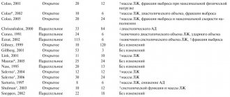
Functional characteristics of the capillary network
The total capacity of the capillary bed is 25–30 liters, while the volume of blood in the human body is 5 liters. Therefore, most of the capillaries are periodically switched off from the bloodstream. In humans, under resting conditions, only 20–35% of capillaries are open at a time. In a muscle at rest, no more than 40% of the capillaries are filled with blood. During physical activity, almost all capillaries of the working muscle are included in the bloodstream. Capillaries themselves are not able to change their lumen. As already mentioned, blood flow in them is regulated by narrowing or dilating blood-bearing arterioles and the use of arteriolovenular anastomoses. Observations indicate that organs are constantly replacing some functioning capillaries with others. High variability of blood flow in the capillaries is a necessary condition for the adaptation of the microcirculatory system to the needs of organs and tissues for the delivery of nutrients.
The structure and work of the heart. Circulation circles
Blood performs its many functions only if it moves. It flows through a giant network of blood vessels at high speed
.
For example, a red blood cell, which delivers oxygen to our cells, travels from the heart to the knee in less than 3 seconds
.
Blood moves through the vessels continuously thanks to the work of the heart,
which is part of the circulatory system.
The continuous flow of blood through a closed system of blood vessels in a strictly defined direction is called
blood circulation
.
The human body is characterized
by a four-chambered heart
and
a closed circulatory system with two circulation circles
.
Blood vessels
are present in almost all tissues.
They are not found
only
in nails, cartilage, tooth enamel, and epithelium
. The nutrition of the cells of these structures occurs due to the movement of necessary substances from nearby tissues.
The structure of the adult human heart. Its dimensions are approximately equal to a hand clenched into a fist. It weighs about 250 grams
for women
and
330 grams for men
.
The heart is located in the center of the chest and is shifted by the lower left edge to the left side
.
asymmetrically
in relation to the midline of the body . Two-thirds of the heart is in the left side of the chest, and one-third is in the right. The upper, expanded part of the heart, from which the vessels depart, is called the base, and the lower, slightly narrowed part is called the apex.
The wall of the heart consists of three layers
.
The outer connective tissue layer is called
the epicardium
.
The middle layer consists of special striated cardiac muscle tissue and is called
the myocardium . The inner layer -
endocardium -
is formed by flat epithelium
.
The heart is surrounded by the pericardial sac
–
pericardium
, which separates the heart from other organs.
Between the epicardium and pericardium there is a closed cavity
filled
with fluid, which reduces friction during heart contractions
.
The human heart consists of right and left halves
(in the picture, the left half is located on the right).
They are separated by a solid partition
and do not communicate with each other.
Each half of the heart contains an atrium and
a ventricle ,
between which there is an atrioventricular opening
.
It is closed on the left side of the heart by the bicuspid valve
, and on the right -
a tricuspid valve
.
The muscular wall of the atria is much thinner than the wall of the ventricles. This is explained by the fact that the atria perform less strenuous work compared to the ventricles. The left ventricle bears a particularly heavy load. Its muscular wall is approximately three times thicker than that of the right ventricle.
2 vena cavae
to the right atrium 4 pulmonary veins
to the left atrium . A large blood vessel departs from the right ventricle -
the pulmonary trunk , and from the left -
the aorta
. The openings from which the pulmonary trunk and aorta begin are closed by semilunar valves in the form of three pockets. They open only during contraction of the ventricles, when blood is released from them under high pressure into the vessels.
Due to the presence of valves, blood moves in only one direction: from the atria to the ventricles, and from the ventricles to the blood vessels.
The heart works continuously throughout a person's life.
Not even the most advanced engine can compare with it in terms of performance. If the heart stops for even a few moments, then loss of consciousness occurs, and if the heart is not immediately forced to contract, death occurs.
The work of the heart is characterized by alternating contraction and relaxation of the atria and ventricles
.
The contraction of the heart is called systole
, and
relaxation -
diastole .
The period that covers one contraction and relaxation of the heart is called the cardiac cycle
.
At rest, the human heart beats an average of 75 times per minute
. Let's calculate the duration of the cardiac cycle for a given rhythm. To do this, divide 60 (there are 60 seconds in one minute) by 75 and get 0.8 seconds - this is the duration of the cardiac cycle. Of this time, atrial systole takes 0.1 seconds, the ventricles at this moment are in a relaxed state. The pressure inside the atria at this time becomes greater than in the relaxed ventricles, and blood flows freely through the atrioventricular openings.
This is followed by ventricular systole, the atria at this moment are relaxed. It lasts 0.3 seconds. At this moment, the pressure inside the ventricles increases, the bicuspid and tricuspid valves quickly slam shut, and the blood is trapped for some time in a closed space inside the ventricles.
Once the pressure in the ventricles exceeds the pressure in the aorta, the semilunar valves open and blood is pumped out of the heart. Contraction of the ventricles is replaced by their relaxation.
The period during which both the ventricles and atria are relaxed is called a general pause.
. Its duration is 0.4 seconds. At this time, the bicuspid and tricuspid valves are open, and the ventricles are filled with blood flowing from the atria.
During the contraction of the heart, blood is released into the vessels leaving the heart
.
All vessels in the human body are divided into arteries, capillaries and veins.
Arteries
-
these are vessels that carry blood from the heart to organs and tissues
.
Oxygen-enriched blood flows through them
.
Such blood is called arterial
. The only exceptions are the pulmonary arteries, which depart from the heart and carry venous blood.
Structure of arteries. Their walls consist of three shells. The outer connective tissue membrane gives the walls elasticity. The middle (two-layer shell) consists of elastic fibers and smooth muscle cells. Let us remember that smooth muscle cells are capable of contracting and relaxing, which causes a change in the diameter of the blood vessel and, accordingly, a change in the amount of blood flowing to the organ. The inner lining is formed by epithelial cells. Like the external one, it gives strength to the arteries.
In the human body, arteries branch many times into smaller vessels.
–
arterioles
.
The smallest arterioles
become
capillaries
.
Capillaries
-
thinnest vessels that penetrate all organs and tissues of the body.
In humans, their number is about forty billion, and their total length reaches one hundred thousand kilometers, that is, they can surround the globe at the equator almost three times.
In capillaries, various substances and gases are exchanged between blood and tissue fluid. These processes are possible because the walls of the capillaries are represented by one layer of cells loosely adjacent to each other. Passing through the capillaries, the blood gives off oxygen and nutrients and is enriched with carbon dioxide and metabolic end products.
Next, blood flows from the capillaries to the venules
–
small veins
. Their walls and the walls of capillaries have a similar structure. This means that venules are also involved in the exchange of substances between blood and tissue fluid.
From the venules, blood collects into larger blood vessels that carry blood to the heart. Blood saturated with carbon dioxide and metabolic products flows through the veins. This type of blood is called venous. The exception is the pulmonary veins, which carry arterial blood to the heart.
The walls of veins are similar in structure to the walls of arteries, but they are much thinner and more elastic. The difference is that the muscle layer in the veins is poorly developed and sometimes completely absent.
In humans, blood flows through two closed systems of vessels, each of which is connected to the heart,
–
these are
the large (systemic) and
small (pulmonary) circles of blood circulation
.
The systemic circulation begins from the left ventricle of the heart by the largest artery - the aorta. It goes up, forming an arc, and then goes down along the spine. Two large arteries branch off from the aortic arch, which carry blood to the head and upper limbs. Below the aortic arch, vessels supply blood to the torso, internal organs and legs. In the organs of the artery, smaller arterioles are divided, which branch and form extensive networks of capillaries. From the capillaries, the blood collects into venules, which merge with each other and form veins. The systemic circulation ends with the superior and inferior vena cava, which flow into the right atrium. The superior vena cava carries blood from the head, neck and arms to the heart, and the inferior vena cava carries blood to the heart from the torso, abdominal organs and lower extremities.
Blood circulating through the systemic circulation
, supplies all cells of the body with oxygen and nutrients and removes carbon dioxide and decay products from them.
From the blood passing through the chambers of the heart, the heart muscle itself cannot extract anything for its own nutrition.
Therefore, just like other organs, it is supplied with arterial blood. Two arteries go to the heart from the aorta. They, like a crown or crown, surround the heart and are therefore called coronary or coronary. The branches of the coronary vessels penetrate into the thickness of the heart muscle, supplying it with nutrients and oxygen. The veins that collect blood from the heart muscle drain directly into the right atrium.
Now let's look at where the blood moves after it is in the right atrium. the pulmonary circulation originates
. From the right ventricle, blood is pushed into the pulmonary trunk, which divides into two pulmonary arteries that enter the left and right lungs.
There they branch into arterioles, then into capillaries, which densely entwine the pulmonary air vesicles. When blood passes through the capillaries of the lungs, it loses carbon dioxide and is saturated with oxygen.
This is where venous blood turns into arterial blood. Further, just as in the systemic circulation, the capillaries merge into venules, which, in turn, form veins, and finally, through the four pulmonary veins, arterial blood enters the left atrium.
It turns out that the left half of the heart is filled with oxygen-rich arterial blood, and the right half is filled with oxygen-poor but carbon dioxide-rich venous blood.
.
Arterial and venous blood do not mix with each other
, since the left and right halves of the heart are separated from each other by a solid septum.
Lesson summary. The movement of blood in the human body (blood circulation) occurs due to the continuous work of the heart, which sequentially distils blood through the systemic and pulmonary circulation. The human circulatory system is closed. The heart is four-chambered, consisting of two atria and two ventricles.
There are three stages in the work of the heart: contraction of the atria, contraction of the ventricles and a general pause. There are three types of vessels: arteries (through which blood moves from the heart), capillaries (the smallest blood vessels in which gas exchange in the lungs and metabolism in tissues occurs) and veins, through which blood returns to the heart.
Features of blood flow in capillaries
Since the capacity of the capillary bed is very large, this leads to a significant slowdown in blood flow in the capillaries. The speed of blood movement through capillaries ranges from 0.3 to 1 mm/s, while in large arteries it reaches 80–130 mm/s. Slow blood flow ensures the most complete exchange of substances between blood and tissues. When blood moves, its cells (erythrocytes) line up in the capillary in one row, since their radius is approximately equal to the radius of the capillary. The significance of such a device becomes clear if we remember that oxygen is carried by red blood cells and its transfer to organ cells will occur most efficiently if the red blood cells are in best contact with the capillary wall. When moving through capillaries, red blood cells are easily deformed, so even the narrowest capillaries are not an obstacle for them. Unlike red blood cells, other blood cells (lymphocytes) have difficulty crossing narrow sections of the capillary bed and can clog the lumen of the capillary for some time.
With a significant decrease in the speed of capillary blood flow, red blood cells can stick together and form aggregates like coin columns of 25–50 red blood cells. Large aggregates can completely clog the capillary and cause blood to stop in it. Increased erythrocyte aggregation occurs in various diseases.
Blood circulation in humans
Arteries
- These are vessels that carry blood from the heart.
They have a thick muscle layer. Veins
are vessels that carry blood to the heart. They have a thin muscle layer and valves.
Capillaries
- These are single-layer vessels in which the exchange of substances between blood and tissues occurs.
Arterial blood
- This is blood saturated with oxygen.
Venous blood
is saturated with carbon dioxide. In the pulmonary circulation, venous blood flows through the arteries, and arterial blood flows through the veins.
The human heart has four chambers
, consists of two atria and two ventricles (in the left half of the heart there is arterial blood, in the right - venous).
The leaflet valves are located between the ventricles and atria
, and between the arteries and ventricles -
semilunar.
The valves prevent blood from flowing backwards (from the ventricle to the atrium, from the aorta to the ventricle).
The thickest wall is at the left ventricle, because it pushes blood through the systemic circulation. When the left ventricle contracts, a pulse wave is created, as well as maximum blood pressure.
Blood pressure:
in the arteries the largest, in the capillaries the average, in the veins the smallest.
Blood speed:
the highest in the arteries, the smallest in the capillaries, the average in the veins.
Big circle
blood circulation: from the left ventricle, arterial blood flows through the arteries to all organs of the body. In the capillaries of the large circle, gas exchange occurs: oxygen passes from the blood into the tissues, and carbon dioxide passes from the tissues into the blood. The blood becomes venous, flows through the vena cava into the right atrium, and from there into the right ventricle.
Small circle:
From the right ventricle, venous blood flows through the pulmonary arteries to the lungs. Gas exchange occurs in the capillaries of the lungs: carbon dioxide passes from the blood into the air, and oxygen from the air into the blood, the blood becomes arterial and flows through the pulmonary veins into the left atrium, and from there into the left ventricle.
You can also read
IF THE INFORMATION IS NOT IN THE MAIN SUMMARY, THEN IT IS IN THE DETAILED SUMMARY:
Blood vessels, Blood pressure and velocity, Heart
ASSIGNMENTS OF PART 2 OF THE USE ON THIS TOPIC
Part 1 tasks
Choose one, the most correct option. Why can't blood get from the aorta to the left ventricle of the heart?
1) the ventricle contracts with great force and creates high pressure 2) the semilunar valves fill with blood and close tightly 3) the leaflet valves are pressed against the walls of the aorta 4) the leaflet valves are closed and the semilunar valves are open
Answer
2
Choose one, the most correct option. Blood enters the pulmonary circulation from the right ventricle through
1) pulmonary veins 2) pulmonary arteries 3) carotid arteries 4) aorta
Answer
2
Choose one, the most correct option. Arterial blood flows through the human body
1) renal veins 2) pulmonary veins 3) vena cava 4) pulmonary arteries
Answer
2
Choose one, the most correct option. In mammals, blood is enriched with oxygen in
1) arteries of the pulmonary circulation 2) capillaries of the systemic circle 3) arteries of the systemic circle 4) capillaries of the systemic circle
Answer
4
Choose one, the most correct option. The vena cava in the human body drains into
1) left atrium 2) right ventricle 3) left ventricle 4) right atrium
Answer
4
Choose one, the most correct option. Valves prevent blood from flowing back from the pulmonary artery and aorta into the ventricles.
1) tricuspid 2) venous 3) bicuspid 4) semilunar
Answer
4
Choose one, the most correct answer. Indicate the sequence of processes occurring in the heart during one full cycle of its work.
1) contraction of the atria and relaxation of the ventricles, relaxation of the atria and contraction of the ventricles, general pause 2) contraction of the atria and ventricles, relaxation of the atria and ventricles, general pause 3) contraction of the right ventricle and atrium, relaxation of the left ventricle and atrium, general pause 4) contraction of the left atrium and right ventricle, general pause, contraction of the right atrium and right ventricle
Answer
1
ARTERIES Choose three correct answers out of six and write down the numbers under which they are indicated. What features are characteristic of human arteries?
A) has a thin muscle layer B) has valves C) carries blood from the heart D) carries blood to the heart E) has elastic elastic walls E) withstands high blood pressure
Answer
112122
ARTERIES - VEINS 1. Establish a correspondence between the signs and blood vessels: 1) vein 2) artery. Write numbers 1 and 2 in the order corresponding to the letters.
A) has a thin muscle layer B) has valves C) carries blood from the heart D) carries blood to the heart E) has elastic elastic walls E) withstands high blood pressure
Answer
112122
2. Establish a correspondence between the structural features and functions and types of vessels: 1) artery, 2) vein. Write numbers 1 and 2 in the order corresponding to the letters.
A) has valves B) the wall contains fewer muscle fibers C) carries blood from the heart D) carries venous blood in the pulmonary circulation D) communicates with the right atrium E) carries out blood flow due to contraction of skeletal muscles
Answer
221122
3. Establish a correspondence between the characteristics and types of blood vessels: 1) arteries, 2) veins. Write numbers 1 and 2 in the order corresponding to the letters.
A) carry blood from the heart B) contain valves throughout C) have a thicker muscle layer in the wall D) can collapse E) resist high pressure E) carry arterial blood in the pulmonary circulation
Answer
121212
4. Establish a correspondence between the sign of human blood vessels and their type: 1) arteries, 2) veins. Write numbers 1 and 2 in the order corresponding to the letters.
A) depart from the ventricles of the heart B) have semilunar valves along their entire length C) carry blood to the heart D) have the fastest blood flow E) have thin and elastic walls, an underdeveloped muscle layer
Answer
12212
ARTERIES - VEINS - CAPILLARIES 1. Establish a correspondence between the features of blood vessels and their types: 1) artery, 2) vein, 3) capillary. Write numbers 1-3 in the order corresponding to the letters.
A) the wall consists of one layer of cells B) endothelial cells fit tightly to each other, forming smooth walls C) the walls have valves D) the walls are thin, elastic, contain muscles E) has the smallest diameter
Answer
31223
2. Establish a correspondence between the characteristic and the blood vessels: 1) arteries, 2) veins, 3) capillaries. Write numbers 1-3 in the order corresponding to the letters.
A) carry out metabolic processes between blood and tissues B) have dense and elastic vascular walls C) blood flows through them to the heart D) have thin single-layer walls E) have semilunar valves throughout their entire length E) have the lowest blood flow rate
Answer
312323
VEINS Select three options. Veins are blood vessels through which blood flows
1) from the heart 2) to the heart 3) under greater pressure than in the arteries 4) under less pressure than in the arteries 5) faster than in the capillaries 6) slower than in the capillaries
Answer
245
VEINS IN EXC. FROM ARTERIES 1. Choose three correct answers out of six and write down the numbers under which they are indicated. Veins, as opposed to arteries
1) have valves in their walls 2) can collapse 3) have walls made of a single layer of cells 4) carry blood from organs to the heart 5) withstand high blood pressure 6) always carry blood that is not saturated with oxygen
Answer
124
2. Choose three correct answers out of six and write down the numbers under which they are indicated. Veins, unlike arteries, are characterized by
1) leaflet valves 2) transport of blood to the heart 3) semilunar valves 4) high blood pressure 5) thin muscle layer 6) fast blood flow
Answer
235
VENOUS BLOOD Choose three correct answers out of six and write down the numbers under which they are indicated. Elements of the human circulatory system containing venous blood are
1) pulmonary artery 2) aorta 3) vena cava 4) right atrium and right ventricle 5) left atrium and left ventricle 6) pulmonary veins
Answer
134
ARTERIAL - VENOUS 1. Establish a correspondence between the type of human blood vessels and the type of blood they contain: 1) arterial, 2) venous
A) pulmonary arteries B) veins of the pulmonary circulation C) aorta and arteries of the systemic circulation D) superior and inferior vena cava
Answer
2112
2. Establish a correspondence between a vessel of the human circulatory system and the type of blood that flows through it: 1) arterial, 2) venous. Write numbers 1 and 2 in the order corresponding to the letters.
A) femoral vein B) brachial artery C) pulmonary vein D) subclavian artery E) pulmonary artery E) aorta
Answer
211121
3. Establish a correspondence between the sections of the human circulatory system and the type of blood passing through them: 1) arterial, 2) venous. Write numbers 1 and 2 in the order corresponding to the letters.
A) left ventricle B) right ventricle C) right atrium D) pulmonary vein E) pulmonary artery E) aorta
Answer
122121
ARTERIAL IN EXC. FROM VENOUS Select three options. In mammals and humans, venous blood, unlike arterial,
1) is poor in oxygen 2) flows in a small circle through the veins 3) fills the right half of the heart 4) is saturated with carbon dioxide 5) enters the left atrium 6) provides body cells with nutrients
Answer
134
HEART Choose three correct answers out of six and write down the numbers under which they are indicated. Which of the following is true to describe the functioning of the human heart?
1) Venous blood is located in the right side of the heart. 2) The superior and inferior vena cava flow into the left atrium. 3) Leaf valves separate the atria and ventricles. 4) The myocardium receives oxygen from the blood in the left ventricle. 5) From the left ventricle, blood enters the aorta. 6) The semilunar valves of the aorta close during ventricular systole.
Answer
135
Analyze the table “Structure of the Heart”. For each cell indicated by a letter, select the corresponding term from the list provided.
1) Contracting, provides blood flow through the systemic circulation 2) Left atrium 3) Separated from the left ventricle by a bicuspid valve 4) Right atrium 5) Separated from the right atrium by a tricuspid valve 6) Contracting, directs blood to the left ventricle 7) Pericardial sac
Answer
451
SIGNATURES 1. Choose three correctly labeled captions for the drawing that depicts the internal structure of the heart. Write down the numbers under which they are indicated.
1) superior vena cava 2) aorta 3) pulmonary vein 4) left atrium 5) right atrium 6) inferior vena cava
Answer
126
2. Choose three correctly labeled captions for the picture that depicts the structure of the human heart. Write down the numbers under which they are indicated.
1) superior vena cava 2) leaflet valves 3) right ventricle 4) semilunar valves 5) left ventricle 6) pulmonary artery
Answer
246
3. Select three correctly labeled captions for the figure “Structure of the Human Heart” and write down the numbers under which they are indicated.
1) vena cava 2) left half of the heart 3) carotid artery 4) pulmonary artery 5) coronary artery 6) bicuspid valve
Answer
146
CHAMBERS 1. Establish a correspondence between the structural and functional features and the chambers of the heart indicated in the figure. Write numbers 1 and 2 in the order corresponding to the letters.
A) is the end of the systemic circulation B) is the beginning of the systemic circulation C) is filled with venous blood D) is filled with arterial blood E) has a thin muscle wall
Answer
12121
2. Establish a correspondence between the chambers of the heart, indicated in the figure by numbers 1 and 2, and their structural features and functions. Write numbers 1 and 2 in the order corresponding to the letters.
A) is the end of the pulmonary circulation B) is the end of the systemic circulation C) is filled with venous blood D) is filled with arterial blood E) is connected to the pulmonary vein
Answer
12211
3. Establish a correspondence between the chambers of the heart, indicated in the figure by numbers 1 and 2, and their structural features and functions. Write numbers 1 and 2 in the order corresponding to the letters.
A) is the end of the pulmonary circulation B) is the beginning of the pulmonary circulation C) is filled with venous blood D) is filled with arterial blood E) has a thinner muscle wall
Answer
12211
LEFT VENTRICLE 1. Select three options. A person has blood from the left ventricle of the heart
1) when it contracts, it enters the aorta 2) when it contracts, it enters the left atrium 3) supplies the cells of the body with oxygen 4) enters the pulmonary artery 5) under high pressure enters the systemic circulation 6) under low pressure enters the pulmonary circulation
Answer
135
2. Choose three correct answers out of six and write down the numbers under which they are indicated. From the left ventricle of the heart
1) blood enters the systemic circulation 2) venous blood comes out 3) arterial blood comes out 4) blood flows through the veins 5) blood flows through the arteries 6) blood enters the pulmonary circulation
Answer
135
RIGHT VENTRICLE Choose three correct answers out of six and write down the numbers under which they are indicated. Blood leaks from the right ventricle
1) arterial 2) venous 3) through arteries 4) through veins 5) towards the lungs 6) towards the cells of the body
Answer
235
LEFT - RIGHT Establish a correspondence between the characteristics and chambers of the human heart: 1) left ventricle, 2) right ventricle. Write numbers 1 and 2 in the order corresponding to the letters.
A) The pulmonary arteries depart from it. B) It enters the systemic circulation. B) Contains venous blood. D) It has thicker muscle walls. D) A bicuspid valve opens into it. E) Contains oxygen-rich blood.
Answer
212111
HEART SEQUENCE 1. Establish the sequence of events that occur in the cardiac cycle after blood enters the heart. Write down the corresponding sequence of numbers.
1) contraction of the ventricles 2) general relaxation of the ventricles and atria 3) blood flow into the aorta and artery 4) blood flow into the ventricles 5) contraction of the atria
Answer
54132
2. Establish the sequence of stages of the human cardiac cycle after a pause, starting with the filling of the chambers with blood. Write down the corresponding sequence of numbers.
1) pause 2) contraction of the atria 3) blood enters the aorta and pulmonary arteries 4) contraction of the ventricles 5) blood from the atria enters the ventricles
Answer
54132
HEART PHASES 1. Establish a correspondence between the processes and phases of the human cardiac cycle: 1) atrial systole, 2) ventricular systole, 3) diastole. Write numbers 1-3 in the order corresponding to the letters.
A) blood moves into the aorta and pulmonary artery B) leaflet valves are open, semilunar valves are closed C) the duration of the phase is 0.4 s D) blood moves from the atria to the ventricles E) leaflet valves are closed, semilunar valves are open E) blood passes from the veins to the atria and ventricles
Answer
213123
2. Establish a correspondence between the positions of the valves in the human heart and the phases of heart function: 1) contraction of the atria, 2) contraction of the ventricles. Write numbers 1 and 2 in the order corresponding to the letters.
A) The bicuspid valve closes. B) The tricuspid valve is open. C) The semilunar valve of the pulmonary trunk (artery) is closed. D) The semilunar valve of the aorta opens. D) The butterfly valve is open. E) The tricuspid valve closes.
Answer
211212
Look at the picture depicting the phase of the cardiac cycle. Determine (A) the name of this phase, (B) its duration, and (C) the direction of blood flow. For each cell indicated by a letter, select the appropriate term or process from the list provided.
1) ventricular systole 2) atrial systole 3) blood flow from the ventricle into the artery 4) 0.1 s 5) 0.8 s 6) blood flow from the atrium into the ventricle 7) blood flow from the veins into the atrium 0.3 s
s
Answer
246
Analyze the table “Human Heartbeat Cycle”. For each letter, select the corresponding term from the list provided.
1) contraction of the atria and ventricles 2) contraction of the ventricles, relaxation of the atria 3) contraction of the atria 4) valves and semilunars are open 5) valves and semilunars are closed 6) valves are closed, semilunars are open 7) from the atria to the ventricles and arteries flows freely from the veins and atria into the ventricles
Answer
368
SPEED SEQUENCE Arrange the blood vessels in order of decreasing speed of blood movement in them
1) superior vena cava 2) aorta 3) brachial artery 4) capillaries
Answer
2314
PRESSURE SEQUENCE 1. Establish the sequence of human blood vessels in order of decreasing blood pressure in them. Write down the corresponding sequence of numbers.
1) inferior vena cava 2) aorta 3) pulmonary capillaries 4) pulmonary artery
Answer
2431
2. Establish the order in which the blood vessels should be arranged in order of decreasing blood pressure in them
1) Veins 2) Aorta 3) Arteries 4) Capillaries
Answer
2341
3. Establish the sequence of arrangement of blood vessels in order of increasing blood pressure in them. Write down the corresponding sequence of numbers.
1) inferior vena cava 2) aorta 3) pulmonary artery 4) alveolar capillaries 5) arterioles
Answer
14532
BIG Choose three correct answers out of six and write down the numbers under which they are indicated. Large circle of blood circulation in the human body
1) begins in the left ventricle 2) originates in the right ventricle 3) is saturated with oxygen in the alveoli of the lungs 4) supplies organs and tissues with oxygen and nutrients 5) ends in the right atrium 6) brings blood to the left half of the heart
Answer
145
Choose three correct answers out of six and write down the numbers under which they are indicated. Which parts of the circulatory system belong to the systemic circulation?
1) pulmonary artery 2) superior vena cava 3) right atrium 4) left atrium 5) left ventricle 6) right ventricle
Answer
235
LARGE SEQUENCE 1. Establish the sequence of blood movement through the vessels of the systemic circulation. Write down the corresponding sequence of numbers.
1) hepatic portal vein 2) aorta 3) gastric artery 4) left ventricle 5) right atrium 6) inferior vena cava
Answer
423165
2. Determine the correct sequence of blood circulation in the systemic circulation, starting with the left ventricle. Write down the corresponding sequence of numbers.
1) Aorta 2) Superior and inferior vena cava 3) Right atrium 4) Left ventricle 5) Right ventricle 6) Tissue fluid
Answer
416235
3. Establish the correct sequence of blood passage through the systemic circulation. Write down the corresponding sequence of numbers in the table.
1) right atrium 2) left ventricle 3) arteries of the head, limbs and torso 4) aorta 5) inferior and superior vena cava 6) capillaries
Answer
243651
4. Establish the sequence of blood movement through the systemic circulation in humans, starting from the ventricle. Write down the corresponding sequence of numbers.
1) left ventricle 2) capillaries 3) right atrium 4) arteries 5) veins 6) aorta
Answer
164253
5. Establish the sequence of processes during the passage of blood in the human body through the systemic circulation, starting with the aorta. Write down the corresponding sequence of numbers.
1) the blood disperses throughout the body and brings oxygen and nutrients to the organs 2) the blood moves through the vessels from the arms, neck and head 3) the blood enters the largest artery 4) in the organs the blood gives off oxygen and collects carbon dioxide 5) along the upper the vena cava carries blood into the right atrium
Answer
31425
GREAT CIRCLE ARTERIES Select three options. Blood flows through the arteries of the systemic circulation in humans
1) from the heart 2) to the heart 3) saturated with carbon dioxide 4) saturated with oxygen 5) faster than in other blood vessels 6) slower than in other blood vessels
Answer
145
SMALL SEQUENCE Establish the sequence of blood movement in a person through the pulmonary circulation. Write down the corresponding sequence of numbers.
1) pulmonary artery 2) right ventricle 3) capillaries 4) left atrium 5) veins
Answer
21354
SMALL CIRCLE ARTERY Select three options. Blood flows through the arteries of the pulmonary circulation in humans
1) from the heart 2) to the heart 3) saturated with carbon dioxide 4) saturated with oxygen 5) faster than in the pulmonary capillaries 6) slower than in the pulmonary capillaries
Answer
135
LARGE - SMALL VESSELS 1. Establish a correspondence between the sections of the circulatory system and the circle of blood circulation to which they belong: 1) Systemic circulation, 2) Lesser circulation. Write numbers 1 and 2 in the correct order.
A) Right ventricle B) Carotid artery C) Pulmonary artery D) Superior vena cava E) Left atrium E) Left ventricle
Answer
212121
2. Establish a correspondence between the vessels and human circulatory circles: 1) pulmonary circulation, 2) systemic circulation. Write numbers 1 and 2 in the correct order.
A) aorta B) pulmonary veins C) carotid arteries D) capillaries in the lungs E) pulmonary arteries E) hepatic artery
Answer
212112
3. Establish a correspondence between the structures of the circulatory system and the human circulation circles: 1) small, 2) large. Write numbers 1 and 2 in the order corresponding to the letters.
A) aortic arch B) hepatic portal vein C) left atrium D) right ventricle E) carotid artery E) alveolar capillaries
Answer
221121
LARGE - SMALL SIGNS Establish a correspondence between the processes and the circles of blood circulation for which they are characteristic: 1) small, 2) large. Write numbers 1 and 2 in the order corresponding to the letters.
A) Arterial blood flows through the veins. B) The circle ends in the left atrium. B) Arterial blood flows through the arteries. D) The circle begins in the left ventricle. D) Gas exchange occurs in the capillaries of the alveoli. E) Venous blood is formed from arterial blood.
Answer
112212
LARGE-SMALL SEQUENCE 1. Establish the sequence of blood movement in the human body, starting with the left ventricle. Write down the corresponding sequence of numbers.
1) left ventricle 2) vena cava 3) aorta 4) pulmonary veins 5) right atrium
Answer
13254
2. Establish the sequence of passage of a portion of blood in a person, starting from the left ventricle of the heart. Write down the corresponding sequence of numbers.
1) right atrium 2) aorta 3) left ventricle 4) lungs 5) left atrium 6) right ventricle
Answer
321645
3. Establish the sequence of circulatory processes, starting from the moment when blood moves from the lungs to the heart. Write down the corresponding sequence of numbers.
1) blood from the right ventricle enters the pulmonary artery 2) blood moves through the pulmonary vein 3) blood moves through the pulmonary artery 4) oxygen enters from the alveoli into the capillaries 5) blood enters the left atrium 6) blood enters the right atrium
Answer
256134
4. Establish the sequence of movement of arterial blood in a person, starting from the moment it is saturated with oxygen in the capillaries of the pulmonary circle. Write down the corresponding sequence of numbers.
1) left ventricle 2) left atrium 3) veins of the pulmonary circle 4) capillaries of the pulmonary circle 5) arteries of the systemic circle
Answer
43215
5. Establish the sequence of movement of arterial blood in the human body, starting with the capillaries of the lungs. Write down the corresponding sequence of numbers.
1) left atrium 2) left ventricle 3) aorta 4) pulmonary veins 5) pulmonary capillaries
Answer
54123
6. Establish the correct sequence of passage of a portion of blood from the right ventricle to the right atrium. Write down the corresponding sequence of numbers.
1) pulmonary vein 2) left ventricle 3) pulmonary artery 4) right ventricle 5) right atrium 6) aorta
Answer
431265
SEQUENCE WITH ORGANS 1. Establish the sequence of movement of the hormone thyroxine through the human circulatory system, starting from the moment of its formation until reaching the target organ. Write down the corresponding sequence of numbers.
1) left ventricle 2) superior vena cava 3) right atrium 4) medulla oblongata 5) pulmonary trunk 6) thyroid capillary
Answer
623514
2. Establish the sequence of blood vessels through which blood passes when supplying the liver, starting from the moment it is saturated with oxygen. Write down the corresponding sequence of numbers.
1) aorta 2) hepatic vein 3) inferior vena cava 4) hepatic artery 5) pulmonary veins
Answer
51423
3. Establish the sequence of blood movement through the vessels, starting with blood saturation with oxygen. Write down the corresponding sequence of numbers.
1) aorta 2) pulmonary veins 3) inferior vena cava 4) hepatic artery 5) left atrium 6) alveolar capillaries
Answer
625143
4. Establish the sequence of movement of vitamin C through the human circulatory system, starting from the moment of its absorption in the gastrointestinal tract. Write down the corresponding sequence of numbers.
1) capillary of the small intestine 2) pulmonary trunk 3) pulmonary vein 4) right ventricle 5) right atrium 6) inferior vena cava
Answer
165423
Choose three correct answers out of six and write down the numbers under which they are indicated. Human pulse
1) is not associated with the speed of blood flow 2) depends on the elasticity of the walls of blood vessels 3) is palpated on large arteries close to the surface of the body 4) accelerates blood flow 5) is caused by the rhythmic oscillation of the veins 6) is not associated with the contraction of the heart
Answer
123
Establish the sequence of carbon dioxide transport from the moment it enters the blood. Write down the corresponding sequence of numbers.
1) left ventricle 2) capillaries of internal organs 3) vena cava 4) capillaries of the alveoli
Answer
2341
Establish a correspondence between human blood vessels and the direction of blood movement in them: 1) from the heart, 2) to the heart
A) veins of the pulmonary circulation B) veins of the systemic circulation C) arteries of the pulmonary circulation D) arteries of the systemic circulation
Answer
2211
When running on a treadmill, a person's heart rate doubled from the original 75 beats per minute. The stroke volume of blood also doubled - from 60 ml to 120 ml. What will be the minute volume of blood in such an intensively working heart? Write down only the number of liters in your answer.
Answer
18
With a pulse of 75 beats per minute, the left ventricle of the heart pumps out 60 ml of blood in one contraction. Calculate the amount of oxygen absorbed by tissues in one minute if the oxygen content in arterial blood is 200 ml/l, and in venous blood - 150 ml/l. In your answer, write down only the amount of oxygen in milliliters.
Answer
225
© D.V. Pozdnyakov, 2009-2020
Regulation of blood microcirculation
How does microcirculation regulation occur? Firstly, microvessels react to stretching: when blood pressure increases, arterioles narrow and limit blood flow into the capillaries, and when pressure decreases, they expand. Secondly, sympathetic nerves approach the largest of the microvessels (but not the capillaries), and when irritated, large arterioles and venules narrow. Thirdly, microvessels are very sensitive to vasoactive substances dissolved in the blood and react even to their concentration, which is 10–100 times less than that required for the narrowing or dilation of large vessels. Thus, skin vessels show high sensitivity to adrenaline (complete closure of the lumen of arterioles occurs when its concentration in the blood is insignificant - the skin turns pale), while the microvessels of internal organs are much less sensitive, and the microvessels of skeletal muscles and the heart can expand under the action of adrenaline. Potassium, calcium, sodium ions, as well as substances that accumulate in tissues during intense activity, lead to the expansion of microvessels. Precapillary arterioles have the greatest sensitivity to the action of vasoactive substances, and large arterioles and venules have the least sensitivity.
Diagnosis of blood microcirculation disorders
Relevant for modern clinical practice, assessment of the state of microcirculation and diagnosis of its disorders in a wide variety of diseases can be done using methods such as capillaroscopy of the skin and mucous membranes, biomicroscopy of conjunctival vessels, laser Doppler flowmetry. The state of microcirculation in any part of the body makes it possible to judge its state in the body as a whole with a high degree of accuracy.
Early signs of capillary blood flow disorders are narrowing of arterioles, congestion in venules, leading to their expansion and significant tortuosity, as well as a decrease in the intensity of blood flow in the capillaries. At later stages, widespread intravascular aggregation of erythrocytes is detected, which inevitably entails a stop of blood flow in the capillaries. The final outcome of microcirculatory disorders is stasis, i.e. complete blockage of blood flow and a sharp disruption of the barrier function of microvessels, which is often accompanied by hemorrhages - the release of red blood cells through the wall of the capillaries, which are the most vulnerable. Arteriovenular anastomoses are more resistant to microcirculatory disorders and tend to maintain blood flow even in conditions of stasis spreading to a significant part of the microcirculatory bed.
Microcirculation disorders underlie a large number of diseases, so their treatment requires restoration of microvascular functions with the help of various medications.
Author: Olga Gurova, Candidate of Biological Sciences, Senior Researcher, Associate Professor of the Department of Human Anatomy of the RUDN University