December 12, 2013
The extensive network of the human circulatory system consists not only of large veins and arteries, but also of the smallest capillaries
The extensive network of the human circulatory system consists not only of large veins and arteries, but also of the smallest capillaries, thanks to which all the substances necessary for optimal life are delivered to each of our cells along with blood. It is not surprising that a person’s health largely depends on the state of his cardiovascular system.
Vessels
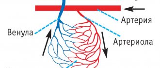
Arteries are vessels that deliver blood from the heart to the organs. The arterial system, which permeates the entire body, includes the arteries of the heart, pulmonary, renal, splenic arteries, aorta, brachiocephalic arteries, arteries of the extremities, etc. Since the arteries need to withstand considerable blood pressure, their walls are initially elastic and elastic. However, as a result of atherosclerosis, the walls of the arteries are weakened. Impaired elasticity of the walls of blood vessels and a decrease in the lumen inside the arteries lead to heart failure and coronary heart disease. As a result of the changes, the arterial walls become stratified and aneurysms form on them. Such ailments are dangerous, because disruption of just one link in the chain of the cardiovascular system will lead to pathological processes inside the body.>
Capillaries are vessels that connect the arterial and venous systems. Thanks to these thinnest vessels, blood and tissues exchange substances with each other. Capillaries permeate tissues that need oxygen.
Veins are vessels that carry blood obtained from capillaries to the heart.
Some numbers
There are more than 95 thousand kilometers of blood vessels in the body of a healthy adult. More than seven thousand liters of blood are pumped through them every day. The size of the blood vessels varies from 25 mm
(aortic diameter)
to eight microns
(capillary diameter).
Down with atherosclerosis!
Atherosclerosis is one of the most common vascular diseases. It is characterized by the formation of fatty plaques on the walls of the arteries. Find out how to prevent atherosclerosis.
Heart
The heart is the organ that pumps blood from the veins to the arteries. Thanks to the work of the heart, blood is supplied to the organs through arteries and capillaries. It is like a powerful pump and consists of muscle tissue - the myocardium. The heart pumps about 5 liters of blood per minute. An abrupt stop in its work is fraught with fatal consequences - myocardial infarction or heart attack often ends in the death of a person.
Foundation of life
The circulatory system consists of more than just the heart, blood, and blood vessels. This is only one of two complementary systems - cardiovascular and lymphatic. The latter serves to transport lymph - a colorless liquid with many lymphocytes. The lymphatic system is also extremely important, because human immunity largely depends on it. It is these two systems - cardiovascular and lymphatic - that make up the largest human circulatory system with a total length of more than 100,000 km. This complex mechanism is driven by the heart. This living motor, consisting of muscles, works with amazing performance, pumping more than 9,500 liters of blood per day. In this way, blood is supplied to every cell.
Circulation circles
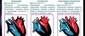
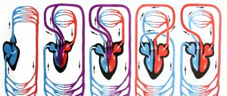
The structure of the human cardiovascular system is characterized by closedness. It forms blood circulation circles. The pulmonary circulation carries blood from the heart to the lungs, and also in the opposite direction. The small circle begins in the right ventricle and ends in the left atrium. In this circle, the blood is saturated with oxygen and removes carbon dioxide. The systemic circulation ensures the movement of blood from the left ventricle to the right atrium, and all organs and tissues are saturated with blood.
What is blood pressure?
Blood pressure is the force with which blood presses against the walls of the arteries. It increases when the heart contracts and pumps out blood, and decreases when the heart muscle relaxes. Blood pressure is stronger in the arteries and weaker in the veins. Blood pressure is measured with a special device - tonometer
.
Pressure readings are usually recorded in two numbers. Thus, normal blood pressure for an adult is considered to be 120/80
.
The first number, systolic pressure
, is a measure of the pressure during a heartbeat.
The second is diastolic pressure
- the pressure during relaxation of the heart.
Such dangerous excess weight
Professor Sergei Boytsov, chief specialist in preventive medicine at the Russian Ministry of Health and Social Development, talks about how extra pounds affect the heart and blood vessels.
Pressure is measured in the arteries and expressed in millimeters of mercury. In the capillaries, the pulsation of the heart becomes invisible and the pressure in them drops to approximately 30 mm Hg. Art. A blood pressure reading can tell your doctor how your heart is working. If one or both numbers are higher than normal, this indicates high blood pressure. If it’s lower, it means it’s reduced. High blood pressure indicates that the heart is working too hard: it requires more effort to push blood through the vessels. It also indicates that a person has an increased risk of heart disease.
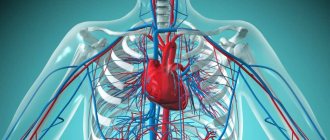
Human circulatory system: structure and functions
Normal life activity is impossible without effective blood circulation: it maintains the constancy of the internal environment, transports oxygen, hormones, nutrients and other vital substances, takes part in the cleansing of toxins, waste, decay products, the accumulation of which would sooner or later lead to the death of an individual organ or the whole organism. This process is regulated by the circulatory system - a group of organs, thanks to the joint work of which blood is consistently moved throughout the human body.
Let's look at how the circulatory system
, and what functions it performs in the human body.
The structure of the human circulatory system
At first glance, the circulatory system is structured simply and clearly: it includes the heart and numerous vessels through which blood flows, successively reaching all organs and systems. The heart is a kind of pump that stimulates the blood, ensuring its smooth flow, and the vessels play the role of guiding tubes that determine the specific path of blood movement throughout the body. That is why the circulatory system is also called the cardiovascular or cardiovascular system.
Let's talk in more detail about each organ that belongs to the human circulatory system.
Organs of the human circulatory system
Like any organismal complex, the circulatory system includes a number of different organs, which are classified depending on their structure, location and functions:
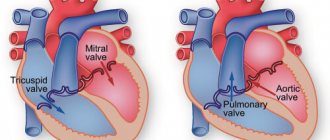
- The heart is considered the central organ of the cardiovascular complex. It is a hollow organ formed primarily by muscle tissue. The cardiac cavity is divided by partitions and valves into 4 sections - 2 ventricles and atria (left and right). Thanks to rhythmic successive contractions, the heart pushes blood through the vessels, ensuring its uniform and continuous circulation.
- Arteries carry blood from the heart to other internal organs. The further from the heart they are located, the thinner their diameter: if in the area of the heart sac the average width of the lumen is the thickness of a thumb, then in the area of the upper and lower extremities its diameter is approximately equal to a simple pencil.
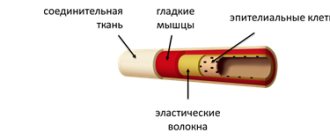
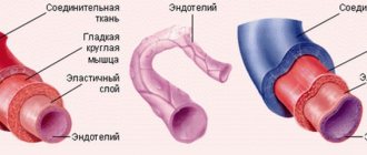
Despite the visual difference, both large and small arteries have a similar structure. They include three layers - adventitia, media and intima. The adventitium, the outer layer, is formed by loose fibrous and elastic connective tissue and includes many pores through which microscopic capillaries pass, feeding the vascular wall, and nerve fibers that regulate the width of the lumen of the artery depending on the impulses sent by the body.
The media, which occupies the median position, includes elastic fibers and smooth muscles, thanks to which the firmness and elasticity of the vascular wall is maintained. It is this layer that largely regulates the speed of blood flow and blood pressure, which can vary within an acceptable range depending on external and internal factors affecting the body. The larger the diameter of the artery, the higher the percentage of elastic fibers in the middle layer. According to this principle, vessels are classified into elastic and muscular.
The intima, or inner lining of the arteries, is represented by a thin layer of endothelium. The smooth structure of this tissue facilitates blood circulation and serves as a passageway for media nutrition.
As the arteries thin, these three layers become less pronounced. If in large vessels the adventitia, media and intima are clearly distinguishable, then in thin arterioles only muscle spirals, elastic fibers and a thin endothelial lining are noticeable.
- Capillaries are the thinnest vessels of the cardiovascular system, which are an intermediate link between arteries and veins. They are localized in the areas most distant from the heart and contain no more than 5% of the total blood volume in the body. Despite their small size, capillaries are extremely important: they envelop the body in a dense network, supplying blood to every cell of the body. This is where the exchange of substances between the blood and adjacent tissues occurs. The thinnest capillary walls easily allow molecules of oxygen and nutritional components contained in the blood to pass through, which, under the influence of osmotic pressure, pass into the tissues of other organs. In return, the blood receives the breakdown products and toxins contained in the cells, which are sent through the venous bed back to the heart and then to the lungs.
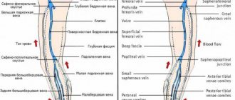
- Veins are a type of vessels that carry blood from internal organs to the heart. The walls of veins, like arteries, are formed by three layers. The only difference is that each of these layers is less pronounced. This feature is regulated by the physiology of the veins: blood circulation here does not require strong pressure from the vascular walls - the direction of blood flow is maintained due to the presence of internal valves. A greater number of them are found in the veins of the lower and upper extremities - here, with low venous pressure, without alternating contraction of muscle fibers, blood flow would be impossible. Large veins, on the other hand, have very few or no valves.
During the circulation process, part of the fluid from the blood seeps through the walls of capillaries and blood vessels to the internal organs. This liquid, visually somewhat reminiscent of plasma, is lymph that enters the lymphatic system. Merging together, the lymphatic pathways form fairly large ducts, which in the area of the heart flow back into the venous bed of the cardiovascular system.
Why does hypotension occur?
Low blood pressure (hypotension) is not dangerous for humans. However, people experiencing hypotension feel weak in the body. Dizziness often occurs, concentration decreases and fatigue increases. Therefore, hypotension can cause great discomfort. The most unpleasant consequences of this phenomenon are considered to be a decrease in intellectual activity. Girls face hypotension, and the World Health Organization states that a low blood pressure level is 100/60 mmHg. Art. for women and 110/70 mmHg. for men. It is worth noting that some feel quite comfortable even with constant hypotension.
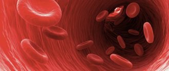
Blood
Blood is a liquid, mobile tissue of the body that penetrates through blood vessels to all tissues and organs, saturating them with oxygen and nutrients.
Blood consists of two main components:
- Plasma is the liquid part of the blood that contains the formed elements. It is a cloudy yellowish liquid.
- Formed elements are special cells that perform the functions of blood: red blood cells, leukocytes and platelets.
Blood with oxygen and nutrients for cells is called arterial, and blood with carbon dioxide and cell metabolic products is called venous.
The importance of veins in the human circulatory system
A feature of the veins is the transfer of red liquid that is not yet saturated with oxygen. They have less strong walls than the largest artery in the body. Accordingly, unlike arterial walls, venous walls experience less pressure. However, among the veins there is also one rather large one. The largest vein reaches a diameter of 2.5 centimeters. Small veins are usually called venules.
The pulmonary vein carries blood that is already saturated with oxygen. Each vein has internal valves that prevent backflow. A person experiencing valve dysfunction may develop varicose veins.