It is believed that heart disease is more relevant to adults and older people. Indeed, as a rule, heart disease often occurs in adulthood. However, in childhood, various heart rhythm disturbances (arrhythmias, extrasystoles, tachycardia, etc.) are often detected, which actually threaten the child’s life. In order to prevent the development of the disease, as well as to begin timely treatment, it is necessary to detect the presence of a problem in time. The First Children's Medical Center has the opportunity to undergo various types of diagnostic examinations using the most modern equipment. The most common method of examination is ultrasound of the child’s heart.
Often, parents of children whose doctor has prescribed an ultrasound of the heart do not know how this procedure is carried out (especially for parents of infants) and how harmful or harmless it is for the child’s body. A lot of questions arise: how to prepare for this type of diagnosis, at what age it can be performed, and so on. In this article, specialists from the First Children's Medical Center will talk about what a heart ultrasound is, in what cases it is prescribed, how a child's heart ultrasound is performed, and what parents need to know about this diagnostic method.
Causes of development of heart pathologies in children
Cardiovascular diseases in children can be either congenital or acquired. If we talk about congenital diseases, they occur in the fetus in the first half of pregnancy - these are congenital heart defects. There are many reasons for their development. This includes hereditary predisposition, ecology, exposure to intrauterine infections, frequent stress, and emotional overload to which pregnant women are exposed. As for acquired cardiovascular diseases, they occur in children, as a rule, due to infections that could not be completely cured.
Content
- Ultrasound examinations of the brain
- Ultrasound of the hip joints
- Abdominal ultrasound
- Ultrasound of the heart
- Audiological screening
- Pediatrician
In the first month, the baby needs regular monitoring; at this time, the slightest developmental disorders can be identified and measures can be taken to eliminate them. The day after discharge, the baby is visited by the local pediatrician and a visiting nurse from the outpatient department. During the examination, the specialist measures height, weight, head circumference, and assesses weight gain. The doctor examines how the navel is healing and whether care is being carried out correctly. The specialist informs the mother which doctors are examined per month and invites her to an appointment for a medical examination when she reaches the age of one month.
In what cases is an ultrasound of the heart prescribed for a child?
An ultrasound of the child’s heart can be prescribed based on a large number of indications. Let's look at some of the most common ones:
- a child gets tired quickly: in infancy - when sucking from a breast or bottle, in an adult - from a normal level of stress;
- the child occasionally develops blue discoloration of the lips and around the mouth;
- shortness of breath (may occur at rest and/or during exercise);
- the child complains of pain in the heart area;
- the child has a rapid or slow heartbeat;
- episodes of loss of consciousness;
- swelling;
- with high blood pressure in a child;
- after suffering severe infectious diseases - after scarlet fever, tonsillitis, which can cause disturbances in the functioning of the heart.
All children at the age of 1 month and one year must undergo a heart ultrasound, and ultrasound diagnostics must be carried out at 3 years, 7 years and in adolescence. Ultrasound of a child’s heart is an opportunity to identify the problem in time and prevent it!
What is a stress echocardiogram?
Stress echocardiography is performed while the heart is beating normally and then again after exercise. In this test, the child is asked to run on a treadmill or pedal an exercise bike to increase their heart rate and the amount of blood and oxygen the heart needs to function.
If your child is too young or unable to run on a treadmill or ride an exercise bike, the doctor may use a medicine called dobutamine to increase the heart rate. A stress echocardiogram may be a more effective way to evaluate arterial stenosis or thrombosis.
What does an ultrasound of the heart show?
An ultrasound of the child's heart can reveal the following:
- valve condition;
- the presence of minor anomalies of heart development;
- the presence of any neoplasms;
- the presence of congenital defects;
- the presence of inflammatory processes in the heart;
- deviations in the size of the heart or its parts from the norm;
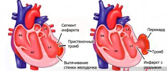
- ischemia;
- myocardial infarction, etc.
An ultrasound of the heart will show quite accurately whether there is cause for concern and how serious the situation is.
Cost of services
| Name of service | Price |
| Ultrasound of the heart (Echo-CG) with Doppler | 2,600 rub. |
Ultrasound diagnostics of the heart is an informative, fast and safe method of studying the condition of a child’s heart, which allows you to accurately establish a diagnosis and carry out the required treatment with maximum efficiency, or get rid of worries and doubts about the state of your baby’s health.
You can get additional information about cardiac ultrasound and make an appointment for diagnostics at a medical center by phone.
Ultrasound of a child’s heart - how is the procedure performed?
An ultrasound of the child's heart is performed in the supine position (on his back). The child's chest is exposed, the doctor applies a hypoallergenic gel, which ensures the sliding of the sensor. When performing ultrasound diagnostics, the device records the reflected waves and the specialist receives the dimensions of the chambers and ventricles of the heart, blood vessels, as well as a complete understanding of the current state of the organ.
If a cardiac ultrasound is scheduled for an infant, it is necessary to ensure that he is not hungry - this is necessary so that the child behaves more calmly during the procedure. Ultrasound diagnostics does not cause any discomfort to the child. No special preparation is required for an ultrasound of a child's heart.
Order of conduct
Echo-CG is performed absolutely painlessly and does not pose any risks to the child’s health, so the study can be repeated as often as the clinical case requires. There are also no age restrictions - the procedure can be carried out from the first days of life.
An ultrasound takes no more than 20 minutes. The child sits on the couch, the doctor applies a special gel to the skin of the chest and a special sensor to improve the conductivity of ultrasound. Then he applies the sensor and moves it across the chest, at which time an image of the heart appears on the monitor. The condition of the organ is assessed visually in real time, and with the help of the program, the necessary changes are made and blood flow is assessed. During the procedure, you may need to change your body position. As a rule, you should lie on your left side or back most of the time. The examination does not cause any discomfort, but for the baby’s peace of mind, the parent can be nearby.
Pathologies that can be detected using cardiac ultrasound
After performing an ultrasound of the heart, a cardiologist analyzes the results, which helps to make the correct diagnosis and prescribe appropriate treatment. Most often, cardiac ultrasound helps identify the presence of the following cardiac pathologies in children:
- the presence of a hole (incomplete fusion) in the septum between the ventricles of the heart;
- the presence of a defect in the form of a hole in the interatrial septum;
- valve defects;
- narrowing of the aorta, etc.
If a child is diagnosed with any inflammatory processes in the heart, then the ejection function will be impaired, decreasing against the norm, and also during diagnosis, changes in the size of all cardiac cavities will be noticeable (they will be enlarged).
Transesophageal ultrasound
The transesophageal echocardiography procedure is more complex and has a number of limitations. An ultrasound sensor is inserted through the esophagus, which makes it possible to see the condition of the organ from all sides. The examination may be indicated for severe heart defects, chest deformities and lung diseases, inflammation of the aorta after prosthetics, etc. Preparation includes abstaining from food at least 6 hours before the examination, and during the procedure special medications may be required to improve the tolerability of the examination. Thus, it is sometimes advisable to perform intravenous anesthesia if a pronounced gag reflex is observed.
Pediatric cardiology in Saratov: what should be done if a child has heart problems?
If you notice that your child is exhibiting certain symptoms characteristic of heart pathologies, we recommend making an appointment with a pediatric cardiologist. A pediatric cardiologist is a doctor involved in the prevention, diagnosis and treatment of diseases of the cardiovascular system and connective tissue. The First Children's Medical Center employs highly qualified and competent specialists - pediatricians and cardiologists, with sufficient experience, who are able to establish the true cause of the dysfunction of the heart and blood vessels, and who are also able to find an approach to a child of any age - from infants to adolescents.
To timely identify a possible pathology of the cardiovascular system in a child, the Clinic carries out the necessary examinations using modern equipment (such as ECG, ultrasound of the heart, exercise tests, daily ECG Holter monitoring, as well as daily blood pressure monitoring). Also in the arsenal of the First Children's Medical Center there are the necessary methods for laboratory diagnosis of diseases of the cardiovascular system.
The First Children's Medical Center has programs for long-term monitoring of your child, here you can get advice from other specialists. For your convenience, the clinic has a day hospital.
What is cardiac ultrasound?
Echocardiograms, also known as cardiac ultrasounds, are used to study a baby's or fetus's heart as it functions. This is a simple and painless procedure, performed according to the same principles as the ultrasound examination of the fetus in the womb of a pregnant woman, which is familiar to all mothers. Echocardiography uses sound waves (ultrasound) to create images of the heart. The Doppler test uses sound waves to measure the speed and direction of blood flow. By combining these tests, the pediatric cardiologist obtains useful information about the anatomy and function of the heart. Echocardiography is the most common test used in children to diagnose or rule out heart disease, and to monitor children who have already been diagnosed with heart problems. This test can be done on children of all ages, even newborns or unborn children.
Question answer
— Why is it worth choosing cardiac ultrasound as a diagnostic method?
This diagnostic method allows you to identify the presence of pathology at an early stage of development, which allows the doctor to prescribe timely treatment and monitor its progress and results. In addition, ultrasound diagnostics is a safe and absolutely painless research method.
— At what age can a child have an ultrasound of the heart?
An ultrasound of a child’s heart can be done even in the first days of life. Ultrasound does not cause any harm to both adults and children, so the frequency of this procedure is not limited.
— We need a good pediatric cardiologist in Saratov. Is it possible to make an appointment and undergo an examination at your center?
Of course, in our Center, a cardiologist of the highest category, candidate of medical sciences, Elena Nikolaevna Shulgina, conducts consultations. You can make an appointment for a consultation and, if necessary, sign up for an examination. You can make an appointment for a consultation from 8.00 to 20.00 by calling (8452) 244-000. Reception is by appointment only.
Make an appointment with a cardiologist
Choose a doctor
What is a transesophageal echocardiogram?
Sometimes a regular echocardiogram does not provide all the images needed. Your doctor may recommend a test that uses special echocardiography probes to take pictures from inside the esophagus. This procedure is called a transesophageal echocardiogram. In this test, a tube with an ultrasound probe is passed down the throat into the esophagus. The esophagus is located directly behind the heart, and images taken there can give a very clear picture of the heart and its structures. This procedure requires anesthesia and takes about 20 minutes.
Routine examinations of the child at 1 month
After patronage, when the baby reaches the age of 1 month, it is necessary to come for a medical examination of the baby to specialists. A healthy child needs to visit a pediatrician, neurologist, surgeon, orthopedist and ophthalmologist. If there are pathologies, a list of specialists is compiled individually.
What to bring to your appointment:
- documents (vaccination certificate, policy, SNILS);
- replacement diapers, 2 diapers, wet wipes;
- toys, spare pacifier;
- spare clothes;
- a notebook with questions for the doctor.
Pediatrician
During the first visit, the doctor will weigh the baby, measure height, conduct an examination and evaluate physical and neuropsychic development. The doctor will ask the mother about the child’s developmental characteristics and give recommendations for care. It is necessary to draw the doctor's attention to any shudders, twitchings and convulsions.
The doctor will measure the circumference of the head and chest, examine the fontanelles, and check the weight gain, which indicates proper development. The mother will be asked to talk about the baby’s skills. By one month, the child can hold objects in his field of vision, follows a toy, turning his head slightly, responds to a voice, briefly raises his head, and smiles. If the child is healthy, he is sent for a second vaccination against hepatitis B (the first is given in the maternity hospital).
Neurologist
To assess neuropsychic and mental development, you must visit a neurologist at the clinic. The doctor checks for the presence of innate reflexes and evaluates muscle tone. If there is increased or decreased tone, a massage is prescribed. The doctor examines the results of an ultrasound scan of the brain and evaluates the baby’s skills and abilities.
Surgeon
The doctor evaluates reflex development, examines the navel, diagnoses or excludes inguinal and umbilical hernias, and checks for the presence of hypo- and hypertonicity of the muscles. In boys, the external genitalia are examined to exclude various pathologies: dropsy, hypospadias, cryptorchidism. The doctor may detect lymphangioma, hemangioma, and vascular lesions. If necessary, a therapeutic massage is prescribed and recommendations are given on which muscles to pay attention to.
Orthopedist
The specialist evaluates the development of bones and muscles. Upon examination, a doctor can diagnose congenital dislocation of the hip joint, clubfoot, and joint dysplasia. The orthopedist, examining the child, actively flexes and extends the limbs and performs many manipulations to determine the condition of the musculoskeletal system.
To eliminate risks, a routine ultrasound of the hip joint is performed, the results of which are reviewed by the doctor at the appointment and determines the exact picture of the baby’s condition. Most pathologies are successfully treated up to 1 year, until the child begins to walk.
The essence of echocardiography
Ultrasound of the heart is the process of studying all the main parameters and structures of this organ using ultrasound.
When exposed to electrical energy, the echocardiograph transducer emits high-frequency sound that travels through the structures of the heart, is reflected from them, captured by the same transducer and transmitted to the computer. He, in turn, analyzes the received data and displays it on the monitor in the form of a two- or three-dimensional image.
In recent years, echocardiography has been increasingly used for preventive purposes, which makes it possible to identify cardiac abnormalities at an early stage.
What does an ultrasound of the heart show:
- heart size;
- integrity, structure and thickness of its walls;
- sizes of the cavities of the atria and ventricles;
- contractility of the heart muscle;
- operation and structure of valves;
- condition of the pulmonary artery and aorta;
- pulmonary artery pressure level (to diagnose pulmonary hypertension, which can occur with pulmonary embolism, for example, when blood clots from the veins of the legs enter the pulmonary artery);
- direction and speed of cardiac blood flow;
- condition of the outer shell, pericardium.
1 ECHO-KG in "MedicCity"
2 Echocardiography at MedicCity
3 Ultrasound of the heart at MedicCity
