Classification
The upper limit of normal platelet count varies from 350,000 to 400,000 per microliter depending on the reference intervals of the specific laboratory performing the analysis. According to the degree of increase, the following types of thrombocytosis are distinguished:
- Soft
: from 350-400 to 700 thousand. - Moderate
: from 700 to 900 thousand. - Heavy
: from 900 to 1000 thousand. - Extreme
: over 1,000,000.
The cause of extreme and severe thrombocytosis is oncohematological pathologies. According to the origin of thrombocytosis there are:
- Primary
(tumor, clonal). They account for approximately 10-15% of all cases of thrombocytosis. The cause is tumor diseases of the blood system. - Secondary
(reactive). The most common variety (about 85%). The cause is infectious, systemic inflammatory processes, and anemia. - False
(pseudothrombocytosis). The reason is an error in the hematology analyzer, which mistakes tumor cell fragments during treatment with chemotherapy drugs, small red blood cells, or red blood cells that have undergone hemolysis as platelets. Pseudothrombocytosis is also observed with cryoglobulinemia. - Hereditary
(family). This is a rare genetic disease, the cause of which lies in a mutation of the genes encoding the synthesis of thrombopoietin and its receptors (THPO, MPL).
Causes of thrombocytosis
Physiological conditions
An elevated platelet level does not always indicate pathology. There is physiological (short-term, transient) thrombocytosis, caused by various circumstances, for example, stress, intense physical activity. The reason is the mobilization of blood platelets, or rather their transition from the marginal position to the central blood flow in the vessels of the spleen and lungs.
In addition, minor physiological thrombocytosis is observed in children from the neonatal period to 11 years. There is also a so-called hemoconcentration thrombocytosis, which is caused by dehydration. This phenomenon is due to a decrease in the volume of the liquid part of the blood (plasma) and a relative increase in formed elements (platelets, erythrocytes, leukocytes). In this situation, it is necessary to focus on the hematocrit - with dehydration it is increased.
Infections
This is the most common cause of thrombocytosis (about 40%). An increase in the level of blood platelets develops when:
- Bacterial infections
. In adults, thrombocytosis occurs mainly with local (pneumonia, pyelonephritis, meningitis, endocarditis) and systemic (sepsis, tuberculosis) bacterial infections. - Viral and fungal infections
. Thrombocytosis occurs less frequently during infection with viruses (hepatitis B, C) and pathogenic fungi (aspergillosis). - Helminthiasis.
In children, parasitic infestations (toxocariasis, ascariasis) are recognized as a common cause of an increase in the number of blood platelets.
There are two pathogenetic mechanisms for the development of thrombocytosis in response to an infectious disease. First, during the fight against pathogens, leukocytes produce a large number of inflammatory mediators, including interleukin-6, which stimulates bone marrow megacaricytopoiesis (the formation of platelet precursors). Secondly, platelets themselves are part of the anti-infective immunity - they are able to produce bactericidal substances, capture, neutralize, and even phagocytose certain types of bacteria, viruses, and foreign particles.
Platelets facilitate the migration of leukocytes to the site of infectious inflammation by interacting with endothelial cells of the vascular wall. Thrombocytosis during infections occurs abruptly, correlates with the severity of the disease, and quickly resolves after the pathogen is eliminated from the body and the inflammatory process subsides. Thrombocytosis is usually mild or moderate; in septic conditions it can reach a severe degree; in children it is somewhat more pronounced than in adults.
Autoimmune diseases
Another common cause of thrombocytosis is considered to be chronic rheumatological pathologies that occur with autoimmune inflammation. The mechanism for increasing the content of blood platelets is the overproduction of substances such as interleukin-6, colony-stimulating factors, which activate bone marrow platelet formation. The degree of thrombocytosis corresponds to the activity of inflammation (minimal during remission, maximum during relapse).
In diseases such as rheumatoid arthritis, systemic lupus erythematosus (SLE), inflammatory bowel diseases (Crohn's disease, ulcerative colitis), a mild or moderate degree is observed. In systemic vasculitis with necrotizing destruction of vessel walls, severe thrombocytosis occurs, since in vasculitis, in addition to other inflammatory mediators, tumor necrosis factor is synthesized in large quantities, which also has a stimulating effect on platelet formation. High platelet counts are especially common in children.
Anemia
Iron deficiency anemia is often the cause of thrombocytosis, especially in children. The exact mechanisms of this phenomenon have not yet been clarified, but an inverse relationship has been clearly established between reduced levels of iron metabolism (ferritin, iron-binding capacity of serum) and an increased level of blood platelets. Iron is thought to have an inhibitory effect on the maturation of megakaryocytes (precursor cells).
In addition, some pluripotent stem cells in the bone marrow are unable to transform into red blood cells under conditions of iron deficiency. As a result, a kind of “bypass” occurs and a larger percentage of stem cells begin to mature along the megakaryocyte pathway. Thrombocytosis in iron deficiency anemia is mild, sometimes moderate. Platelets quickly return to normal after correction of iron deficiency and an increase in hemoglobin.
However, as anemia worsens, platelet levels drop to a state of thrombocytopenia. The cause of iron deficiency can be a lack of iron in food, increased iron consumption (growth period in children, pregnancy, lactation) or chronic blood loss (long menstruation, bleeding from the gastrointestinal tract due to gastric ulcer).
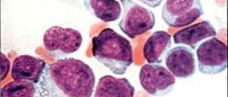
Malignant blood diseases
The cause of approximately 15% of all thrombocytosis is hemoblastosis - chronic myeloid leukemia, Ph-negative myeloproliferative pathologies (essential thrombocythemia, polycythemia vera, and primary myelofibrosis). The increase in the number of blood platelets in these diseases is due to clonal (tumor) transformation of the megakaryocyte lineage of the bone marrow due to various mutations, which leads to hyperproduction of platelets.
These diseases are more common in adults and older people, in children - only in exceptional cases. Initially, thrombocytosis is moderate, as it progresses it increases, reaching a severe or extreme degree, which is why microcirculation disorders, arterial and venous thrombosis of various localizations often occur. The concentration of blood platelets normalizes very slowly, only after courses of specific myelosuppressive treatment.
Splenectomy
The spleen, being an organ that deposits blood, retains a large number of formed elements, including platelets. The spleen is also directly involved in thrombocytopoiesis, secreting the hormones thrombocytopenin and splenin, which suppress bone marrow maturation of megakaryocytes. Therefore, thrombocytosis after splenectomy is caused by two mechanisms: the release of platelets, normally located in the splenic depot, into the circulating blood, and the phenomenon of “disinhibition of the bone marrow,” i.e. increased platelet production.
An increase in the number of blood platelets does not occur immediately, but approximately a week after splenectomy, reaches a maximum by the 13-14th day (up to 700-800 thousand), often becoming the cause of venous thrombosis of the portal vein, and then slowly returns to normal over several weeks or months .
Injuries and surgeries
Massive tissue damage (wound during abdominal surgery, fracture, extensive burns) causes activation of the blood coagulation system, namely the vascular-platelet unit, which is the first stage of hemostasis. It involves vasospasm, as well as adhesion and aggregation of platelets at the site of damage to the vascular wall. The consumption of platelets stimulates their active release from the depot and a compensatory increase in their bone marrow production. The extent of damage correlates with the degree of thrombocytosis. This type of thrombocytosis usually does not require treatment.
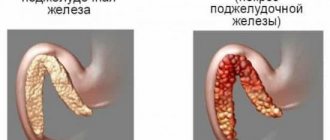
Oncological diseases
The cause of thrombocytosis in solid (non-hematopoietic) tumors is the ability of cancer cells to produce interleukin-6, which stimulates thrombocytopoiesis. This feature was found in small cell lung cancer, colon adenocarcinoma, and malignant mesothelioma. In addition, tumor disintegration often causes bleeding, leading to iron deficiency anemia. The degree of thrombocytosis is usually moderate, in children it can be severe, and regresses after long-term treatment with chemotherapy.
Rare causes
- Functional asplenia
: sickle cell anemia, chronic alcoholism, celiac disease. - Use of medications
: vincristine, adrenaline. - Rebound phenomenon
: development of thrombocytosis 1-2 weeks after treatment of thrombocytopenia or discontinuation of medications that cause thrombocytopenia (methotrexate, vitamin B12, prednisolone).
Thrombocytosis in children
In children, thrombocytosis is often observed and occurs in 13% of newborns, in the first month - in 36%, and thrombocytosis in children under one year (6 to 11 months) occurs in 13% of children. This is a physiological feature of blood tests in children under one year of age. The maximum increase that can be observed is 1200 x 10 in 9/l. In this case, if the child is in absolute health, you need to recheck the blood test several times. In normal health, elevated platelets in infants are a variant of the norm, so this condition does not require treatment.
If blood cell levels remain high for a long time, certain diseases need to be ruled out. If we consider the causes, then chronic myeloproliferative diseases in children and cases of familial thrombocytosis associated with mutations of the thrombopoietin gene or receptor gene are very rare. Reactive thrombocytosis occurs quite often and is an excessive physiological reaction to any primary problem. This condition is transient and tests return to normal when the cause is eliminated.
A common cause of this condition in children is bacterial and viral infections, which in 85% of cases cause such changes in the blood. In the first place of increased level are bronchitis , pleurisy and pneumonia , infections of the urinary system ( pyelonephritis ), bacterial meningitis , inflammation of the gastrointestinal tract and skeletal system. In secondary thrombocytosis, platelets are usually mildly elevated, except for severe infections between 9 months and 2 years of age or Kawasaki disease. In this case, the level can exceed 1000 × 10 in 9/l and most often in premature babies.
A common hematological cause is iron deficiency, so if thrombocytosis is present in a child, serum ferritin must be determined. Overproduction of platelets is observed in hemolytic anemia , acute bleeding, and after spenectomy.
Inflammatory diseases. During inflammatory processes, the amount of interleukin 6 increases, which causes an increase in the synthesis of thrombopoietin . Such diseases in children include juvenile arthritis , chronic hepatitis , ankylosing spondylitis beginning in adolescence, inflammatory enteropathy , sarcoidosis , Kawasaki disease . Most often in pediatric practice, thrombocytosis occurs in Kawasaki disease, which develops in the second week of the disease.
Drug-induced thrombocytosis. Associated with the use of corticosteroids and adrenaline , which leads to an increase in these formed elements due to release from the spleen. Also, the use of antibiotics ( carbapenems , cephalosporins ) is the cause of this condition. However, this is observed after a course of antibacterial treatment and is a response of the bone marrow to an increase in thrombopoietin synthesis. In the neonatal period, an increase in platelets occurs when mothers use drugs or when a woman is treated with psychotropic drugs during pregnancy.
Thus, in children, the state of thrombocytosis is secondary and the most common causes are:
- infectious;
- inflammatory;
- hematological
Reactive thrombocytosis in children does not require treatment and only sometimes leads to thrombosis. They are not an indication for the prescription of antiplatelet agents even at levels of 1000 × 10 in 9/L, if there are no risk factors for thrombosis. It is necessary to establish the cause and treat the underlying disease.
Diagnostics
Thrombocytosis is detected in a clinical blood test. Although very high platelet counts are more common in hematologic diseases, platelet levels alone cannot determine the cause of thrombocytosis. Therefore, if it is detected, you should visit a therapist. The doctor carefully asks about the patient’s complaints, how long ago the symptoms occurred, and conducts a general examination of the patient. Then, based on the data obtained, an additional examination is prescribed, including:
- Blood tests
. In a general blood test, the content of other formed elements (erythrocytes, leukocytes) is determined, and the leukocyte formula is calculated. The concentration of inflammatory markers (ESR, CRP) is measured. The indicators of serum iron, TBC, ferritin are assessed. The presence of autoantibodies (RF, ACCP, antibodies to the cytoplasm of neutrophils) is checked. In case of endocarditis and sepsis, an analysis for procalcitonin and presepsin is performed. - Pathogen identification
. To identify the pathogen, microscopy, bacterial culture of urine and sputum are performed. If tuberculosis is suspected, an intradermal test with tuberculin is prescribed. Using an enzyme immunoassay, antibodies to viruses, parasites, and fungi are detected, and using the polymerase chain reaction method, their DNA and RNA are detected. To diagnose meningitis, a cerebrospinal fluid analysis is informative. - Genetic research
. In patients with myeloproliferative pathologies, mutations of Janus kinase (JAK2V617F), thrombopoietin receptors (MPL), and erythropoietin are determined using fluorescent hybridization (FISH) and PCR. Sometimes chromosomal abnormalities are detected - trisomies, deletions. In chronic myeloid leukemia, cytogenetic analysis reveals the Philadelphia chromosome (Ph). - X-ray
. On an X-ray of the lungs, in case of pneumonia, foci of darkening and infiltrates are noted, in case of tuberculosis - enlargement of the mediastinal lymph nodes, expansion of the roots of the lungs, rounded shadows (cavities) of the upper lobes of the lungs. In patients with arthritis, x-rays of the joints show a narrowing of the joint space, areas of erosion, and marginal osteoporosis. - Ultrasound
. Ultrasound of the abdominal organs in case of pyelonephritis determines compaction and expansion of the pyelocaliceal system, and in case of blood diseases - splenomegaly. In bacterial endocarditis, cardiac echocardiography reveals vegetations of the valves and sometimes effusion into the pericardial cavity. - Endoscopy
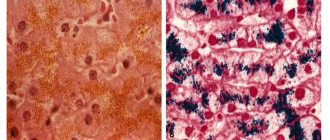
. In patients with inflammatory bowel pathologies, fibrocolonoscopy is performed, which reveals hyperemia of the mucous membrane, lack of vascular pattern, erosion, and ulcerative defects. Crohn's disease is characterized by the "cobblestone pavement" symptom - alternating deep ulcers with unchanged mucous membrane. - Histological studies
. In bone marrow aspirate for malignant hematological pathologies, hyperplasia of the megakaryocyte lineage of hematopoiesis is noted (in polycythemia vera - all three lineages), a large number of blast cells (in myeloid leukemia), proliferation of reticulin and collagen fibers (fibrosis). In case of vasculitis, a biopsy of a vessel reveals pronounced perivascular infiltration with lymphocytes and plasma cells.
Platelet count according to Fonio
Symptoms of thrombocytosis
Typically, symptoms develop gradually. At the initial stages of the disease they may be completely absent. The main manifestations of thrombocytosis are provoked by two factors: increased bleeding or the occurrence of blood clots in the blood vessels. If thrombocythemia is secondary, then the likelihood of such disorders occurring is much lower: the number of platelets in this form of the disease is lower than in the primary one.
The main symptoms of thrombocytosis include:
- pain in the feet and hands, their numbness;
- headache;
- irritability and weakness;
- bleeding gums;
- visual impairment;
- blood in stool;
- nosebleeds.
Correction
Conservative therapy
In most cases, to correct thrombocytosis, it is enough to eradicate the cause, i.e. treatment of the underlying disease. Short-term thrombocytosis that develops due to stress or drug administration does not require intervention. In case of persistent long-term thrombocytosis, consultation with a hematologist is necessary to identify the cause and prescribe appropriate treatment. Therapy for thrombocytosis has several areas, including:
- Fighting infection
. To eliminate the infectious agent, antibacterial (amoxicillin), antifungal (fluconazole), and antiparasitic agents (mebendazole) are used. Treatment of viral hepatitis requires long-term use of peligated interferon in combination with antiviral drugs. - Treatment of iron deficiency anemia
. Correction of iron deficiency is carried out with tablet preparations (iron sulfate). For children, there are forms of syrup and drops for oral administration. The addition of ascorbic acid promotes better absorption. - Therapy of autoimmune diseases
. Treatment of autoimmune diseases is carried out using medications that suppress inflammation - glucocorticosteroids (prednisolone), immunosuppressants (cyclophosphamide). - Targeted therapy
. For myeloproliferative diseases, specific targeted treatment is prescribed to slow down the progressive growth of the malignant tumor. These drugs include Janus kinase inhibitors (ruxolitinib), tyrosine kinase inhibitors (imatinib, dasatinib). - Symptomatic treatment
. To relieve high thrombocytosis, medications are used that suppress the activity of the megakaryocyte lineage, and, consequently, the production of platelets - anagrelide, interferon-alpha, hydroxyurea. For polycythemia, regular bloodletting is successfully used as a treatment method to remove excess formed elements. - Blood thinning
. In case of high thrombocytosis, antiplatelet agents (acetylsalicylic acid) are prescribed to prevent thrombosis. In case of contraindications (peptic ulcer of the stomach, duodenum), platelet receptor blockers (clopidogrel, ticagrelor) are used. In people at high risk of thrombosis (elderly, patients with diabetes mellitus or atrial fibrillation), anticoagulants (warfarin, dabigatran) are used.
Specialized treatment
The only method that allows achieving complete recovery from a malignant hematological disease is allogeneic bone marrow transplantation. This requires HLA typing to select a compatible donor. However, due to the high risk of developing life-threatening complications, this method is used only if conservative treatment is ineffective.
Forecast
The outcome depends on both the underlying pathology and the degree of thrombocytosis. For example, acute viral infection and iron deficiency anemia are characterized by a benign course. Patients with essential thrombocythemia, with proper selection of pathogenetic and symptomatic treatment, can live more than 80 years. People with chronic myeloid leukemia, on the other hand, live about 5-10 years from diagnosis.
Since reactive thrombocytosis almost always occurs in children, their prognosis is favorable. Thrombosis is not typical for mild to moderate thrombocytosis. In extreme or severe cases, there is a very high probability of fatal thrombosis leading to myocardial infarction, pulmonary infarction, and ischemic stroke.
Diet
Diet for essential thrombocythemia
- Efficacy: therapeutic effect after 3 weeks
- Timing: constantly
- Cost of products: 1400-1500 rubles per week
Nutrition is an auxiliary factor in this condition. Taking into account the tendency to blood clots, your diet should include foods that help thin the blood: figs, beets, lemon, prunes, garlic, raspberries, cranberries, strawberries, artichokes, oranges, olive oil, raisins, blueberries, as well as spices and herbs (chili pepper, thyme, paprika, turmeric, oregano, curry, mint, cinnamon).
The diet needs to be balanced according to the main nutrients and vitamins and microelements must be introduced. Iodine, which is rich in seaweed and seafood, iron contained in red meat and liver, magnesium in cereals, bran bread, vegetables, honey, fruits, as well as calcium, are useful.
A varied diet containing sea fish, squid, shrimp, seaweed, vegetable oils, chicken and beef liver, oatmeal, dairy products, eggs, nuts (cashews and almonds), fresh vegetables and fruits will maintain metabolic processes at a normal level.
It is important to adhere to the drinking regime - drinking fluid within 2-2.5 liters per day. This improves its fluidity. If there is no indication for taking diuretics due to the underlying disease, they should be abandoned since this group of drugs helps to thicken the blood. The same applies to diuretic herbs and laxatives that are taken uncontrolled.
In addition, there are foods that contribute to blood thickening that should be avoided. This is sugar, white bread, bananas, mangoes, potatoes, buckwheat and semolina, fatty meats, any smoked meats, rosehip infusions, alcohol, legumes, walnuts, sorrel, lettuce, chokeberry.