The term "hypoplasia" means "underdevelopment". Such defects of intrauterine development are also found in the arteries of the brain. In the arteries of the carotid artery basin, these anomalies exist in approximately 4% of people, and in the arteries of the vertebrobasilar basin - in every tenth person.
Despite the fact that the brain is the most energy-intensive organ and requires massive blood supply, hypoplasia of the arteries that supply it can go unnoticed for a long time. This is due to the characteristics of the cerebral circulatory system.
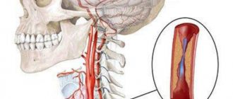
Most of the hemispheres receive blood from the system of internal carotid arteries, which form the anterior and middle cerebral arteries, as well as the anterior communicating artery. The posterior part of the hemispheres, the brainstem and the cerebellum are fed from the basin of two vertebral arteries, which merge in the cranial cavity into the main artery, from which the posterior cerebral and posterior communicating arteries are formed.
Due to the anterior and posterior communicating arteries, a connection is formed between the arterial basins of the brain, forming a circle. It is called Willis, and within its limits a compensatory flow of blood into areas with reduced arterial blood flow is possible, including due to hypoplasia of any segment of the artery. It is this feature of the brain’s blood circulation that ensures the long-term asymptomatic course of most hypoplasias, which often become an accidental discovery.
Clinical manifestations of hypoplasia of the cerebral arteries occur against the background of another pathology that worsens the conditions for compensation of cerebral circulation. Most often this is atherosclerosis of cerebral vessels or diseases of the cervical spine. As a result, transient ischemic attacks and ischemic strokes may occur.
results
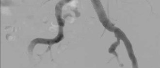
A 19-year-old patient came to the SSMU clinic on the referral of a neurologist from the district clinic for a triplex scan of the brachiocephalic arteries (TS BCA) with complaints of frequent severe dizziness, recurrent headaches, and lack of coordination.
Similar symptoms have been bothering him for 5 years; he denies any connection with the injury. According to the results of TS of the BCA, bilateral narrowing of the diameter of both VAs was revealed. On the left, the diameter in the V1 segment is no more than 1.8 mm, then at the level of the V2 segment it is in the range of 1.5-1.7 mm, in the V3 segment it is 1.5 mm.
On the right, the diameter of the V1 segment does not exceed 1.5 mm, the diameter of the V2 segment is in the range of 1.3-1.6 mm, in the V3 segment the diameter reaches 1.4 mm. During a transcranial study at the level of V4 segments, the VA is located only on the left side.
There is a pronounced decrease in the linear velocity of blood flow (LBV) on both sides - at the level of the 2nd segment of the LSV, no more than 15-18 cm per second. At the level of the V4 segment on the left, the BSC is reduced to 10 cm per second. The basilar artery is not visualized during TS BCA.
When performing a rotation test to the left side, the LSC along the PA decreases to 4-6 cm per second. Both VAs originated from the anterior wall of the subclavian arteries in segment 1. No ultrasound signs of pathological tortuosity were detected on either side.
On both sides, the PA entered the bone canal at the level of the C6 vertebrae. The above-described ultrasound changes were regarded as signs of hypoplasia of both VAs with severe hypoperfusion in the vertebrobasilar region.
For the purpose of additional verification and detailed examination of the intracranial bed, computed tomography (CT) with contrast was performed, which revealed a sharp narrowing of both VAs at the level of the V4 segment with signs of hypoperfusion in the basilar artery. The patient was sent for planned hospitalization to the neurological department of the SSMU clinic.
Symptoms
Symptoms of chronic cerebral circulatory disorders depend on the vascular pool in which ischemia of brain tissue develops. All patients may be concerned about:
- headache;
- dizziness;
- noise in the head;
- memory impairment;
- sleep disorders.
Hypoplasia of the carotid arteries is also characterized by weakness and/or numbness in the limbs and speech impairment. For ischemia in the vertebral artery basin - impaired coordination, gait instability.
To diagnose hypoplasia of cerebral arteries and determine treatment tactics, the following are carried out:
- cerebral angiography - X-ray examination of the arteries of the brain with a contrast agent injected into them;
- CT angiography;
- MR angiography;
- ultrasound examination (duplex scanning) of the vessels of the neck and head;
- PAT;
- single photon emission computed tomography.
Conservative therapy with drugs that improve blood supply and metabolism of the brain is carried out if the examination data suggests that it can prevent further worsening of cerebral ischemia and ischemic stroke.
conclusions
TS BCA is the leading available non-invasive method for visualizing VA hypoplasia. The technique makes it possible to assess with great accuracy the diameter, course and condition of the walls of the arteries, and also, unlike other imaging methods, to study the speed indicators of blood flow and objectively judge the signs of hypoperfusion in the vertebrobasilar region.
Among additional examination methods for detailed visualization of intracranial arteries, we consider CT with contrast to be the most appropriate.
- Views: 4614
- Comments:
Did you like the post? Do you find it useful or interesting? Support the author!
Hypoplasia of the vertebral artery
If you complain to a neurologist about dizziness and instability, then most likely you will be advised to undergo an ultrasound examination of the cerebral vessels. And now you are reading a medical report, where it is written in black and white “small diameter vertebral artery” or “vertebral artery hypoplasia.” It sounds alarming, and the attending physician is in no hurry to allay the concern. What actually lies behind the incomprehensible diagnosis and what to do about it now?
What it is
Hypoplasia of the vertebral artery (small diameter artery) is a congenital narrowing of the vertebral artery, most often the right one. A person normally has two vertebral arteries (sometimes there are more), which flow into the basilar artery, which is part of the circle of Willis.
The circle of Willis is, in fact, the connection of the branches of all large vessels of the brain. Normally, this circle is closed, which ensures blood supply and brain function when any of the arteries is “switched off” (for example, due to a blood clot). Therefore, if one of the vertebral arteries is narrowed and the blood flow through it is changed, then the second takes on part of the load.
Symptoms of hypoplasia
Vertebral artery hypoplasia usually has no symptoms if other vessels are functioning normally. In this case, blood circulation is compensated by the second vertebral artery and larger carotid arteries (remember the circle of Willis?). If, nevertheless, compensation does not occur, then patients complain of blurred vision, unsteady gait, and impaired coordination of movements.
Causes
The following reasons give rise to it:
- Hereditary factors. For example, one of the parents may carry a recessive gene, which, due to consanguineous marriages, manifests itself in the child. Characteristic of closed communities where incest is permitted. A striking example is cerebellar hypoplasia due to disruption of the VLDLR gene, which manifests itself in cases of consanguineous blood mixing.
- Teratogenic factors: physical, biological and chemical effects on the body of the mother and child. For example, neuroinfections, living in areas with high levels of radiation, drugs that have not passed the teratogenicity test.
- Injuries during pregnancy.
- Maternal toxicosis.
- Smoking, alcoholism and drug addiction of parents.
- Pathologically reduced amount of amniotic fluid.
Left artery lesion
The disease is not easy to recognize in the early stages. It develops for several years and does not make itself felt. Impaired blood circulation provokes stagnation of blood in the cervical region, causing pain syndromes in the neck area. Sudden movements are accompanied by unpleasant sensations.
Damage to the left-sided aorta leads to abnormal blood pressure. Due to improper blood supply, the body changes, and the disease begins to gradually develop. The clinical picture of damage to the left transverse sinus occurs due to the immature left vertebral artery.
In mild cases, the problem cannot be noticed, only with the help of MRI images. At night there are severe headaches, nausea and even vomiting. There is a risk of having a heart attack or stroke.
It is more dangerous for health when the work of both the left and right arteries is disrupted. This leads to lifelong disability.
How it develops
The disease appears as a result of a disorder in the structure of the aorta. Defects develop during embryonic maturation. Pontocerebellar hypoplasia occurs due to a lack of CDG (carbohydrate glycoprotein). This deviation is clearly visualized.
The pathology refers to an autosomal recessive disease. There is a delay in the child's development and obvious microcephaly.
The cervical vessels are responsible for supplying blood to the brain; they form a dense circle. If the lumen narrows, life-threatening diseases appear.
Even with minor damage, the cerebellum ceases to receive normal nutrition. The child’s fine motor skills suffer and handwriting cannot be developed. If the brain stem is damaged, facial expressions of the entire face are disrupted, and the swallowing process fails.
With the development of microcephaly, the patient loses weight and volume of the head. The development of internal elements changes and defects appear.
The convolutions lag behind in development and are not deep. The visual tubercle does not protrude and looks smaller. The trunk and pyramid of the oblong part are also much smaller, which affects the size of the skull.
During the process of growth, the facial part of the child grows quickly and the brain part of the skull grows more slowly. As a result, the deformation of the head can be seen with the naked eye.
Causes of pathology
The problem may begin to form in the womb. When planning pregnancy, it is recommended to study all the factors that influence the occurrence of hypoxia. Many factors can provoke hypoplasia of the right transverse sinus. Main reasons:
- bruises and injuries;
- ionization and radiation waves;
- genetic predisposition.
Cases have been recorded when changes appeared without clearly defined reasons. Experts could not explain this phenomenon. In adults, pathology can occur as a result of:
- osteochondrosis in acute or chronic form;
- dislocation of the vertebrae;
- overgrowth of the nape membrane with bone tissue;
- thrombosis;
- a strong blow to the head;
- spinal bruise;
- atherosclerosis.
Age-related changes provoke progression of the disease. Doctors often make incorrect diagnoses, since the disorder is influenced by many factors.
Hypoplasia/aplasia of the transverse/sigmoid sinus
Hypoplasia and aplasia of the transverse/sigmoid sinuses on the right or left are common findings, in which 2D MR venograms indicate a decrease in the flow signal or its complete absence in case of aplasia. These types of venous sinus structures are normal variants of the anatomical structure.
In most cases, these options should not cause difficulties in the absence of clinical data, the absence of changes in the brain substance of the corresponding area and normal characteristics of the MR signal in the area of the corresponding sinus, in addition, it should be taken into account that on the left these “anomalies” are quite common.
However, sometimes there are situations that confuse not only beginners, but also specialists with a trained eye. For example, if there is a history of ipsilateral headaches or if there is a history of trauma, testing may reveal cortical hemorrhage, which may represent either contusional changes or venous infarction.
Key points
- frequency and location: hypoplasia and aplasia are more common on the left than on the right (Figure 1).
- sinus dimensions: look at the sinus in cross section; with hypoplasia/aplasia, the transverse and sigmoid sinuses will be smaller (Figure 2)
- MR signal: no pathological increase in signal in the sinus area on T2 and FLAIR. A thrombosed sinus will be "bloated" and have abnormally high signal levels on T2 images, instead of normal flow loss (Figure 3)
- sagittal T1 images: can also be used to assess whether an increase in the T1 signal in the sinus region due to methemoglobin in the acute stage of thrombosis excludes hypoplasia and aplasia (Fig. 4).
- sinus territory: in case of venous infarction, it should be on the side and territory of the corresponding sinus or adjacent veins.
- post-contrast study: firstly, you can see normal enhancement of the meninges in the sinus area, secondly, in the case of hypoplasia or aplasia there will be no increase in the MR signal in the sinus area; in the case of sinus thrombosis, the thrombus will show marked enhancement.
- jugular foramen: compare the diameter of the jugular foramina. The jugular foramen on the side of the hypoplastic sinus will be smaller in diameter compared to the opposite side. CT images in the bone window are better suited for this purpose.
Differential diagnosis
- Chronic sinus thrombosis... This is a real trap for the radiologist! In this situation, only analysis of previous MRI studies can help. Always insist on providing previous studies if you are in doubt.
Imaging for chronic sinus thrombosis
- Chronic sinus thrombosis produces a high signal on FLAIR and an abnormally elevated heterogeneous signal on T2, instead of the usual “void flow.”
- The sinus is usually smaller on cross-sectional imaging.
- eccentric pinpoint zones of T2 flow void in the sinus region indicate recanalization and give a weak signal on MR venograms.
- in some cases, contrast enhancement may be necessary to demonstrate enhancing thrombus on cross-sectional images. In the case of the transverse sinus on sagittal sections, and in the case of the superior sagittal sinus on coronal and axial images.
Treatment methods
Treatment may be necessary if severe deformation occurs, patency is impaired and blood flow begins to flow poorly. The patient requires surgical treatment if there is a threat of ischemic stroke.
Drug treatment
Glycine tablets Phezam (capsules) Mexidol injections
Complex therapy is prescribed with the help of pharmacological drugs. They help blood circulation function normally, strengthen the walls of blood vessels, and stimulate metabolism in cells. Medicines prescribed by a specialist:
- nootropics;
- blood flow regulators;
- metabolism accelerators;
- vasodilators.
If the tissues are affected, the patient has severe hypoxia, then the following drugs are suitable: Actovegin, Reomacrodex.
Surgery
Surgery is necessary for people whose large aortas are damaged. The size and type of deformity is diagnosed, and then specialists decide whether surgery is necessary. The decision is influenced by the patient’s well-being and possible risks.
During the operation, deficiencies are corrected. After about 3 months, it is necessary to check the operated area. Doctors do this using the instrumental method.
Folk remedies
Recipes for folk remedies are used for symptomatic treatment at the initial stage of pathology.
In severe cases, traditional medicine methods do not bring the desired effect. For additional therapy that will improve immunity, you can prescribe decoctions or tinctures, lemon, olive oil and chamomile flowers help. Folk remedies have sedative, antihypertensive and vasodilating properties. Suitable:
- infusions and decoctions of rose hips and hawthorn;
- lemon balm and mint teas;
- extract of valerian, motherwort.
Causes of the disease and its manifestations
Hypoplasia of the vertebral arteries is of congenital origin. Unfortunately, it is impossible to foresee and influence its development. A connection has been established between the occurrence of this vascular anomaly and the intrauterine effect on the body of the fetus and the pregnant woman of the following environmental factors:
- Ionizing radiation and radiation;
- Infections of viral and bacterial origin;
- Toxic substances and chemicals;
- Drug effects;
- Bad habits.
The hereditary factor plays a very important role in the origin of hypoplasia of the vertebral arteries. The presence of this vascular anomaly has been noted in relatives, especially first-degree relatives.
Very rarely, hypoplasia manifests itself in children. It usually makes itself felt in young and middle age. The main provocateurs of cerebral circulation disorders in the initially narrowed vertebral artery can be the following reasons:
- Osteochondrosis of the spine, leading to the appearance of bone growths that compress the artery;
- Spondylolisthesis and subluxations of the cervical vertebrae, deforming the spinal canal with blood vessels;
- Ossification of the vertebral-occipital membrane through which the vertebral artery penetrates into the cranial cavity;
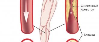
- Vascular atherosclerosis;
- Formation of blood clots in the lumen of an anomalous artery.
In a child, hypoplasia of the vertebral artery can manifest itself only in the case of its critical narrowing against the background of a disconnected circle of Velisius. In this case, the body is deprived of the ability to compensate for the lack of blood flow through connections of the affected artery with other cerebral vessels.
Vertebral artery hypoplasia is an abnormality of this vessel that a person is born with. But it manifests itself only after some time, when age-related changes in the spine or vascular wall occur. This leads to its critical narrowing with signs of cerebrovascular accident.
Underdevelopment of the central nervous system. Brain hypoplasia
Intrauterine underdevelopment of the structures of the central nervous system is one of the most serious defects affecting the functioning of the entire organism.
You can guess about the underdevelopment of the central nervous system structures from the symptoms, which are detected almost from the moment of birth. Thus, with hypoplasia of the cerebellum, which is responsible for the coordination of movements, the child experiences incoordination and deterioration in muscle tone. And when he begins to walk, a balance disorder is revealed. Hypoplasia of the corpus callosum, which is responsible for the coordinated work of both hemispheres, can lead to a variety of consequences. First of all:
- muscle spasms;
- convulsions;
- impaired skin sensitivity and body temperature regulation;
- deterioration of auditory and visual memory.
Brain hypoplasia (or microcephaly) is a proportional underdevelopment of all its structures. In early childhood, such a disorder manifests itself as delayed growth and development, and in later childhood – serious intellectual impairment.
Treatment of hypoplasia: general principles
There is no pathognomonic treatment (directed against provoking factors and slowing down the development of the disease) - medicine is taking the first steps in curing fetal diseases during intrauterine development. Rather, it is necessary to take preventive measures (in particular during pregnancy), which will help prevent failure in the formation of organs and systems, leading to disruption of tissue formation.
In the treatment of hypoplasia after the birth of a child and in adulthood, replacement therapy is practiced. So, with hypoplasia of the thyroid gland, accompanied by its hypofunction, the patient is injected with gland hormones.
You can read more about the tactics and methods of treating hypoplasia on our clinic’s website https://dobrobut.com.
Related services: Ultrasound examination Consultation with an obstetrician-gynecologist during pregnancy