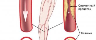
Every fourth stroke develops in the posterior parts of the brain in the area of responsibility of the vertebral arteries. Atherosclerosis and narrowing of the vertebral artery
can occur in any part of it and can cause ischemic stroke. Unlike narrowing of the carotid arteries, the role of which in the development of stroke has been known for a long time and methods for their treatment have been developed, the pathology of the vertebral arteries has not yet been sufficiently studied. However, modern technologies for diagnostic testing and endovascular treatment methods have opened up new opportunities for intervention in this disease.
Vertebral artery syndrome is a condition associated with obstruction of the patency manifested by symptoms of cerebrovascular insufficiency in the posterior part of the brain.
The causes of impaired patency can be very different, so the main task in the management of patients is an accurate diagnosis, so that as soon as the identification of the exact cause will allow it to be eliminated and the symptoms that are painful for patients to be removed. Every year the disease becomes younger, which is associated with an increase in the number of young people who sit for long periods at computers and have a sedentary lifestyle.
Timely diagnosis guarantees a favorable result of treatment of vertebral artery syndrome in our clinic.
General information
What is vertebral artery hypoplasia?
Hypoplasia of the right vertebral artery and hypoplasia of the left vertebral artery are an abnormal decrease in the lumen of these arteries (approximately to a diameter of less than 2 mm).
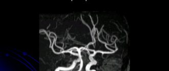
But there is no single criterion for narrowing the diameter of the vessel, and a number of authors include a decrease in the diameter of the vessel of less than 3 mm as signs of VA hypoplasia. The right vertebral artery is a branch of the right subclavian artery, which arises from the brachiocephalic trunk, and the left one arises from the left subclavian artery, which originates from the aortic arch. Next, both vertebral arteries (VA) rise in the bone canal to the brain and merge in the cranial cavity, forming the basilar artery. The vertebral arteries supply 15–30% of blood to the structures of the brain stem, the temporal/occipital lobes, the cerebellum, the hypothalamic region, and segments of the spinal cord, that is, they play a significant role in ensuring blood flow to the brain structures. Below is a picture of hypoplasia of the right/left VA.
Hypoplasia is one of the most common pathologies of the vertebral arteries. Its occurrence in the population, according to various sources, varies widely between 2.34% and 26.5% and is predominantly congenital in nature. Hypoplasia of one of the arteries occurs predominantly; cases of bilateral hypoplasia of the VA are relatively rare. The main place of narrowing of the lumen of the vertebral arteries is the place of entry into the bone canal (in the cranial cavity), which leads to a significant decrease in blood flow mainly to the posterior parts of the brain with the gradual development of non-vertebrogenic PA syndrome .
Hypoplasia of the vertebral artery is a factor in a significant reduction in blood circulation in the vertebrobasilar region, which disrupts the hemodynamics of the brain as a whole, increasing the risk of developing ischemic stroke (acute cerebrovascular accident). The situation is sharply worsening against the backdrop of the development of cerebral atherosclerosis , which is extremely common in the population due to poor lifestyle and especially dietary habits.
Compensation mechanisms that delay the manifestation of pathology
The following mechanisms for replenishing cerebral blood flow have been most clearly studied:
- Vertebral arteries are able to compensate for the impaired inflow in one of the symmetrical vessels due to the redistribution and reverse flow of blood through a short common basal trunk. Thus, each artery “helps out” the other when necessary.
- Vascular collaterals extend in the form of thin branches. But in conditions of insufficiency of the main trunk, they take on a large mass of blood and try to bypass the area of narrowing.
- Development of anastomoses - most often the subclavian artery serves as a “donor vessel” for the vertebral branches; the pressure in it is much higher, it “pushes” blood into the bypass network and fills the vertebral arteries.
- Local arterial hypertension is formed to ensure the transit of blood flow through a narrowed passage. In this case, hypertension is considered secondary and is opportunistic in nature in order to maintain the level of cerebral circulation.
Pathogenesis
Currently, hypoplasia of the vertebral arteries is considered as one of the manifestations (during the formation of the fetal vascular system) of undifferentiated connective tissue dysplasia. Also in the pathogenesis of clinical manifestations, the development of vasospastic reflex reactions caused by irritation of the sympathetic plexus of the vertebral arteries is of significant importance. The resulting flow of afferent impulses irritates the overlying centers of vascular-motor regulation, which is manifested by local/diffuse reactions that predominantly affect the vessels of the vertebrobasilar system.
Classification of the disease
Vascular disease affects and damages all areas of the brain. They provoke slow tissue development. The pathology may be congenital or appear with age.
Hypoplasia of the cervical artery most often begins to develop in the fetus in the womb.
The circle of Willis ensures normal blood supply to the brain. It is formed by large aortas of the spine with two branches. Usually the vessels develop evenly. A branched artery is responsible for natural blood supply. Vascular hypoplasia can be left-sided, right-sided or bilateral.
Hypoplasia of neck vessels disrupts blood supply and prevents normal nutrition of brain cells.
If you do not undergo diagnosis in time, cardiovascular failure and serious malfunction of the vestibular apparatus may develop.
First, you should undergo certain therapy, due to which the blood flow will return to normal.
Causes
Hypoplasia is predominantly congenital in nature, the development of which can be caused by both chromosomal mutations and factors with an adverse effect on the mother’s body during pregnancy. These factors include:
- Bruises/injuries during pregnancy .
- Infectious diseases that have a negative impact on the fetus, especially in the early stages of development ( influenza , rubella ).
- Bad habits (smoking, alcoholism, drug addiction).
- Prolonged contact with harmful substances (work in hazardous industries, toxic chemicals, radiation).
- Drug abuse.
Prevention
Simple preventive measures will help prevent negative consequences for the body. If you regularly play sports, before training you should drink some water and perform a light back massage. This simple tactic helps to speed up the blood, improve metabolism and absorb the necessary amount of moisture into the intervertebral discs.
Try not to lift or hold heavy objects with outstretched arms. You need to learn how to carry weights correctly so as not to injure your back. With an even posture, you need to squat down, take the load and straighten your legs with it. At the same time, place your hands as close to your body as possible. Distribute the load evenly, so you don't have to carry it in one hand if you can carry two bags in both hands. Use bags, carts and rolling suitcases to transport heavy items.
When doing heavy physical work, which is inherently associated with lifting weights, wear a special support belt or corset. Do not lift loads weighing more than 10 kg. Use comfortable shoes in everyday life, giving preference to models made from natural raw materials that securely and securely support your foot.
Women, especially during pregnancy, should avoid wearing high-heeled shoes, as they are very harmful to the spine. After being in a bent position for a long time, you can hang on the bar or stretch up.
Vessels
Anterior Cerebral Artery: Segments, Resistance Index, Normal, Aneurysm, Branches
Feb 03, 2021 Kokh V. A.
13612
Vessels
Duplex Scanning of Brachiocephalic Arteries: What is BCA, Explanation
Feb 03, 2021 Kokh V. A.
10244
Vessels
Symptoms
The initial signs of VA hypoplasia are nonspecific, and they can be mistaken for manifestations of encephalopathy , cervical osteochondrosis or vegetative-vascular dystonia . PA hypoplasia syndrome is clinically manifested by three groups of symptoms:
- A group of vertebral symptoms (pain in the cervical spine/occipital region).
- Local symptoms - pain in the vertebral artery/structures of the corresponding vertebral segment (during palpation) with pronounced irradiation to the head.
- Symptoms at a distance from the vascularization zone of the VA, as well as due to irritation of the sympathetic plexus of the vertebral artery (increased blood pressure, migraine -type pain , body instability when walking).
The specificity of the clinical manifestations of VA hypoplasia is determined by a number of factors: the localization/extension of the lesion, the functional state of the arterial-venous system of the brain - the presence of anastomoses, collaterals, the state of the vascular wall.
Standard patient complaints include frequent, severe headaches ; dizziness ; surges in blood pressure ; inability to concentrate; memory loss; increased weather sensitivity ; deterioration of vision/appearance of “spots” in the field of vision; unsteady gait, decreased performance; difficulty making sudden movements/turns.
The presence of neurological complaints is traditionally considered to be indirect signs of stenosis . The symptoms of left-sided and right-sided VA hypoplasia are generally similar, however, the pathology of the vertebral artery on the right is often localized higher (in the intracranial area), which causes a more rapid development of symptoms and their more pronounced manifestation (a sharp drop in vision, loss of consciousness, severe coordination disorder).
The left vertebral artery initially has a larger working diameter, so symptoms do not appear so sharply and later. With left-sided hypoplasia, the characteristic symptoms are sharp, throbbing occipital pain; shots to the head; pain on palpation of the 1st and 2nd vertebrae of the neck; feeling of sand/double vision; tinnitus/hearing impairment. With bilateral VA hypoplasia, due to the lack of a compensatory effect, symptoms develop quickly, and there is a high risk of developing serious complications.
Treatment
Treatment is required only when signs of decompensation begin. In some patients, adaptation is quite sufficient and does not cause any symptoms. Increased physical activity or additional vascular and cardiac diseases reveal anatomical insufficiency of the vertebral vessels.
In mild cases, patients are helped by vasodilators. The drugs allow you to expand the vessel and increase blood flow. These include the well-known nitro group (Nitroglycerin, Sustak, Erinit). These medications may be accompanied by headache, tachycardia, nasal congestion, fever, dizziness, and vomiting.
It is possible to reduce the negative effect by simultaneous administration of antiplatelet agents (Curantyl, Trental).
Any conservative treatment has the following goals:
- support of compensatory mechanisms;
- prevention of thrombotic complications.
It is impossible to change the structure of the vessel with drugs.
Drugs are used that replenish brain hypoxia and support cell metabolism (nootropics, Piracetam, Cortexin).
Folk remedies do not treat hypoplasia. Patients should learn this and not waste time on unnecessary waits. You can prevent subsequent atherosclerosis with the help of dietary recommendations, the use of herbs and plants to strengthen the vascular wall.
Consequences and complications
In general, VA hypoplasia significantly impairs quality of life. The most serious complications of VA hypoplasia primarily include a high risk of cerebrovascular accident/ stroke , which is caused by a decrease in blood supply to brain structures. In addition, frequent complications of the pathology of the vertebral arteries include neurological symptoms: headaches ( migraine ), irritability, depression, severe fatigue, disorders of the autonomic nervous system, impaired auditory/visual function, weakened cognitive abilities, dementia, decreased ability to work, arterial thrombosis
Diagnostics
In order for the doctor to make a correct diagnosis, neurological examinations should be completed, the results of which determine the presence of the disease. The patient will need to consult a neurologist, therapist, cardiologist and ophthalmologist.
The doctor prescribes an MRI, then ultrasound scanning, Dopplerography and angiography. All methods will help determine the shape, size and structure of blood vessels. Experts also look at the state of the circulatory system.
The diagnosis is confirmed if:
- the internal lumen in diameter is less than 2 mm;
- blood flow moves at a speed of less than 40 ml/min;
- there is no stenosis.
As a result of the examination, patients observed:
- stroke anomaly in approximately 9% of cases;
- stenosis – 20%;
- deformation – 40%;
- extravascular compression 10%;
- blockage at 4%.
Basically, damage to the blood supply to the left side is diagnosed when a person experiences cardiovascular failure. Even if you regularly visit a specialist, it is impossible to notice the disorder.
Damage to the left side appears in adults, and symptoms should be closely monitored. The first sign is pain in the neck. Other symptoms may be absent and making an accurate diagnosis difficult.
About 25% of people have a disorder in the circulatory system, half of them can live their whole lives and not know about it. The initial stage of the pathology does not cause serious manifestations.
Forecast
The prognosis is determined by many factors, in particular, unilateral/bilateral VA pathology, since with unilateral VA hypoplasia, blood flow compensation mechanisms develop, the functional state of the arterial-venous system of the brain, and the condition of the vascular wall (in particular, the presence of atherosclerotic plaques). After surgical treatment and drug rehabilitation, in most cases the lumen of the vertebral arteries can be normalized, and neurological symptoms regress.
How is hypoplasia detected?
Existing methods make it possible to make a correct diagnosis after checking the shape, direction, and diameter of the vertebral arteries in each segment.
Read about the features of the anatomical structure and location of the vertebral arteries in this article.
Ultrasound techniques, including conventional ultrasound, Dopplerography of the vessels of the neck and head, duplex scanning, make it possible to check the direction of the vertebral arteries, internal diameter, nature of tortuosity, and wall structure.
One of these signs is a deviation from the norm of the vessel lumen in the direction of decrease. The average diameter is considered to be 3.6–3.8 mm.
A narrowing of up to 2 mm is regarded as significant hypoplasia, especially if the area is localized in the upper segment of the artery
Tomography is carried out using a magnetic resonance (MR) machine and computer technology. MRI signs allow us to assess blood flow and the capabilities of the circle of Willis. In conclusion, information is given to the doctor about all vascular components. Congenital abnormalities ranging from hypoplasia to aplasia (complete absence of part of the artery) are possible.
The most informative is MR angiography, which combines contrasting of blood vessels with the possibility of resonant reflection of waves.
List of sources
- Shmidt E.V. Classification of vascular lesions of the brain and spinal cord // Journal. neuropathology and psychiatry named after. S.S. Korsakov. 1985. T. 85, No. 9. p. 1281-1288.
- Kurtusunov B. T. Variant anatomy of the vertebral arteries at the stages of human ontogenesis. Author's abstract. diss. Doctor of Medical Sciences Volgograd, 2011.
- Pizova N.V., Druzhinin D.S., Dmitriev A.N. Hypoplasia of the vertebral arteries and cerebrovascular accidents // Journal of Neurology and Psychiatry. 2010. No. 7. P. 56–58.
- Kometov A.V., Zhukova M.V. Variants of the structure of the vessels of the arterial circle of the cerebrum depending on the type of Kimmerle anomaly. Congress of Manual Therapists of Russia, 1st: Materials. M 1999; 32.
- Odinak M.M., Mikhailenko A.A., Ivanov Yu.S. and others. Vascular diseases of the brain. St. Petersburg: Hippocrates 1997.
Underdevelopment of the genitourinary system
Underdevelopment of the genitourinary system manifests itself in the form of:
- kidney hypoplasia (possible underdevelopment of one kidney and absence of the second);
- underdevelopment of the testicles in men and the uterus in women, manifested by reproductive dysfunction.
During intrauterine development, a combination of both disorders is possible - from the urinary tract and the reproductive system.
In men, testicular hypoplasia can be accompanied by a reduction in the penis and scrotum. And in women with uterine hypoplasia, the ovaries, fallopian tubes and external genitalia (clitoris, labia) may not undergo such changes.
A woman’s reproductive function depends on the degree of endometrial hypoplasia of the uterus - if the inner layer of the uterus is underdeveloped, a fertilized egg will not be able to attach to the inner surface of this organ.