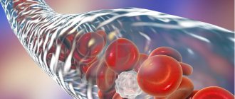
According to the World Health Organization, in most developed countries, deep vein thrombosis and pulmonary embolism have become one of the leading causes of mortality. Millions of people around the world die every year from acute blockage (embolism) of a blood vessel by a thrombus. The danger of thrombosis is that a clot can reach a size comparable to the diameter of a venous vessel and completely block the blood flow in it. Or, if it ruptures, the clot can travel with the blood into the vessels of the lungs and lead to pulmonary thromboembolism. Often with fatal outcome.
Related article Catch it if you can. How to detect and destroy a blood clot
“A blood clot has broken off,” they sometimes say in this case about the cause of death. Even fairly young people often die from detached blood clots. According to specialists from the Moscow State Medical and Dental University named after A. I. Evdokimov, more than 90% of deaths from pulmonary embolism occur in patients who were not diagnosed with embolism and who did not receive treatment.
What causes blood clots?
Thrombi, in the form of blood clots, form in blood vessels in response to damage to the vascular wall, slowing of blood flow and changes in blood composition. “Thrombosis does not occur out of nowhere; there must be some prerequisites. This is facilitated by the presence of varicose veins in the lower extremities. If it is, you should pay attention to compactions that may appear in the veins,” says phlebologist, surgeon Fedor Shpachenko .
According to the phlebologist, compactions may indicate that blood clots have already formed in the veins. “Often, these blood clots may not occur in the superficial veins that we see, but in deep veins, and it is impossible to visualize them without special research methods,” warns Shpachenko.
If the blood clot comes off
The thrombus is not always firmly attached to the wall of the vessel. When a clot is superficially attached, a simple surge in pressure, a blow, or a careless movement is enough for the clot to come off and travel through the circulatory system. Moving through the vessels, the detached thrombus sooner or later reaches the most important organs, and usually the pulmonary vein, if the thrombus was venous, or the left atrium, if arterial.
Pulmonary thrombus
In the most dangerous cases, blockage of the large vessels of the lung occurs, after which death occurs within a few minutes. A less severe option is infarction-pneumonia, when a blocked vessel bursts and the lungs begin to fill with venous blood, which is accompanied by acute pain and hemoptysis. In this case, the patient has every chance of survival.
The mildest case, when a blood clot enters the pulmonary veins, is accompanied by an increase in blood pressure in the lungs, pain, shortness of breath, and suffocation. If you call an ambulance right away, the patient is likely to survive.
Coronary thrombus
If a blood clot located in the left atrium or coronary arteries breaks off, the patient is at risk of: myocardial infarction, stroke if the blood clot enters the brain through the bloodstream, infarction of the intestines or kidneys if the arteries leading to them are blocked, as well as blood stagnation and gangrene with subsequent loss limbs if the blood clot gets into the large arteries of the arm or leg.
Who is at risk?
Those who are most likely to develop thrombosis are people who are obese and have varicose veins, women who take oral contraceptives, and those who take many long flights. But a high risk of blood clots occurs when all these factors are combined.
“Patients with obesity and varicose veins are at risk. As for medications, in particular oral contraceptives, now most of them are well balanced and we rarely see the development of thrombosis and thromboembolism in women taking these drugs. People with a sedentary lifestyle, impaired hemostasis, namely blood clotting disorders, also fall into this group. If the patient has all these provoking factors, then the risk of developing thrombosis increases. Age also makes it worse. Over the years, the vessels lose elasticity and become brittle, plus a number of concomitant diseases cause swelling of the lower extremities,” says Shpachenko.
How do blood clots occur?
If a blood clot has formed on the wall of an artery, its appearance can be described in the following stages:
- Some process damages the artery wall.
- The body notices the violation and begins to build protection against blood loss, forming a large number of special blood cells - platelets, which, attaching to the damaged area, form a kind of patch.
- In case of coagulation disorders or changes in the hematopoietic system, platelet formation does not stop on time and continues longer than expected. This causes too much growth to form on the wall. Or platelets, which are in small quantities in the blood, floating past in the bloodstream, stick to the resulting accumulation.
The causes of damage to the walls of blood vessels can be:
- mechanical disruption of the structure due to injury;
- infectious lesion;
- high levels of glucose molecules in the blood;
- immune system dysfunction.
If there are no factors that contribute to the formation of blood clots, any injury or other damage will not lead to a large accumulation of blood cells. Under the layer of platelets, the artery wall will heal and recover, and the crust will resolve over time.
There are several stages of thrombus formation:
- disruption of the structure of the inner surface of the artery;
- activation of blood clotting factors;
- platelet adhesion at the site of injury;
- the appearance of substances that trigger a chain of reactions that form fibrin threads, which contribute to thrombus formation;
- a kind of network of fibrin threads is formed, into which blood cells enter, creating a large clot;
- Over time, the clot thickens, forming a thrombus.
When a blood clot breaks off under the influence of any factors, it begins to move through the bloodstream. Once it hits the nearest bottleneck, the blood flow will be blocked. If a similar situation happens outside a medical facility, it is impossible to save the person.
How to diagnose thrombosis at an early stage?
Thrombosis at an early stage can be detected with timely diagnosis of the veins of the lower extremities. To confirm or refute the diagnosis, phlebologists often recommend laboratory and ultrasound duplex scanning (USD) of the veins. This diagnostic method allows you to see the walls and lumen of the veins, the presence of a blood clot in them, its size, and even roughly judge how long ago the process is. You can also do angiography: a contrast study of blood vessels. And the predisposition to the appearance of blood clots can be identified using a coagulogram: a comprehensive analysis of blood clotting indicators.
Regulation of the coagulation system
Figure 6. Contribution of extrinsic and intrinsic tenase to fibrin clot formation in space. We used a mathematical model to investigate how far the influence of a clotting activator (tissue factor) could extend in space. To do this, we calculated the distribution of factor Xa (which determines the distribution of thrombin, which determines the distribution of fibrin). The animation shows the distributions of factor Xa produced by extrinsic tenase (VIIa–TF complex) or intrinsic tenase (IXa–VIIIa complex), as well as the total amount of factor Xa (shaded area). (The inset shows the same thing on a larger concentration scale.) It can be seen that activator-produced factor Xa cannot travel far from the activator due to the high rate of inhibition in the plasma. On the contrary, the IXa–VIIIa complex works far from the activator (since factor IXa is inhibited more slowly and therefore has a greater effective diffusion distance from the activator), and ensures the distribution of factor Xa in space.
[9]
Let's take the next logical step and try to answer the question - how does the system described above work?
Cascade device of the coagulation system
Let's start with the cascade - a chain of enzymes that activate each other. A single enzyme operating at a constant speed produces a linear dependence of product concentration on time. For a cascade of N enzymes, this dependence will have the form tN, where t is time. For the effective operation of the system, it is important that the response is of precisely this “explosive” nature, since this minimizes the period when the fibrin clot is still fragile.
Triggering of coagulation and the role of positive feedbacks
As mentioned in the first part of the article, many clotting reactions are slow. Thus, factors IXa and Xa themselves are very poor enzymes and require cofactors (factors VIIIa and Va, respectively) to function effectively. These cofactors are activated by thrombin, a device where the enzyme activates its own production is called a positive feedback loop.
As we have shown experimentally and theoretically, the positive feedback of factor V activation by thrombin forms the activation threshold - the property of the system not to respond to small activation, but to quickly respond when a large one appears. This ability to switch seems to be very valuable for folding: it helps prevent “false positives” of the system.
The role of the intrinsic pathway in the spatial dynamics of folding
One of the intriguing mysteries that haunted biochemists for many years after the discovery of the essential coagulation proteins was the role of factor XII in hemostasis. Its deficiency was detected in simple clotting tests, increasing the time required for clot formation, but, unlike factor XI deficiency, was not accompanied by coagulation disorders.
One of the most plausible options for unraveling the role of the internal pathway was proposed by us using spatially inhomogeneous experimental systems. Positive feedbacks have been found to be important specifically for the propagation of coagulation. Effective activation of factor X by external tenase on the activator will not help form a clot away from the activator, since factor Xa is rapidly inhibited in the plasma and cannot move far from the activator. But factor IXa, which is inhibited an order of magnitude slower, is quite capable of this (and is helped by factor VIIIa, which is activated by thrombin). And where it is difficult for him to reach, factor XI, also activated by thrombin, begins to work. Thus, the presence of positive feedback loops helps create the three-dimensional structure of the clot.
Protein C pathway as a possible localization mechanism for thrombus formation
Activation of protein C by thrombin itself is slow, but accelerates sharply when thrombin binds to the transmembrane protein thrombomodulin, synthesized by endothelial cells. Activated protein C is capable of destroying factors Va and VIIIa, slowing down the coagulation system by orders of magnitude. The key to understanding the role of this reaction was spatially inhomogeneous experimental approaches. Our experiments suggested that it stops the spatial growth of the thrombus, limiting its size.
How to determine deep vein thrombosis?
Thrombosis often occurs asymptomatically or with mild symptoms. The nature of patient complaints may vary depending on the location of the blood clot, the duration of the disease and the nature of the lesion. The main symptom of deep vein thrombosis is pain in the leg, namely in the calf muscle. Swelling may also be a concern. When a blood clot forms in the deep veins of the lower extremities, the skin may take on a reddish or blue tint.
Article on the topic
Dissolve the clot. What foods improve blood quality
“Signs of thrombosis can manifest themselves as swelling and pain in the lower extremities, and the location of this pain can be very diverse. Sometimes you can’t guess what the pain is due to: either osteochondrosis, or blood clots, or other diseases. Therefore, if your legs swell, or there are varicose veins in your legs, then you should pay attention to the possible development of blood clots,” says Shpachenko. An ultrasound duplex scan or angiography will help you understand the exact picture.
Summarizing
In recent years, the complexity of the coagulation system has gradually become less mysterious. The discovery of all essential components of the system, the development of mathematical models and the use of new experimental approaches made it possible to lift the veil of secrecy. The structure of the coagulation cascade is being deciphered, and now, as we saw above, for almost every significant part of the system, the role it plays in the regulation of the entire process has been identified or proposed.
Figure 7 shows the most recent attempt to reconsider the structure of the coagulation system. This is the same diagram as in Fig. 1, where parts of the system responsible for different tasks are highlighted with multi-colored shading, as discussed above. Not everything in this scheme is securely established. For example, our theoretical prediction that activation of factor VII by factor Xa allows clotting to respond in a threshold manner to flow rate remains as yet untested experimentally.
Figure 7. Modular structure of the coagulation system: the role of individual coagulation reactions in the functioning of the system.
[1]
It is quite possible that this picture is not yet completely complete. However, progress in this field in recent years gives hope that in the foreseeable future, the remaining unsolved regions of the coagulation circuitry will gain meaningful physiological function. And then it will be possible to talk about the birth of a new concept of blood coagulation, which replaced the old cascade model, which faithfully served medicine for many decades.
The article was written with the participation of A.N. Balandina and F.I. Ataullakhanova and was originally published in Priroda [10].
How is pulmonary embolism diagnosed?
As a result of pulmonary embolism, cardiac function, pulmonary blood flow and gas exchange are disrupted. This condition is accompanied by characteristic symptoms, primarily a rapid heartbeat. There may also be a stabbing pain in the chest that gets worse while breathing. Patients may experience severe shortness of breath, and the respiratory rate may increase to 30-40 per minute. In addition, cyanosis or pallor of the skin, decreased blood pressure, cough, and hemoptysis may be observed.
“In the case of pulmonary embolism, difficulty breathing and chest pain may occur. In people with a disease of the vascular-cardiac system, for example, atrial fibrillation, pulmonary embolism can occur quite often,” says Shpachenko.
If thromboembolism is suspected, urgent hospitalization is required. To confirm the diagnosis, the hospital will order electrocardiography, chest x-ray and echocardiography. A radionuclide scan of the lungs, probing of the right side of the heart, and computed tomography with contrast of the pulmonary arteries can also be performed.
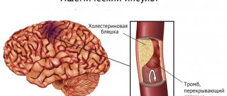
What is thrombosis?
Thrombosis can be venous or arterial. Venous is considered an acute disease; it appears due to disruption of the structure of the venous wall during injury, surgery, radiation or chemotherapy, due to increased blood clotting, due to a slowdown in the speed of blood flow.
Arterial thrombosis is a pathological condition. Patients have atherosclerotic plaques - cholesterol deposits that narrow the lumen of the artery and provoke circulatory deficiency. Over time, the plaques become clotted.
There is also pulmonary embolism (PE), a fatal disease when the lumen of the pulmonary artery is completely or partially blocked by a blood clot. According to statistics, almost 50% of patients with massive pulmonary embolism die within 30 minutes after its occurrence.
Diagnostics
Problems associated with various pathologies of blood vessels, veins and arteries are dealt with by a phlebologist surgeon. If the doctor suspects a blood clot in the leg, then, taking into account the corresponding symptoms, he will prescribe examination and treatment. Diagnostics includes examination, laboratory tests and instrumental examination.
Inspection
The examination begins with interviewing the patient, collecting anamnesis and direct examination. A phlebologist uses special methods to determine pathology:
- Lowenberg's symptom
(phlebotonometry), using a tonometer; - marching test
, with application of an elastic bandage; - Mayo test
, with a tourniquet applied.
The appearance or worsening of pain, as well as severe distension of the veins and their clear pattern that appears as a result of manipulation, is a sign of the presence of thrombosis in the legs.
Laboratory samples
To clarify the causes of the disease, the doctor prescribes a series of tests:
- general and biochemical blood tests, which allow you to evaluate blood clotting and check for the presence of an inflammatory process;
- coagulogram to determine the level of D-dimer (the residual amount of fibrin after breakdown).
Instrumental examination
Diagnosis of thrombosis should be as accurate as possible. This is facilitated by the use of such modern methods:
- Dopplerography
. It allows you to assess the speed of blood flow and detect its disturbances. - Duplex ultrasound scanning
determines the size of the lumens of blood vessels, their tortuosity, and wall thickness. This method also reveals abnormalities in the structure of blood vessels and determines the presence of blood clots. - Digital subtraction venography
. Examines the condition of blood vessels and identifies blood clots. It is carried out using a contrast agent (iodine), which is administered intravenously. The resulting X-ray images (with computer processing) clearly show the affected areas, as well as the size of the blood clot and the nature of its location. - Radionuclide scintigraphy
.
Before performing phlebography, make sure that you are not allergic to the drug used for the procedure.
So, what is next?
At the first suspicion of thrombosis, you should consult a doctor. In acute cases, the patient is immediately sent to the operating table for vein ligation, installation of a vena cava filter, or surgical removal of a blood clot. But more often thrombosis is treated on an outpatient basis. The doctor will conduct an initial diagnosis, confirm the conclusion with an ultrasound examination (duplex scanning of veins) and prescribe anticoagulants - blood thinners, the task of which is to clear the lumen of the vessel. Be patient! The duration of taking anticoagulants can range from 3 months to conditionally lifelong use. And all this time you will need to visit the clinic so that the doctor evaluates the effectiveness (lack of relapse), safety (lack of bleeding) of therapy and determines the required duration of treatment.
Treatment
After diagnosing thrombosis of the lower extremities, a blood clot in the leg requires urgent treatment. In order to get rid of problematic pathology, a set of therapeutic measures is used.
Drug treatment
It is necessary in both acute and chronic stages of development:
Thrombolytic drugs
. They are prescribed for urgent dissolution of a blood clot. The medicine is injected into the vessel, in the immediate vicinity of the fibrin clot. The procedure is carried out inpatiently, as it is an emergency medical intervention.
| Generation | Name | Type | Action |
| I | Streptokinase Streptodecase Urokinase Fibrinolysin | Fibrin-selective, created by combining human and bacterial material | Act throughout the bloodstream |
| II | Alteplase Actilyse Prourokinase (recombinant) | Systemic, isolated from activated human plasmin | Show activity on the surface and in thrombus structures |
| III | Tenecteplase Lanoteplase Urokinase (plasminogen) | Combined | Combined action |
Anticoagulant therapy. Taking these drugs normalizes the properties of the blood, preventing the formation of new and growth of existing platelet clots. The drugs are:
- Direct
. Provide a rapid effect on blood composition (Heparin and Enoxaparin sodium). - Indirect
. Prescribed to prevent further development of leg thrombosis. They have a long-term effect (Warfarin, Acenocoumarol).
Antiplatelet drugs
. Drugs in this group improve the flow properties of blood, reduce cohesion, and disaggregate platelets (Aspirin, Pentoxifylline, Methylethylpyridinol).
Absorbable anti-inflammatory ointments
, helping to relieve swelling and pain (Heparin, Venorutin).
Bandaging the affected area with an elastic bandage
.
Physiotherapy
Hardware procedures are prescribed to eliminate inflammation, reduce pain and restore hemodynamics. To do this, the following procedures are used:
- electrophoresis;
- darsonvalization;
- UHF;
- magnetotherapy.
Operative method
Surgery is performed when there is a high risk of pulmonary embolism. The complex of surgical measures includes thrombectomy, ligation of the great deep vein, and introduction of a vena cava filter.
Folk remedies
Herbal medicine has an effective effect on blood circulation and helps strengthen the walls of veins and blood vessels. Herbs such as chamomile, calendula, and horse chestnut can be used as tinctures, teas, and compresses. Alcohol tincture of birch buds is especially helpful for vascular diseases. It is used externally.
Hirudotherapy (treatment with leeches) reduces blood clotting and stops the development of the disease.
Regular medical examination, therapeutic exercise and other preventive measures will help avoid the development of thrombosis in the legs.