Tricuspid regurgitation is the reversal of blood flow from the right ventricle back into the atrium, but is not a diagnosis in itself. This is not even a disease, but a consequence of a malfunction of the tricuspid valve, which closes the passage from the right atrium to the corresponding ventricle.
The condition can be primary or secondary, depending on the origin of the pathological process. Recovery is carried out surgically.
The prospects for a complete cure are good, but only in the early stages, when there are no anatomical defects in the heart or distant systems yet.
Fortunately, the duration of the initial phase is sufficient for a thorough diagnosis. The intervention is planned, except for exceptional cases.
The approximate time frame from the moment the deviation occurs to the formation of a clear clinic is 3-6 years.
Development mechanism
The essence of the pathological process is the disruption of hemodynamics at the local level and the formation of a persistent anatomical defect.
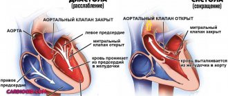
In the normal state of affairs, blood in the cardiac structures moves strictly in one direction, ending the cycle in the left ventricle and being transported to the aorta, and from there to its branches in a large circle.
The heart is represented by a group of chambers, each separated from the other by valves, which does not allow fluid connective tissue to move in the opposite direction.
The tricuspid structure closes the gap between the right atrium and ventricle. In case of weakness, insufficiency, or defects of the connective tissue, a reverse flow of blood or regurgitation occurs, which is called according to the name of the valve that causes the condition.
The result of the deviation is, firstly, a disruption in the transport of blood in the small circle, and secondly, an insufficient amount of it, which is released into the aorta.
This leads to generalized hemodynamic deviations, tissue hypoxia, and multiple organ failure in the future.
Acquired heart defects
Acquired heart defects are a group of diseases accompanied by disruption of the structure and function of the heart valve apparatus and leading to changes in intracardiac circulation.
Acquired heart defects develop as a result of acute or chronic (long-term) diseases and injuries that impair the function of the valves and cause changes in intracardiac hemodynamics (blood movement through the vessels).
More than half of all acquired heart defects are due to lesions of the mitral (located between the left atrium and the left ventricle) valve, about a third are lesions of the aortic (separates the left ventricle and aorta) valve, the rest are represented by combined (changes affecting several valves) defects.
Symptoms of acquired heart disease
- Dyspnea.
- Marked weakness.
- Changes in skin color - constant pallor or, conversely, pinkishness.
- Feeling of heartbeat.
- Possible pain in the heart area during physical activity.
- Headaches, dizziness, fainting (loss of consciousness).
Forms
Acquired heart defects are classified into several categories.
According to etiology (cause of occurrence) there are:
- rheumatic (occurs as a result of rheumatism - a systemic inflammatory disease of connective tissue with predominant damage to the heart);
- endocardial (due to endocarditis - inflammation of the inner lining of the heart);
- syphilitic (due to syphilis - a systemic disease that is predominantly sexually transmitted and affects many organs and systems) and so on.
Based on the type of valve affected, the following are distinguished:
- aortic;
- mitral;
- tricuspid valve defect;
- valve defect of the pulmonary artery trunk.
By the number of affected valves:
- isolated, or local (damage to 1 valve),
- combined defect - insufficiency and stenosis (narrowing of the lumen) simultaneously occur on one valve;
- combined defect - changes affect several valves.
Functionally:
- stenosis - narrowing of the lumen of the opening as a result of post-inflammatory (occurs after the inflammatory process) cicatricial adhesions of the valve leaflets;
- insufficiency - incomplete closure of the heart valve leaflets;
- prolapse - protrusion, protrusion or eversion of the valves into the cavity of the heart.
According to the severity of the defect and the degree of hemodynamic disturbance (blood movement through the vessels) of the heart:
- does not have a significant effect on intracardiac circulation;
- moderately expressed;
- pronounced.
According to the state of general hemodynamics:
- compensated heart defects - without circulatory failure (a condition in which the heart is not able to adequately supply blood to all organs and tissues);
- subcompensated - with transient (temporary) decompensation (inability to compensate for impaired blood flow) caused by excessive physical exertion, elevated body temperature, pregnancy, and so on;
- decompensated - with developed circulatory failure.
Reasons for purchasing
The most common causes of acquired heart defects are:
- rheumatism is a systemic inflammatory disease of connective tissue primarily affecting the heart;
- infective endocarditis (inflammation of the inner wall of the heart);
- atherosclerosis is a chronic disease characterized by hardening and loss of elasticity of the walls of the arteries, narrowing of their lumen due to the so-called atherosclerotic plaques (formations consisting of a mixture of fats (primarily cholesterol (a fat-like substance that is a “building material” for the body’s cells) and calcium ));
- heart injuries (bruises and wounds of the heart muscle);
- syphilis is a systemic disease, transmitted primarily through sexual contact and affecting many organs and systems;
- sepsis (blood poisoning) and others.
Diagnostics
- Analysis of the medical history and complaints - when (how long ago) and what kind of complaints appeared, whether the patient consulted a doctor, underwent examination and treatment, with what results, and so on.
- Analysis of life history - past infectious diseases and chest injuries are clarified.
- Analysis of family history - it is found out whether any of the close relatives have heart diseases, what kind of diseases they are, and whether there have been cases of heart defects in the family.
- Medical examination. Wheezing in the lungs, murmurs in the heart are determined, the level of blood pressure is measured, the boundaries of the heart are determined by percussion (by tapping) (to determine hypertrophy (increase in size)), heart sounds and sounds are listened to to determine the type of defect, the lungs are auscultated and the size of the liver is determined ( to diagnose heart failure, a condition in which the heart cannot provide adequate blood flow to all organs).
- General blood test - allows you to detect signs of inflammation in the body (increased levels of leukocytes (white blood cells), increased levels of ESR (erythrocyte sedimentation rate (red blood cells), a nonspecific sign of inflammation)) and identify complications and possible causes of heart disease.
- A general urine test can detect complications of heart defects.
- Biochemical blood test - determination of the level of total cholesterol (a fat-like substance that is a “building material” for the body’s cells), “bad” (promotes the formation of atherosclerotic “plaques” (cell clots)) and “good” (prevents the formation of “plaques”) cholesterol, level of triglycerides (fats, the source of cell energy), blood sugar.
- Electrocardiography (ECG) is a method of recording the electrical activity of the heart on paper. Allows you to diagnose changes in heart rhythm, determine the type of arrhythmia and signs of ischemia (insufficient blood supply to the heart muscle).
- Phonocardiography is the recording of sound signals of the beating heart: murmurs and tones. The method allows you to assess the duration, intensity, nature, origin of heart murmurs and tones, and record the third and fourth heart sounds, which are indistinguishable by ear, which ultimately allows you to determine the type and nature of the heart defect.
- Echocardiography (EchoCG) is an ultrasound examination of the heart. Allows you to diagnose the defect itself, the area of the atrioventricular orifice (connecting the atrium and ventricle of the heart), the severity of regurgitation (backflow of blood), the condition and size of the valves, and determine the pressure in the vessels.
- Chest X-ray with intravenous contrast agent (angiocardiography) - assesses the condition of the lungs, the size of the heart and its chambers. The method helps to identify specific changes in the vascular bed in such patients.
- Multislice computed tomography - cardiography (MSCT of the heart) is a method of layer-by-layer scanning of heart structures, based on recording an X-ray beam passing through tissue using several rows of ultra-sensitive detectors. Cardiac MSCT enables 3-dimensional reconstruction of the heart and is used to identify valve defects.
- Cardiac magnetic resonance imaging (MRI) is a method of obtaining diagnostic images based on the physical phenomenon of nuclear magnetic resonance, thus it is safe for the body.
- It is also possible to consult a therapist or cardiac surgeon.
Treatment of acquired heart disease
Conservative (drug) treatment of acquired heart disease is prescribed only with the goal of stabilizing the heart rhythm, preventing heart failure (a condition in which the heart is unable to provide normal blood flow to all organs), complications and relapses (recurrences) of the underlying disease that caused the heart disease.
The main treatment method for acquired heart defects is surgery.
Valve defect correction:
- valvotomy (dissection of fused heart valves);
- valvuloplasty (restoration of valve function by cutting the valve walls and subsequent suturing of new leaflets).
- Prosthetic valve replacement (replacement with an artificial one).
Complications and consequences
- The development of heart failure (a condition in which the heart is unable to adequately supply blood to all organs and tissues).
- Heart rhythm disturbance (any heart rhythm other than normal).
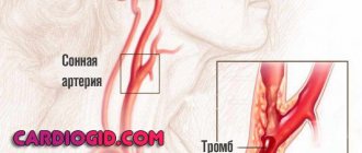
- Thromboembolic complications (complications in which blood clots (blood clots in a vessel) can enter any vessel in the body through the bloodstream, block its lumen and cause organ dysfunction).
- Disability of patients.
- Lethal outcome (death).
Prevention of acquired heart disease
Prevention of all acquired heart defects consists in preventing the underlying disease that causes the defect (for example, timely treatment of tonsillitis (an infectious disease primarily affecting the palatine tonsils) prevents the development of rheumatism (a systemic inflammatory disease of connective tissue primarily affecting the heart)).
Forms of violation
Typification of the pathological process is carried out on two grounds.
Based on the origin of the anatomical defect, they talk about:
- Primary form. It develops spontaneously, against the background of cardiac problems themselves. Including aortic insufficiency, previous inflammatory, infectious conditions and others.
It is characterized by greater complexity from the point of view of cure and prospects for recovery, since correction requires not only the symptomatic component, but also the acquired defect.
This group also includes congenital factors caused by genetic defects and spontaneous deformations of the tricuspid valve.
- Secondary variety. Against the background of current pathologies of distant organs and systems.
Degrees of regurgitation
Another basis for classification is the degree of deviation from the norm. Also called stages of the pathological process.
Accordingly, they distinguish:
- Weakly expressed type. 1st degree. The amount of blood returning is not known exactly. The volumes of the jet do not exceed 1 cm in diameter. The intensity of manifestations with minimal tricuspid regurgitation is insignificant or completely absent, which makes early diagnosis a matter of luck. This is the best time to start therapy under the supervision of cardiac surgeons.
- Moderate type. 2nd degree. Characterized by disruption of normal blood flow in a volume of 2 cm, no more. Recovery is carried out surgically. The clinical picture is minimal, characterized by chest pain, shortness of breath during intense physical activity. There is a chance for a complete cure, the likelihood of the formation of persistent cardiac and extracardiac defects is present, but it is not yet great. Even if these occur, the likelihood of a high-quality, long life is maximum.
- Expressed type. 3rd degree. A stream of blood more than 2 cm in diameter. Chronic heart failure of congestive type develops. There are prospects for recovery, but they are not complete, and long-term, lifelong maintenance therapy is required.
- Terminal phase. 4th degree. Surgical assistance does not make much sense, since the heart, kidneys, liver, and brain are significantly changed. Recovery is impossible; palliative care is required to ensure an acceptable quality of life for the short remaining period of life. Death occurs from acute heart failure.
Classifications are used to accurately assess the patient’s condition, prospects for treatment, and determine diagnostic and therapeutic tactics.
Discussion
After eliminating the mitral defect through prosthetics, an improvement in myocardial contractility was noted, as well as regression of the phenomena of chronic heart failure and pulmonary hypertension in the majority of patients. Despite this, patients experienced right ventricular dilatation and relatively low TAPSE and TAPSV scores over time, suggesting progression or persistence of right ventricular dysfunction. According to A.A. Magizova et al. [2], right ventricular dilatation is a predictor of recurrence or progression of tricuspid regurgitation in the postoperative period. In our study, we also revealed progression of tricuspid regurgitation in the mid-term period in patients who did not undergo annuloplasty (0.4±0.5 versus 1.3±0.8, p
<0.001). In patients after annuloplasty, the diameter of the annulus fibrosus decreased and remained relatively stable over time, which cannot be said for patients without annuloplasty. Despite the improvement in some echocardiographic parameters after tricuspid annuloplasty, no statistically significant differences in the functional status of patients were observed in the mid-term period. When analyzing various parameters, a statistically significant relationship was revealed between the tricuspid valve annulus fibrosus index and indicators such as tricuspid regurgitation, right ventricular dilatation and the severity of heart failure. Thus, as a derivative of right ventricular dilatation, the tricuspid annulus index may also characterize the degree of right ventricular dysfunction, like TAPSE and TAPSV.
Functionally, tricuspid regurgitation is an indicator of right ventricular dysfunction. The diameter of the annulus fibrosus (and its index) is a more stable echocardiographic parameter than tricuspid regurgitation, which is known to depend on a number of factors (pre- and afterload of the right ventricle, its contractility). Therefore, the indexed diameter of the annulus fibrosus can be recommended as a more adequate parameter for making a decision about tricuspid annuloplasty and as an indicator that indirectly characterizes the dysfunction of the right and left ventricles. G. Dreyfus et al. [7] also suggested performing annuloplasty of the tricuspid valve when the diameter of the fibrous ring, measured intraoperatively, is more than 70 mm. The American Heart Association/American College of Cardiology guidelines [10] recommend tricuspid valve annuloplasty when the annular fibrous diameter index is more than 21 mm/m2. Many authors [4, 5, 7] emphasize that tricuspid regurgitation is an independent predictor of progression of chronic heart failure and increased mortality after adequate correction of valvular defects of the mitral and/or aortic valves. The average follow-up period was 4.9 years (58.5 months), there were no deaths during this time in the study group. In this regard, it is impossible to say how progressive tricuspid insufficiency affects the survival of patients with operated mitral disease of rheumatic origin. This can be judged by studying long-term results.
How dangerous is the disease?
Complications arise starting from the third, less often the second stage of the pathological process. Tricuspid valve regurgitation determines the following consequences for health and life:
- Acute heart failure. Disruption of the normal functioning of cardiac structures. It is characterized by a triad of signs: a decrease in blood output, a drop in local and generalized hemodynamics, and arrhythmic processes. It has a short period of development in an acute case; in a latent course, the duration of formation of a full-fledged picture is 2-4 weeks; death occurs as a result of stopping the work of a muscular organ.
- Cardiogenic shock. The condition is lethal in almost 100% of cases. There is no prospect of cure. Even with partial recovery, there is a guarantee of a repeat episode.
- Heart attack. Myocardial nutritional disturbances, acute tissue necrosis and, as a result, decreased functional activity. Heart failure develops with all its consequences.
- Stroke. Cerebral ischemia.
- Dangerous forms of arrhythmia leading to cardiac arrest.
Minor regurgitation provokes fatal complications in 0.3-2% of cases, often the result of a random coincidence.
Hemodynamically significant forms determine the risk of death in a wide range: from 10 to 70% and higher.
The main cause of death is not regurgitation, but organic defects of the heart and systems developing against its background.
Material and methods
A retrospective analysis of the results of treatment of 56 patients with rheumatic mitral valve disease, operated on in the cardiac surgery department No. 1 of the State Budgetary Institution of Healthcare with Special Clinical Clinical Hospital No. 1 in the period from 2005 to 2010 was carried out. Patients with concomitant interventions, such as operations on the aortic valve and ascending aorta, myocardial revascularization, rhythm restoration interventions were not included in the study.
The majority of patients were women ( n
=47; 84%), average age 55.9±8.2 years. The average NYHA functional class was 3.4±0.5. In 25 (45%) patients, long-term rhythm disturbances in the form of atrial fibrillation or atrial flutter were detected. 21 (38%) patients had a previous history of closed mitral commissurotomy. The left ventricular ejection fraction averaged 58.5±9.7%. The table shows the main clinical characteristics of patients before surgery.
Clinical characteristics of patients before surgery
All patients underwent mitral valve replacement using standard techniques under conditions of artificial circulation, normothermia, cold blood cardioplegia, with cannulation of the ascending aorta and vena cava. In 50 (89%) patients a mechanical prosthesis was implanted, in 6 (11%) - a biological one. In 37 (66%) cases, tricuspid valve annuloplasty was performed: 7 patients underwent support ring repair, and 30 patients underwent DeVega annuloplasty. The average degree of tricuspid regurgitation before surgery was 0.8±0.8: in patients with annuloplasty - 0.9±0.9, without annuloplasty - 0.4±0.5 ( p
=0.029).
The average index of the diameter of the fibrous ring in patients was 18.6±2.4 mm/m2 (in patients with annuloplasty - 18.8±2.4 mm/m2, without annuloplasty - 18.2±2.4 mm/m2; p
=0.38); systolic pressure in the pulmonary artery - 42.7±12.3 mmHg. The decision to perform tricuspid valve repair and its variant was made by the operating surgeon.
Between November 2013 and January 2014, all patients underwent an examination, including examination by a cardiologist, electrocardiography, questionnaires, and echocardiography. The median follow-up period was 58.5 months (36–105 months). To assess right ventricular dysfunction, the echocardiographic protocol additionally included measurement of the amplitude of tricuspid annulus motion (TAPSE) and its peak systolic velocity (TAPSV) [8, 9].
Statistical processing of the data was carried out using the SPSS Statistics 17.0 program. To compare qualitative characteristics, Fisher's test or χ2 was used. When comparing quantitative characteristics, analysis of variance or Student's test was used. If the data did not conform to a normal distribution, Mann–Whitney analysis was used for comparison. To determine the relationship between characteristics, use the Spearman rank correlation method. Differences were considered statistically significant at p
<0,05.
Causes
Factors of formation are divided into primary and secondary, according to the main forms of the pathological process.
Primary factors
- Burdened heredity. Leads to the development of tricuspid valve insufficiency. Problems arise during the prenatal period. In this case, there is a genetic predisposition. The exact mechanism, however, is not known.
One thing has been proven: in the presence of a sick parent, children are born with the defect in question and regurgitation in 12-15% of cases. Spontaneous defects of the perinatal period are possible, caused by internal and external factors.
- Spikes in the heart. These are small fibrin strands that disrupt the normal anatomical structure of the organ. They develop as a result of inflammatory processes of any type, especially infectious. This is a kind of protective mechanism, as well as further deposition of calcium salts to isolate the affected area.
- Previous heart attack. It ends with the replacement of functionally active tissues with weak, scarred ones, incapable of contraction, signal transmission, or spontaneous excitation.
If the process affects the tricuspid valve, the following options are possible: its complete fusion, stenosis, or functional failure, immediately leading to severe regurgitation. Recovery is urgent, surgical.
- Inflammatory pathologies of the heart (myocarditis and others). Accompanied by rapid destruction of tissue of cardiac structures. Treatment is urgent, in a hospital, with the use of antibiotics and NSAIDs, as well as steroids and diuretics.
- Rheumatism. Inflammatory pathology of a chronic nature, with frequent relapses and short periods of remission. Therapy is lifelong, using supportive tactics. If necessary, surgical correction of the consequences is performed.
Secondary factors
The secondary pathological process is caused by cardiac problems and extracardiac issues:
- Pulmonary hypertension and the development of specific abnormalities in the anatomical development of the heart. Requires urgent treatment in the early stages, since in the later stages there is no longer any sense. The main risks are smokers, alcoholics, asthmatics and patients with long-term COPD.
- Cardiomyopathy.
- Endocrine pathologies: hyperthyroidism, excess of adrenal hormones, their deficiency, diabetes mellitus and others.
Risk factors
They do not directly cause tricuspid regurgitation, but lead to the onset of the pathological process:
- Long-term smoking.
- Consuming alcohol in immoderate quantities.
- A long period of immobilization, without the possibility of vigorous activity. Development takes a long time, from six months or more.
- Drug addict.
- Excessive use of “dangerous” drugs: glycosides, antiarrhythmics, progestin agents, also hormonal medications, broad-spectrum antibiotics.
- Harmful working conditions affect chemical, hot production, and mines.
The reasons are considered as a whole; a system of development factors is possible.
Causes of development of tricuspid valve defects
Experts identify a number of main causes of acquired tricuspid valve defects, including:
- Rheumatic endocarditis (the most common cause of both insufficiency and stenosis);
- Infectious endocarditis of non-rheumatic origin;
- Calcification (deposition of calcium salts) on the valve leaflets and in the area of the valve ring;
- Conditions accompanied by hypertrophy and/or dilation of the right heart (pulmonary hypertension due to damage to the mitral valve, cardiomyopathy). In this case, we are talking about secondary insufficiency resulting from the expansion of the valve ring and the inability of the valves to compensate for the increase in the resulting lumen;
- Tumors of the heart, as well as tumors of the mediastinum, exerting mechanical pressure on its right parts from the outside;
- Consequences of penetrating injuries and surgical interventions.
Characteristic symptoms
Manifestations depend on the stage of the pathological process. A hemodynamically insignificant variety has no signs at all.
Typical signs in other situations include:
- Liver lesions. They make themselves known in the later stages. They are determined by pain in the right hypochondrium, an increase in the size of the organ, and yellowness of the skin due to excess bilirubin. A gradual formation of insufficiency is possible.
- Abdominal pain of unknown localization. Wandering, radiating to the iliac regions. Acute discomfort is not typical, therefore it is impossible to confuse it with the clinical picture of appendicitis.
- Shortness of breath for no apparent reason. It develops first against the background of intense physical activity, then occurs in a state of complete rest. Significantly reduces quality of life.
- Polyuria. As a result of developing renal failure. At later stages (3-4), with primary damage to the excretory system, it is replaced by a reverse process. Daily diuresis is 500 ml or less.
- Tachycardia. The heart rate reaches 120-150 beats. They are full-fledged, regular. Type - sinus. Less often paroxysmal.
- Weakness, lack of ability to work.
- Feeling of constant cold. The patient freezes because the intensity of peripheral circulation decreases.
- Increased pressure in the veins. Objectively, the symptom is manifested by swelling of the cervical vessels, their intense pulsation, and visible tension. Not only the doctor, but also the patient himself or the people around him can determine the sign. However, blood pressure drops in most cases. Not significant, however, clinical significance is present.
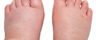
- Swelling of the lower extremities. As a logical continuation of increasing renal failure.
- Breathing problems.
As a result, the patient has a whole range of symptoms from both distant organs and systems, and the cardiac structures themselves. The reason for all the sensations lies in the disruption of blood circulation, both in the large and in the small circle.
Clinical picture of tricuspid insufficiency and stenosis
Since the main cause of the development of tricuspid valve defects is rheumatic endocarditis, the development of the disease is often preceded by an increase in temperature and clinical signs of streptococcal sore throat. Valve damage manifests itself as signs of heart failure:
- Fatigue and decreased tolerance to physical activity;
- Labored breathing;
- Cyanosis (blueness) of the skin and mucous membranes;
- Enlarged liver;
- Peripheral edema, mainly in the lower extremities, as well as fluid accumulation in the abdominal cavity (ascites);
- Heart rhythm disturbances.
Mild forms of tricuspid insufficiency can occur for a long time without significant clinical manifestations, causing complaints only in the stage of decompensation. Symptoms of heart failure due to tricuspid stenosis are usually much more pronounced and are diagnosed at the onset of the disease.
Diagnostics
The examination is carried out under the guidance of a cardiologist, and if the process is proven, the specialized surgeon continues to work. He is also responsible for prescribing treatment.
Scheme of events in the correct order:
- Oral questioning of the patient regarding complaints, their duration, as well as collecting anamnesis. This way the doctor understands the direction of further examination.
- Blood pressure measurement. Usually it is slightly reduced. Heart rate is higher than normal. The rhythm is correct; as it progresses, spontaneous premature beats (extrasystoles) occur.
- Listening to sound (auscultation). A sinus noise of reverse blood flow is detected. Tones can be either normal or dull.
- Daily monitoring. To record cardiac performance indicators over 24 hours in dynamics. It is most often used as the first method, after a routine examination. Provides comprehensive information about the movement of blood pressure and heart rate during the day.
- Electrocardiography. Assessment of the functional state of the heart.
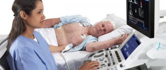
- Echocardiography. Method of visualization of cardiac structures. It is carried out as a matter of priority, since it allows one to detect organic abnormalities on the part of the tricuspid valve.
- MRI or CR (much less often). It is carried out to detail the image of the heart and surrounding tissues.
- Pulmonary artery pressure measurement.
- Load tests. At an early stage, later not used due to significant danger.
The methods are aimed both at establishing the fact of an anatomical defect and at verifying the alleged diagnosis.
Diagnosis of tricuspid valve defects
In the cardiology department, patients undergo a thorough modern examination by leading Israeli specialists. In addition to the history, characteristic complaints and clinical signs, tricuspid valve defects cause pathological noises, determined by the doctor during auscultation of the heart. If valve damage is suspected, patients undergo a number of additional tests, the main of which are:
- Electrocardiography (recording of electrical potentials of the conduction system of the heart). The method allows you to diagnose myocardial hypertrophy and heart rhythm disorders. Patients with tricuspid defects often experience atrial fibrillation and intraventricular conduction disorders;
- X-ray examination - overview of the chest and computed tomography of the heart;
- Magnetic resonance imaging, which has important diagnostic value for accurate assessment of the size of the right ventricle
- Ultrasound diagnostics of the heart – echocardiography;
- Invasive methods, such as cardiac catheterization, are often used in preparation for surgery, as well as to exclude ischemia (coronary circulatory insufficiency).
Treatment methods
Therapy is carried out under the full supervision of a cardiac surgeon. Methods of exposure depend on the stage of the pathological process.
Grade 1 tricuspid regurgitation is the best time to start therapy. But there are no symptoms yet, identification is incidental (random), and does not pose any difficulties during a targeted search.
At this stage, dynamic observation for 3-5 years is indicated. In the absence of progression, with stagnation of the process, there is no need for treatment. Sometimes patients can live a quality life without knowing about their condition, without major restrictions.
Tricuspid regurgitation of grade 2 and higher is corrected strictly surgically. There are several intervention options.
But before the treatment stage, it is necessary to stabilize the patient’s condition, if there is time for that (planned operations).
Drugs used:
- Antiarrhythmics in the minimum dosage to restore an acceptable heart rate (Amiodarone, Hindin).
- Beta blockers (Metoprolol).
- Glycosides. In order to normalize myocardial contractility.
- Cardioprotectors.
- Anticoagulants. To prevent the formation of blood clots, which cause frequent premature death of patients.
- Diuretics in the treatment of early manifestations of renal conditions.
The duration of the preparatory period varies from 2 to 4 months, possibly more.
By the time of surgery, the rhythm should be stable, correct, blood pressure within the reference value or close to it.
Depending on the stage of the pathological process and the nature of the changes, plasty or prosthetics of the tricuspid valve is indicated. Both methods are generally equivalent.
Correction of pathologies and defects of distant organs is carried out under the supervision of specialized specialists. The list of techniques is wide and is determined based on the severity of the process.
Attention:
The use of folk remedies is impossible. Since the effect of them in case of organic deviation on the part of the cardiac structures is zero.
Changing your lifestyle will also not play a key role. It makes sense to give up smoking, alcohol and drugs. When carrying out severe therapy for third-party pathologies, correction by a treating specialist is recommended.
An integrated approach to the treatment of tricuspid valve defects
Conservative treatment of tricuspid defects caused by rheumatic endocarditis includes a course of antibacterial and anti-inflammatory therapy. Modern antiarrhythmic drugs and drugs that improve the condition of patients suffering from heart failure are widely used. Anticoagulants and antiplatelet agents are indicated for high risk of thrombotic complications in patients with atrial fibrillation, as well as after operations to replace the valve with a mechanical implant.
Surgical treatment is recommended for significant hemodynamic disturbances. It includes the following procedures:
- Percutaneous balloon commissurotomy is used in some cases of tricuspid stenosis. This minimally invasive procedure can dilate a moderately deformed annulus fibrosus without the need for open surgery;
- Tricuspid valve annuloplasty. An operation that restores the anatomical integrity and mobility of the valve leaflets. This intervention is preferable for most tricuspid defects. Successful restoration of a natural valve prevents the need for valve replacement'
- Surgery to replace the tricuspid valve is currently used for severe forms of the defect that are not subject to an organ-preserving procedure. High-quality mechanical and biological prostheses take over the function of the affected valve and restore normal blood circulation.
Cardiac surgeons choose the type of operation depending on the type of tricuspid defect, the degree of valve damage and the severity of hemodynamic disorders. High professionalism and extensive experience of specialists contribute to the optimal choice of treatment method.
One of the advantages of treatment at Herzliya Medical Center is the opportunity to undergo a full range of necessary diagnostic, therapeutic and rehabilitation procedures within one hospital. The individual approach of the clinic’s specialists to the problems of each patient allows us to achieve the desired result in an extremely short time.
Forecast
{banner_banstat9}
Depends on the stage and nature of therapy.
- At the first stage, the survival rate is 100%, especially if there is no progression of the condition.
- The second is associated with a probability of 85%.
- Third - 45%.
- The fourth or terminal puts an end to the patient, giving no chance. The median is 1-2 years, often even less.
When carrying out complex therapy, it is possible to stabilize the conditions of even the most severe patients, prolonging life for several years.
Favorable prognostic factors:
- Period of youth.
- Absence of somatic pathologies, bad habits, complications after the operation.
- Good family history.
- Response to treatment.
- Reduction of symptoms.
Determining the possible outcome falls on the shoulders of the cardiologist. In order to say anything concrete, you need to at least carry out a full diagnosis.
