Erythremia is a tumor clonal disease of the hematopoietic system, in which there is proliferation of erythroid, granulocytic and megakaryocytic lineages of hematopoiesis with predominant activation of erythropoiesis. At the same time, an increase in the level of red blood cells and hemoglobin, thrombocytosis and leukocytosis is noted in the blood. Almost all patients with erythremia are carriers of the JAK2V617F mutation.
- How does erythremia develop?
- Symptoms of erythremia
- Stages of erythremia
- Diagnosis of erythremia
- Treatment of erythremia
- Prognosis for erythremia
How does erythremia develop?
The causes of erythremia are still unknown. It is believed that this is a multi-stage disease in which, under the influence of external factors, the genome of a normal cell is damaged, which leads to its malignant transformation and the formation of a tumor cell clone that replaces normal hematopoiesis.
Almost all patients have a mutation in the JAK2 gene. Exon 14 is usually affected; 90-96% of patients have the V617F mutation. 2% of patients have mutations in exon 12. Damage to other genes, in particular MPL, CALR, is very rarely detected. All of these genetic pathologies are specific to erythremia, so their determination is necessary to confirm the diagnosis.
So, molecular genetic disorders cause activation of the JAK-STAT signaling pathway, which leads to increased proliferation of hematopoietic sprouts and an increase in the number of blood cells.
Monocytes and megakaryocytes (platelet precursors) produce a variety of cytokines - biologically active molecules that stimulate fibrotic changes, the formation of new blood vessels, which ultimately leads to osteosclerosis and bone marrow fibrosis. In addition, massive production of cytokines contributes to the development of tumor intoxication, which aggravates the general condition of patients.
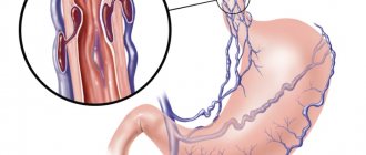
There is also a disruption in the connection of stem cells with the microenvironment. This provokes the formation of foci of hematopoiesis outside the bone marrow. The liver and spleen are primarily affected.
Symptoms of erythremia
Clinically, erythremia manifests itself in two syndromes:
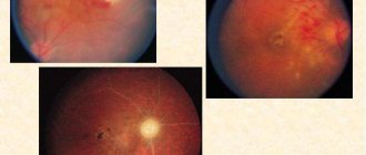
- Plethora (plethora). This syndrome is characterized by an increase in the number of circulating red blood cells. Symptomatically manifested by headaches, dizziness, attacks of rapid heartbeat, itchy skin and visual disturbances. The skin and mucous membranes have a bluish tint. Vascular complications are also possible: thrombosis, erythromelalgia (redness of the fingers, pain and burning sensation in them).
- Myeloproliferative syndrome - develops as a result of hyperplasia of hematopoietic germs. Symptomatically manifested by weakness, fever, sweating, itchy skin, bone pain. With the breakdown of granulocytes, a disturbance in urate metabolism is observed, which leads to the development of gout, kidney stones and urate diathesis.
Stages of erythremia
During its development, erythremia goes through several stages. The first, also known as initial, can last more than 5 years. During this period, moderate manifestations of plethoral syndrome are mainly present. The blood test shows moderate erythrocytosis, and in the bone marrow there is an increase in the proliferation of all hematopoietic lineages with the exception of the lymphocytic lineage. The spleen is not enlarged, complications rarely develop.
The second stage of erythremia is polycythemia. It is divided into 2 substages A and B. Stage A lasts 5-15 years. It is characterized by an increase in the number of formed elements in the blood. As a result, a pronounced plethoric syndrome is formed, which is complicated by thrombosis, bleeding, and an increase in the size of the liver and spleen. In the blood test, cytosis increases; in the bone marrow, in addition to the proliferation of erythropoietic, granulocytic and thrombopoietic lineages, cicatricial changes are noted.
With erythremia in stage B, cytosis and clinical symptoms continue to increase. Foci of tumor growth form in the spleen, and cicatricial changes progress in the bone marrow.
Stage 3 of erythremia - anemic. Here, bone marrow fibrosis already develops, which leads to its depletion and a decrease in the level of blood cells. The disease can transform into acute leukemia.
Myocarditis
Myocarditis is a lesion of the heart muscle of a predominantly inflammatory nature, caused by direct or indirect effects through immune mechanisms of infection, parasitic or protozoal invasion, chemical or physical factors, as well as those arising from allergic and autoimmune diseases.
Myocarditis of non-rheumatic etiology is classified into several groups:
- caused by viruses: coxsackie B, coxsackie A, ECHO, influenza, infectious mononucleosis, polio, etc.;
- parasitic (with trichinosis),
- protozoans (for trypanosomiasis (Chagas disease), syphilis, toxoplasmosis, amebiasis),
- bacterial (for bacterial endocarditis, septicemia, diphtheria),
- myocarditis in autoimmune diseases (immune complex vasculitis),
- allergic (drug reactions, serum sickness),
- myocarditis caused by physical and chemical influences.
According to the variant of the clinical course, they are distinguished: fulminant or fleeting (up to 1 month), acute (up to 3 months), subacute (3-6 months), chronic (active or persistent); by severity – light, moderate and severe; by prevalence - focal and diffuse myocarditis.
Clinical variants of the course: pseudocoronary (painful), decompensatory, pseudovalvular, arrhythmic, thromboembolic, mixed, asymptomatic (30%).
The frequency of occurrence of various clinical manifestations varies: cardialgia - 80%, shortness of breath - 50%, interruptions in heart function - 40%, increase in heart size (may be insignificant), muffled first tone - 8-90%, splitting, bifurcation of the first tone - 10 %, accent of the second tone over the pulmonary artery - 30%, systolic murmur at the apex - 50%, tachycardia - 50%, bradycardia - 10%, congestive heart failure - 30%, hypotension is possible.
Diagnosis of myocarditis is based on clinical and anamnestic data. The diagnostic rule for diagnosing myocarditis is: a combination of a previous infection with two major or one major and two minor criteria. The presence of previous infection must be proven by clinical and laboratory data. Major signs of myocardial damage include: signs of congestive circulatory failure, cardiogenic shock, pathological changes on the ECG, an increase in the concentration of cardioselective enzymes in the blood, an increase in the size of the heart according to ECHO-CG or radiography. Minor signs of myocardial damage include tachycardia (sometimes bradycardia), weakening of the first sound, protodiastolic gallop murmur, moderate systolic murmur at the apex of the heart.
In addition, there are methods for objective confirmation of myocarditis:
- study of biopsy material from the myocardium of the right ventricle and IVS,
- MRI of the heart with contrast (detection of inflammatory interstitial edema in the myocardium),
- radioisotope study of the myocardium with a radiopharmaceutical tropic for inflammation.
When treating this pathology, whenever possible, treatment of the underlying disease and elimination of etiological factors are carried out. There is currently no specific treatment for myocarditis itself. Treatment is symptomatic: therapy for heart failure, rhythm and conduction disorders. For mild myocarditis, drug therapy is not needed. For moderate and severe myocarditis, bed rest (at least the first 2-3 weeks) and symptomatic drug therapy are required. The effectiveness of NSAIDs for myocarditis has not been proven. Their use is possible with concomitant pericarditis. There is also no convincing evidence of the effectiveness of aminoquinolone derivatives. Currently, the possibilities of using immunomodulators, specific antiviral drugs, monoclonal antibodies against T-lymphocyte antigens, and antithymocyte globulin are being studied. And only in extremely severe cases is an attempt to use prednisolone with azathioprine justified.
Diagnosis of erythremia
As part of the diagnosis of erythremia, the patient undergoes a comprehensive examination, which includes the following measures:
- Anamnesis and physical examination, during which the color of the skin of the face and extremities is assessed. The size of the liver and spleen must be determined.
- A detailed blood test, including counting the number of blood cells and erythrocyte indices.
- Trephine biopsy of the bone marrow followed by histological and histochemical examination.
- Determination of erythropoietin level.
- Molecular genetic testing for the presence of specific V617F mutations.
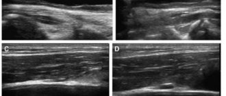
- Ultrasound of the liver and spleen to accurately determine their size.
The diagnosis of erythremia is made according to the 2021 WHO criteria, which distinguishes three major criteria and one minor one.
Major criteria for erythremia:
- The hemoglobin level is more than 160 g/l in women and more than 165 g/l in men.
- Hyperplasia of three myelopoiesis lineages in bone marrow trephine.
- The presence of a mutation in the JAK2 gene.
A minor criterion is a decrease in erythropoietin levels.
To make a diagnosis of erythremia, it is necessary either to have all three major criteria, or 1-2 major and one minor.
Treatment of erythremia
The main goals of treatment for erythremia are:
- Preventing the formation of blood clots and treating already developed thrombosis and thromboembolism.
- Reducing the level of tumor intoxication and controlling associated symptoms (fever, itching, weight loss).
- Reducing the risk of transformation into acute myeloid leukemia or myelofibrosis.
- Preventing the development of complications if surgical interventions or pregnancy are necessary.
To assess the likelihood of developing thrombosis during erythremia, risk stratification is carried out according to the following criteria:
- Age over 60 years.
- Presence of thrombosis in the past.
- Cardiovascular risk factors: arterial hypertension, excess weight, diabetes, physical inactivity.
If the patient does not have the listed factors, then he is classified as a low-risk group, in the presence of cardiovascular factors - as an intermediate one, and if he is over 60 years old and/or has a history of thrombosis, he is considered to be at high risk of thromboembolic complications. The platelet level does not affect the risk of thrombosis, but it plays a certain role in the development of bleeding. To prevent the development of thrombosis, it is necessary to eliminate cardiovascular risk factors, as well as prescribe antiplatelet agents.
In addition, the following methods are used to treat erythremia:
- Removing excess red blood cells in the blood. It can be carried out either using hemoexcursion (regular bloodletting) or erythrocytepheresis (removal of red blood cells directly).
- Cytoreductive therapy. Cytostatics and interferons are used.
- Treatment of complications of already developed thrombosis.
Hemoexcursions (bloodletting) for erythremia
Bloodletting is used to reduce the volume of circulating blood. The volume of hemoexcursion is 250-500 ml per procedure, after which the missing volume of fluid is replenished with saline solution. Or the second option is a preliminary infusion of antiplatelet agents together with saline in a volume exceeding the volume of blood removed. Sessions are carried out every other day until the hematocrit reaches 40-45%. For elderly patients, sessions are performed 2 times a week, or the volume of blood excursion is reduced.
Erythrocytopheresis
Erythrocytapheresis refers to methods of extracorporeal detoxification, which replaced bloodletting. It is based on the removal of red blood cells, followed by the return of plasma and replenishment of the volume with solutions of crystalloids or colloids. In one session, up to 400 ml of red blood cells can be removed.
Acetylsalicylic acid preparations
Acetylsalicylic acid, or aspirin, is prescribed to prevent thrombotic complications. Aspirin should be prescribed to every patient with erythremia unless there are contraindications. If they are present, clopidogel or ticlopidine is prescribed.
Cytoreductive therapy
Hydroxyurea
Theoretically, hydroskiurea can be used as part of first-line therapy in patients of any age. But due to the fact that it is genotoxic and can provoke a leukemic effect, such first-line treatment is not recommended for patients under 50 years of age and pregnant women. It is also necessary to discontinue treatment in case of intolerance and ineffectiveness of therapy. The criterion for intolerance to hydroxyurea is the presence of at least one of these symptoms:
- Exceeding the hematocrit by more than 45% 3 months after treatment with hydroxyurea at a dosage of 2000 mg/day.
- Lack of control of myeloproliferation. This may be indicated by a platelet level of more than 400×10 9 and a leukocyte level of more than 10×10 9 after 3 months of treatment.
- Presence of an enlarged spleen (more than 10 cm below the costal margin) or the inability to eliminate the symptoms of splenomegaly.
- Myelopenia when using minimal dosages of drugs - platelet level below 100×10*9 or hemoglobin below 100 mg/l.
- Ulcers on the legs.
- Damage to the skin and mucous membranes.
- Disruption of the gastrointestinal tract.
- Pneumonitis.
- Fever.
Interferon alpha
Interferon alfa is highly effective against erythremia and allows a molecular response to be achieved in some patients. It also relieves plethoral syndrome well, reducing the severity of itching. However, its widespread use is limited by poor tolerability. It is mainly recommended for patients under 50 years of age.
Busulfan
With the help of busulfan, active control of erythremia is possible, however, with long-term use, the risk of developing secondary leukemia increases. Therefore, it is used in patients over 70 years of age with intolerance to hydroxyurea and interferons.
Erythremia (polycythemia vera, Vaquez disease)
Erythremia (ER) is a myeloproliferative disease, chronic
Icical, benign current leukemia, in which there is
increased formation of erythrocytes, as well as neutrophilic leukocytes
ov and platelets. The source of tumor growth is the progenitor cell.
Ca myelopoiesis.
The incidence of erythremia is about 0.6 per 10,000 population. Both men and women get sick equally often. Erythremia is a disease of older people: the average age of those affected is 55-60 years, but the disease can occur at any age.
Etiology. The reasons for the development of the disease are unknown.
Pathogenesis. Erythremia is based on tumor clonal proliferation of all three hematopoietic lineages - red, granulocytic and megakaryocytic, but the growth of the red lineage dominates. In this regard, the main substrate of the tumor is red blood cells maturing in excess. Foci of myeloid hematopoiesis appear in the spleen and liver (which never happens normally). An increased number of red blood cells and platelets in the peripheral blood reduces the speed of blood flow, increases the viscosity and coagulability of the blood, which causes the appearance of a number of clinical symptoms.
Classification. The stage of the disease, the involvement of the spleen in the pathological process and the subsequent transformation of erythremia into other diseases of the blood system are taken into account.
Stage I - initial:
hemoglobin content is at the upper limit of normal, a slight increase in the mass of circulating red blood cells, the spleen is slightly enlarged (due to blood overflow) or without changes. Blood pressure is normal or slightly elevated, and focal bone marrow hyperplasia is noted in the iliac trephine specimen. The duration of stage I can exceed 5 years.
Stage II - expanded:
phase A - without myeloid metaplasia of the spleen (simple variant of plethora without splenomegaly). Total three-fold hyperplasia of the bone marrow. Absence of extramedullary hematopoiesis; phase B - with myeloid metaplasia of the spleen. Major myeloproliferative syndrome: pancytosis in the peripheral blood, in the bone marrow there is panmyelosis with or without focal myelofibrosis, myeloid metaplasia of the spleen with or without fibrosis.
Stage III - terminal:
degeneration of a benign tumor into a malignant one (myelofibrosis with anemization, chronic myelo-leukemia, acute leukemia). Myelofibrosis develops in almost all patients for more than 10-15 years; it reflects the natural evolution of the disease. A sign of myelofibrosis is cytopenia (anemia, thrombocytopenia, and less commonly, leukopenia). The development of chronic myeloid leukemia is manifested by an increase in leukocytosis, an increase (or appearance) in the peripheral blood of myelocytes, promyelocytes, as well as the detection of the Ph chromosome in blood and bone marrow cells.
Acute leukemia usually develops in patients treated with cytostatics and radioactive phosphorus.
Anemia in patients with erythremia may be associated with frequent bloodletting, increased deposition of red blood cells, as well as their hemolysis.
Clinical picture. Erythremia manifests itself in two major syndromes.
Plethoric syndrome
caused by an increased content of red blood cells, as well as leukocytes and platelets (plethora - plethora). This syndrome is caused by: 1) the appearance of subjective symptoms; 2) disorders of the cardiovascular system; 3) changes in laboratory parameters.
1. Subjective symptoms of plethoric syndrome include headaches, dizziness, blurred vision, angina pectoris, skin itching, erythromelalgia (sudden onset of hyperemia with systemic
a pleasant shade of the skin of the fingers, accompanied by sharp pain and burning), possible sensations of numbness and chilliness of the limbs.
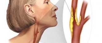
2. Disorders of the cardiovascular system are manifested in changes in the color of the skin and visible mucous membranes, such as erythrozzanosis, peculiarities of the color of the mucous membrane at the point of transition of the soft palate to the hard palate (Kuperman's symptom), hypertension, the development of thrombosis, and, less often, bleeding. In addition to thrombosis, swelling of the legs and erythromelalgia are possible. Circulatory disorders in the arterial system can lead to serious complications: acute myocardial infarction, strokes, visual impairment, and renal artery thrombosis.
3. Changes in laboratory parameters: an increase in the content of hemoglobin and red blood cells, an increase in hematocrit and blood viscosity, moderate leukocytosis with a shift in the leukocyte formula to the left, thrombocytosis, a sharp slowdown in ESR.
Myeloproliferative syndrome
caused by hyperplasia of all three hematopoietic lineages in the bone marrow and extramedullary. It includes: 1) subjective symptoms, 2) splenomegaly and (or) hepatomegaly, 3) changes in laboratory parameters.
1. Subjective symptoms: weakness, sweating, increased body temperature, bone pain, heaviness or pain in the left hypochondrium (due to
splenomegaly).
2. Splenomegaly is explained not only by myeloid metaplasia of the organ (the appearance of foci of extramedullary hematopoiesis), but also by blood stagnation. Less commonly, liver enlargement is observed.
3. Among the laboratory indicators, deviations from the physiological norm in the peripheral blood have the greatest diagnostic significance: pancytosis, often with a shift in the leukocyte formula to the left; trephine biopsy reveals three-line hyperplasia of the bone marrow, and in the puncture of the spleen there are foci of myeloid metaplasia of the organ.
The different severity of syndromes at different stages of the disease causes extreme variability in the clinical picture. It is possible to observe patients with undoubted erythremia, who show almost no complaints and are fully able to work, and patients with severe damage to internal organs who require therapy and have lost their ability to work.
At stage I of the diagnostic search in the initial stage of the disease, patients may not present any complaints. As the disease progresses, complaints are associated with the presence and severity of plethora and the myeloproliferative process. The most common complaints are of a “pletoric” nature, caused by increased blood supply to the vessels and functional neurovascular disorders (headaches, erythromelalgia, blurred vision, etc.). All these symptoms may be associated with other diseases, which must be clarified during further examination of the patient.
Complaints caused by the presence of myeloproliferative syndrome (sweating, heaviness in the left hypochondrium, bone pain, increased body temperature) are also nonspecific for erythremia. The skin itching that appears after taking water treatments is quite typical. This symptom is observed in 55% of patients in the advanced stage and is explained by overproduction of basophils and histaminemia. The nature of urticaria, observed in 5-7% of patients, is similar.
The listed symptoms are important for determining the stage of erythremia: usually they indicate the transition of the disease to full-blown
or the terminal stage with the development of myelofibrosis as the most common outcome of erythremia.
Patients may have a history of complications of the disease such as strokes and myocardial infarction. Sometimes the disease debuts with precisely these complications, and the true cause of their development - erythremia - is revealed when examining a patient for a stroke or myocardial infarction
Indications of previous treatment with radioactive phosphorus, cytostatics or bloodletting may suggest the presence of some kind of tumor blood disease. A decrease in the symptoms of plethoric syndrome during treatment with these drugs suggests erythremia.
At stage II of the diagnostic search, it is possible to identify distinct symptoms only in stage II (advanced) of the disease. The main signs of plethoric syndrome are found: erythrocyanosis, injected conjunctival vessels (“rabbit eyes”), a distinct color border at the transition point of the hard palate to the soft palate. You can identify symptoms of erythro-melalgia: swelling of the tips of the fingers, feet, lower third of the leg, accompanied by local hyperemia and a sharp burning sensation.
When examining the cardiovascular system, hypertension and enlargement of the left ventricle are diagnosed; in the advanced stage of the disease, “variegated legs” (changes in the color of the skin of the legs, mainly their distal part) in the form of areas of pigmentation of varying intensity, caused by impaired venous circulation.
When palpating the abdomen, one can detect an enlarged spleen, which is one of the characteristic signs of the disease. Enlargement of the spleen may be due to: 1) increased deposition of blood elements; 2) “working” hypertrophy due to an increase in its sequestering function; 3) extramedullary hematopoiesis (myeloid metaplasia with a predominance of erythropoiesis). These reasons are often combined. Liver enlargement is due to similar reasons, as well as the development of fibrosis and nonspecific reactive hepatitis. It should be borne in mind that hepatomegaly can be observed with a malignant liver tumor with the development of secondary erythrocytosis.
Complications of erythremia in the form of cerebral vascular thrombosis are expressed by a number of focal symptoms identified during the study
cns.
However, even at stage II it is impossible to definitively diagnose erythremia, since many of its symptoms can be associated with symptomatic erythrocytosis. In addition, symptoms such as hypertension, splenomegaly and hepatomegaly are characteristic of a wide variety of diseases.
In this regard, III of the diagnostic search becomes crucial, as it allows: a) to make a final diagnosis; b) clarify the stage of erythremia; c) identify complications; d) monitor treatment.
Peripheral blood analysis
detects erythrocytosis, an increase in hemoglobin content and hematocrit, which, however, also occurs with symptomatic erythrocytosis.
An increase in hemoglobin levels in combination with erythrocytosis, leukocytosis and thrombocytosis is of diagnostic significance. When examining the leukocyte formula, a shift to the left to immature forms of granulocytes is detected. If changes in peripheral blood are minor or the data are inconclusive (for example, erythrocytosis is not combined with thrombocytosis), then a bone marrow examination (trephine biopsy) must be performed.
Presence of total-442 in trepanate
loy three-line hyperplasia of the bone marrow with a predominance of formen-Hbix elements of erythropoiesis, replacement of adipose tissue with a red line of bone marrow make it possible to make a final diagnosis. The expansion of the “bridgehead” of hematopoiesis is also detected using radionuclide bone scanning with 32P. Histochemical study
reveals increased activity of neutrophil alkaline phosphatase.
Complications. The course of erythremia is complicated by: 1) vascular thrombosis (cerebral, coronary, peripheral arteries); 2) hemorrhagic syndrome: bleeding after minor surgical interventions (tooth extraction), from the vessels of the digestive tract, hemorrhoids, which is caused by poor retraction of the blood clot due to changes in the functional properties of platelets; 3) endogenous uricemia and uricosuria (due to increased cell death at the nuclear prestages of their maturation), which is manifested by symptoms of urolithiasis and gouty arthritis.
The outcomes of the disease are the situations indicated in stage III of the disease (myelofibrosis, chronic myelogenous leukemia, acute leukemia, anemia).
Diagnostics. Erythremia can be suspected in individuals with persistent erythrocytosis in combination with neutrophilic leukocytosis, thrombocytosis in the absence of diseases (or conditions) that could cause erythrocytosis.
Diagnostic criteria for erythremia (in the advanced stage) are:
Category "A".
• Increase in the mass of circulating red blood cells.
• Normal arterial blood oxygen saturation (more than 92%).
• Enlarged spleen. Category "B".
• Leukocytosis more than 12,109/l (in the absence of obvious reasons for the appearance of leukocytosis).
• Thrombocytosis more than 400-109/l.
• Increased levels of neutrophil alkaline phosphatase (in the absence of infection).
• Increase in unsaturated vitamin B12-binding capacity of blood serum.
The diagnosis of ER is reliable in the presence of three signs of category A or two signs of category A and one sign of category B.
Difficulties in making a diagnosis are due to the development of so-called symptomatic erythrocytosis in a number of diseases. Absolute and relative erythrocytoses are distinguished. With absolute erythrocytosis, an increase in the mass of circulating erythrocytes and increased erythropoiesis are noted. Relative erythrocytosis is characterized by a decrease in the volume of circulating plasma and a normal mass of circulating erythrocytes. Relative erythrocytosis is often detected in men suffering from hypertension, obesity, neurasthenia, and taking diuretics. Secondary absolute erythrocytosis develops in smokers; it is caused by an increase in the content of carbon monoxide in the blood.
Reasons for the development of symptomatic erythrocytosis: 1) generalized tissue hypoxia (pulmonary pathology, heart disease, hemoglobinopathies, obesity, etc.); 2) paraneoplastic reactions (Nochek tumors, tumors of the cortex and medulla of the adrenal glands, pituitary gland, ovaries, vascular tumors, tumors of other organs); 3) renal ischemia
(renal artery stenosis, hydronephrosis, polycystic disease and other kidney anomalies); 4) unknown causes (CNS disease, portal hypertension).
Relative erythrocytosis is observed during exicosis (dehydration due to diarrhea, vomiting, excessive sweating, etc.). Differential diagnosis is based on taking into account the entire clinical picture. In difficult cases, it is necessary to examine the content of erythropoietin in the blood; with erythremia it does not increase.
The formulation of a detailed clinical diagnosis includes information about 1) the stage of the disease; 2) presence of complications; 3) phase of the process (exacerbation or remission); 4) the presence of pronounced syndromes (portal hypertension, hypertension, etc.).
Treatment. The entire complex of therapeutic measures for ER appears to be as follows.
In the advanced stage of the disease, in the presence of plethoric syndrome, but without leuko- and thrombocytosis, bloodletting is used as an independent method of therapy, and it is necessary to achieve a reduction in the hematocrit level to normal values (less than 45 %).
400-500 ml of blood are taken every other day (in a hospital setting) or after 2 days (in a clinic setting). To prevent thrombosis (develop as a result of bloodletting, and also as a complication of erythremia), acetylsalicylic acid is prescribed at a dose of 0.5-1 g/day the day before and on the day of bloodletting, and then for 1-2 weeks after the end of bloodletting. In addition to acetylsalicylic acid, other disaggregants are prescribed - ticlid, plavike, pentoxifylline. Before bloodletting, to prevent pulmonary embolism, it is advisable to administer 5000 units of heparin intravenously (through a Dufault needle), as well as 5000 units of heparin under the skin of the abdomen 2 times a day for several days after bloodletting. In case of poor tolerance to bloodletting, observed in persons with severe cerebral atherosclerosis, exfusion is limited to 350 ml (2 times a week). When bleeding, it is necessary to reduce hemoglobin to 150 g/l.
If bloodletting is not effective enough, as well as in forms of the disease that occur with pancytosis and splenomegaly, cytostatic therapy is prescribed. The age of patients over 55 years expands the indications for the use of cytostatics. Indirect indications for cytostatic therapy are other signs of myeloproliferative syndrome (itching), as well as the severity of the disease, visceral vascular complications (stroke, myocardial infarction), and exhaustion.
Contraindications to cytostatic therapy: young age of patients, refractoriness to treatment at previous stages, as well as excessively active cytostatic therapy in the past due to fear of the disease transitioning to the anemia phase. The effect of cytostatic therapy should be assessed 3 months after the end of treatment; this is explained by the fact that red blood cells produced before treatment live on average about 2-3 months. The decrease in the number of leukocytes and platelets occurs much earlier, according to their life span. The criterion for the effectiveness of cytostatic therapy is the achievement of hematological remission (complete, when all blood parameters are normalized, or partial, in which the number of red blood cells, leukocytes and/or platelets remains slightly increased).
Of the cytostatic drugs at the first stage, hydroxyurea (hydrea) is usually prescribed at a dose of 30-50 mg/(kg day) (2-3 capsules per
day). During treatment, it is necessary to monitor the number of leukocytes. Hydrea is combined with a-interferon at a dose of 3-5 million IU subcutaneously 3-7 times a week for a long time (at least a year), which allows to stop thrombocytosis, plethora, and skin itching.
For hyperthrombocytosis, anagrelide is used.
• The outcomes of erythremia (myelofibrosis, acute leukemia, chronic myeloid leukemia) are influenced according to the principles of treatment of these diseases: for myelofibrosis, anabolic steroids, nitostatics and red blood cell transfusions are used; for acute leukemia, polychemotherapy is indicated, for chronic myeloid leukemia - cytostatic drugs.
• Symptomatic therapy for attacks of erythromelalgia is carried out with the help of antiplatelet agents, non-steroidal anti-inflammatory drugs (acetylsalicylic acid, indomethacin). Arterial hypertension and angina attacks are eliminated in accordance with the rules for the treatment of these conditions.
• For complications of erythremia by vascular thrombosis, anticoagulant and antiplatelet therapy is used.
• Patients with erythremia are registered at the dispensary with the frequency of visiting a doctor and the appointment of peripheral blood tests once every 3 months.
Forecast. With uncomplicated erythremia, life expectancy can reach 15-20 years (further complications arise). If complications from the cardiovascular system develop early enough or the disease progresses, life expectancy is reduced. Timely initiation of therapy extends life expectancy, although this is not observed in all cases.
Prevention. There are no radical measures to prevent the disease, and therefore we can only talk about secondary prevention, which consists of dynamic monitoring of patients and anti-relapse therapy.
Prognosis for erythremia
In general, the prognosis for erythremia is relatively favorable. The overall 10-year survival rate is about 75%. But the disease can transform into acute myeloid leukemia (5% risk) or myelofibrosis (slightly less than 10% risk).
Symptoms of erythremia (headaches, bone pain, paresthesia) can worsen the quality of life of patients. The main cause of death in patients is thrombosis, bleeding and severe infections, the risk of which increases when the pathology transforms into acute myeloid leukemia or myelofibrosis.
Treatment of erythremia at the Euroonko clinic is carried out using modern treatment protocols. In difficult cases, the decision is made collegiately by a council of specialists. We attach great importance to monitoring the patient during the period of remission. This also makes it possible to achieve an increase in the effectiveness of the treatment.
Book a consultation 24 hours a day
+7+7+78
Leading Israeli oncologists
- Professor Aron Sulkes
Oncologist - Professor Moshe Inbar
Oncologist
- Professor Ofer Merimsky
Specialist in the treatment of sarcomas
- Dr. Yair Bar
Oncologist
Depending on in which vessels thrombosis occurs, a clinical picture of acute abdomen, gangrene of the lower or upper extremities, pulmonary artery thrombosis with hemoptysis, myocardial infarction, or cerebral stroke may develop.
“Like any other leukemia, erythremia is characterized by the development of secondary gout, accompanied by the development of urolithiasis and arthritis with severe pain, most often affecting the big toe joint.”