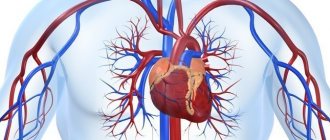
Congenital heart defects (CHD) are developmental anomalies that lead to disruption of the morphological structure of the heart, including the valve apparatus and great vessels.
CHDs occur during the period of intrauterine development (usually at 2-8 weeks) as a result of disruption of embryogenesis processes. These anomalies can occur either alone or in combination with each other.
The overall prevalence of this group of diseases is up to 5-8 cases per 1000 births. Congenital defects can be associated with chromosomal abnormalities, however, non-chromosomal congenital heart defects are often diagnosed.
The overall incidence of non-chromosomal congenital heart disease is up to 7 cases per 1000 births, of which up to 3.5% are perinatal losses, 20% are diagnosed prenatally, 5.6% of pregnancies are terminated due to a detected fetal anomaly. Complex non-chromosomal heart defects are less common, approximately 2 cases per 1000 births. The outcome in 8% of cases is perinatal death, 40% are diagnosed in utero, and 14% cause termination of pregnancy.
Causes of congenital heart disease
The main leading causes in the formation of defects are most often structural and quantitative chromosomal abnormalities and mutations, i.e.
primary genetic factors. It is also necessary to pay attention to potentially teratogenic environmental factors: various intrauterine infections (rubella viruses, cytomegalovirus, coxsackie, infectious diseases in the mother in the first trimester), medications (vitamin A, antiepileptic drugs, sulfasalazine, trimethoprim), constant contact with toxic substances ( paints, varnishes). In addition, it must be remembered that maternal factors have a negative impact on intrauterine development: reproductive problems preceding a given pregnancy, the presence of diabetes mellitus, phenylketonuria, alcoholism, smoking, age, but also factors from the father - age, drug use ( cocaine, marijuana).
The leading role belongs to the multifactorial theory of the development of congenital heart defects (up to 90%).
Types of congenital heart defects
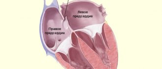
- Atrial septal defect (ASD) or patent foramen ovale is diagnosed when one or more holes are identified in the interatrial septum. One of the most common congenital heart defects. Depending on the location of the defect, its size, and the strength of blood flow, more or less pronounced clinical signs are determined. ASD is often combined with other cardiac anomalies and is associated with Down syndrome.
- Ventricular septal defect (VSD) is diagnosed when the interventricular septum is underdeveloped at various levels with the formation of a pathological communication between the left and right ventricles. It can occur either alone or together with other developmental anomalies. With a small defect, there is often no pronounced lag in physical development. VSD is dangerous because it can lead to the development of pulmonary hypertension, and therefore must be promptly corrected surgically.
- Coartation of the aorta is a segmental narrowing of the aortic lumen with disruption of normal blood flow from the left ventricle to the systemic circulation. Up to 8% of all cases of congenital heart disease are detected, more often in boys, and are often combined with other anomalies.
- Patent ductus arteriosus is diagnosed when the duct of Batallus is not closed, which is detected in newborns and becomes overgrown in the future. As a result, a partial discharge of arterial blood from the aorta into the pulmonary artery occurs. With this congenital heart disease there are often no severe clinical manifestations, however, the pathology requires surgical correction, since it is associated with a high risk of sudden cardiac death.
- Pulmonary atresia - underdevelopment (full or partial) of the pulmonary valve leaflets with the development of backflow of blood from the pulmonary artery into the cavity of the right ventricle is diagnosed. Subsequently leads to insufficient blood supply to the lungs.
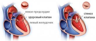
- Pulmonary valve stenosis is an anomaly in which narrowing of the pulmonary valve opening is diagnosed. As a result of pathology, most often of the valve leaflets, normal blood flow from the right ventricle to the pulmonary trunk is disrupted.
- Tetralogy of Fallot is a complex combined congenital heart disorder. Combines ventricular septal defect, pulmonary stenosis, right ventricular hypertrophy, and aortic dextraposition. With this pathology, a mixture of arterial and venous blood occurs.
- Transposition of the great vessels is also a complex congenital disorder. With this pathology, the aorta originates from the right ventricle and carries venous blood, and the pulmonary trunk originates from the left ventricle and carries arterial blood, respectively. Paroxysm is severe and is associated with high mortality in newborns.
- Dextrocardia is an anomaly of intrauterine development characterized by the right-sided placement of the heart. Often, there is a “mirror” arrangement of other unpaired internal organs.
- Ebstein's anomaly is a rare congenital heart defect, diagnosed when the location of the tricuspid valve leaflets changes. Normally - from the atrioventricular fibrous ring, with an anomaly - from the walls of the right ventricle. The right ventricle is smaller, and the right atrium is elongated, including abnormal valves.
How will pregnancy proceed if the mother has a heart defect?
The prognosis of pregnancy depends on the degree and combination of damage, as well as on the activity of the rheumatic process (in other words, on whether it is currently exacerbating) and on the severity of circulatory disorders.
The issue of maintaining or terminating a pregnancy is decided collectively by a cardiologist and an obstetrician-gynecologist in each case individually. If pregnancy occurs after heart surgery, you need to consult a cardiac surgeon. You should know that corrective heart surgery does not always lead to the elimination of organic changes in the valve apparatus or the elimination of congenital developmental anomalies. Often, after surgical treatment, there is a relapse of the underlying disease, for example, in the form of restenosis (re-narrowing) after some operations.
It is extremely difficult to decide whether pregnancy is permissible in women with prosthetic heart valves. They are at high risk of developing blood clots, so pregnant women with mechanical valves constantly receive anticoagulant (anti-clotting) therapy.
Of course, the issue of continuing pregnancy in women with cardiovascular diseases is best decided in advance, before its onset. The basis for proper management and treatment of such pregnant women is an accurate diagnosis that takes into account the cause of the disease.
Defects with blood discharge to the right serve as a contraindication to pregnancy, as well as any type of defect with decompensation, in which circulatory failure has already formed.
Methods for diagnosing congenital heart disease
Diagnosis of congenital heart disease is based on collecting medical history data (presence of developmental defects, including congenital heart defects, genetic diseases in close relatives; information about pregnancy and the presence of etiological factors in parents).
When collecting complaints, attention is paid to the child’s developmental delay, poor weight gain, poor appetite, sluggish sucking from the breast or bottle, breast refusal, cyanosis, and frequent respiratory infections.
Physical examination
During a physical examination, pay attention to the color of the skin, determine pulse and blood pressure (on the right arm and either leg), perform auscultation of the heart and lungs, pay attention to the presence of peripheral edema, perform pulse oximetry, and determine diuresis.
Instrumental diagnostics
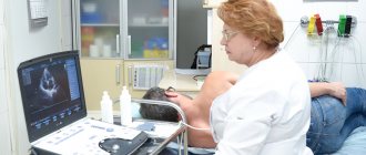
However, the leading role in the diagnosis and confirmation, differential diagnosis of congenital heart disease is played by instrumental examination methods: x-ray examination of the chest organs, electrocardiography, echocardiography, MRI, CT, catheterization of the cardiac cavities.
Sign up for diagnostics To accurately diagnose the disease, make an appointment with specialists from the Family Doctor network.
At-risk groups
Taking into account the causes of fetal pathologies, scientists have identified risk groups. Women from these groups come under the close attention of doctors due to the high likelihood of developing anomalies.
These include:
- women over 35 years of age;
- patients who already have children with congenital pathology;
- women with a history of miscarriage, premature birth, stillbirth or antenatal fetal death;
- couples with a family history;
- patients exposed to radiation or exposed to toxins at work.
In such cases, ultrasound is performed repeatedly - the doctor needs to examine the fetus in detail, evaluate its development over time in order to exclude congenital anomalies.
Deviations from the norm
During ultrasound examination, there are cases of identifying deviations from the norm, which may be an indicator of deterioration in the condition of the fetus and its development process. So, deviations are considered:
- The number of beats per minute is less than 100.
- Increase in the number of heartbeats over 200.
- Complete absence of heartbeat.
During the first ultrasound, the doctor may not detect a fetal heartbeat. When its size exceeds 0.8 cm, there is a possibility of a non-developing pregnancy. However, this is not a definitive diagnosis. After five to seven days, a control ultrasound examination is performed, which confirms or refutes the diagnosis.
Also, the reason for the absence of heartbeat in the early stages may indicate a short period of time. After all, no one knows exactly when conception occurred and how many days the embryo is. It is necessary to undergo a re-test.
There is also a third reason for deviations in the number of heartbeats from the norm - the presence of pathological processes in the development of the internal organs of the embryo. To make such a diagnosis, the doctor examines the “four-chamber section.” An ultrasound image shows all four chambers of the heart at once. Ultrasound can detect more than 70% of heart defects. To clarify the diagnosis, echocardiography is performed.
Having discovered pathologies of the fetal cardiovascular system, doctors do not always suggest terminating the pregnancy. Today, surgery is well developed: highly qualified pediatric surgeons, high-tech equipment, innovative methods of treating pathologies of heart formation. The operation is carried out only with the consent of the parents and after a thorough examination. In practice, doctors have more than a dozen cases of saving babies born with heart defects.
In later stages, deviations from the norm depend on:
- Motor activity of the child.
- Physical activity of the mother.
- Presence of diseases during pregnancy.
Sources
- Pereira SP., Tavares LC., Duarte AI., Baldeiras I., Cunha-Oliveira T., Martins JD., Santos MS., Maloyan A., Moreno AJ., Cox LA., Li C., Nathanielsz PW., Nijland MJ., Oliveira PJ. Sex dependent Vulnerability of Fetal Nonhuman Primate Cardiac Mitochondria to Moderate Maternal Nutrient Reduction. // Clin Sci (Lond) - 2021 - Vol - NNULL - p.; PMID:33899910
- Ben Abdelaziz A., Bchir A., Ben Salah A., Ben Salem K., Mansour N., Hsairi M., Ennigrou S., Nacef T., Dhidah L., Bellaj R., Mehdi F. Professor Mohamed Soussi Soltani : Leader, Innovator and Researcher in Public Health. // Tunis Med - 2021 - Vol99 - N1 - p.5-11; PMID:33899170
- Saavedra LPJ., Prates KV., Gonçalves GD., Piovan S., Matafome P., Mathias PCF. COVID-19 During Development: A Matter of Concern. // Front Cell Dev Biol - 2021 - Vol9 - NNULL - p.659032; PMID:33898461
- Alexanderson-Rosas E., Antonio-Villa NE., Sanchez-Favela M., Carvajal-Juarez I., Oregel-Camacho D., Gopar-Nieto R., Flores-Garcia AN., Keirns C., Hernandez-Sandoval S. ., Espinola-Zavaleta N. Assessment of Atypical Cardiovascular Risk Factors Using Single Photon Emission Computed Tomography in Mexican Women. // Arch Med Res - 2021 - Vol - NNULL - p.; PMID:33896676
- Bhasin D., Arora GK., Isser HS. Young woman with recurrent pregnancy loss. // Heart - 2021 - Vol107 - N10 - p.821-854; PMID:33893215
- Lee K., Laviolette SR., Hardy DB. Exposure to Δ9-tetrahydrocannabinol during rat pregnancy leads to impaired cardiac dysfunction in postnatal life. // Pediatr Res - 2021 - Vol - NNULL - p.; PMID:33879850
- Richards C., Sesperez K., Chhor M., Ghorbanpour S., Rennie C., Ming CLC., Evenhuis C., Nikolic V., Orlic NK., Mikovic Z., Stefanovic M., Cakic Z., McGrath K ., Gentile C., Bubb K., McClements L. Characterization of cardiac health in the reduced uterine perfusion pressure model and a 3D cardiac spheroid model, of preeclampsia. // Biol Sex Differ - 2021 - Vol12 - N1 - p.31; PMID:33879252
- Song Y., Xu J., Li H., Gao J., Wu L., He G., Liu W., Hu Y., Peng Y., Yang F., Jiang X., Wang J. Application of Copy Number Variation Detection to Fetal Diagnosis of Echogenic Intracardiac Focus During Pregnancy. // Front Genet - 2021 - Vol12 - NNULL - p.626044; PMID:33868367
- Khanna R., Chandra D., Yadav S., Sahu A., Singh N., Kumar S., Garg N., Tewari S., Kapoor A., Pradhan M., Goel PK. Maternal and fetal outcomes in pregnant females with rheumatic heart disease. // Indian Heart J - 2021 - Vol73 - N2 - p.185-189; PMID:33865516
- Kirby A., Curtis E., Hlohovsky S., Brown A., O'Donnell C. Pregnancy Outcomes and Risk Evaluation in a Contemporary Adult Congenital Heart Disease Cohort. // Heart Lung Circ - 2021 - Vol - NNULL - p.; PMID:33863666
Heartbeat monitoring using ultrasound
So, when is a fetal heartbeat visible on an ultrasound? Since the first contractions occur already in the fifth or sixth weeks of pregnancy, the first beats can be heard using ultrasound.
To do this, use a special sensor that is inserted into the vagina. Using a sensor with which the doctor examines the fetus through the abdomen, the heartbeat is heard in the seventh week.
Each stage of pregnancy has its own heart rate:
- Between the sixth and eighth weeks, the fetal heart beats 110-130 times per minute.
- In the ninth and tenth weeks, the heart rate is 170-190 beats.
- Starting from the eleventh week and until birth, the baby’s heart in the womb makes 140-160 beats per minute.
The difference in indicators is affected by the process of formation and development of the fetal nervous system.
With the help of ultrasound examination of the fetal heart, not only the number of contractions is monitored, but also its location, size, nature of contractions, and their rhythm.