Heart wounds
Classification.
Heart wounds are divided into non-gunshot (knife, etc.) and gunshot: into those penetrating into the cavities of the heart and non-penetrating. Penetrating ones, in turn, are divided into blind and through ones. This is the localization of injuries in relation to the chambers of the heart: injuries to the left ventricle (45-50%), right ventricle (36-45%), left atrium (10-20%) and right atrium (6-12%). They, in turn, with and without damage to intracardiac structures.
Currently, cardiac injuries account for 5 to 7% of all penetrating chest wounds, including gunshot wounds - no more than 0.5-1%. In case of stab wounds of the heart and pericardium, isolated pericardial injuries account for 10-20%. Pericardial injuries themselves do not pose a threat to the life of the victim, however, bleeding from the crossed pericardial vessels can lead to cardiac tamponade.
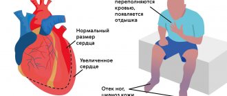
Cardiac tamponade is a condition in which blood entering the pericardial cavity “strangles” the heart.
Acute cardiac tamponade occurs in 53-70% of all cardiac injuries. The degree of tamponade is determined by the size of the heart wound, the rate of bleeding from the heart into the cavity of the cardiac membrane, as well as the size of the pericardial wound. Small stab wounds to the pericardium are quickly closed by a blood clot or adjacent fat, and cardiac tamponade quickly ensues. The accumulation of more than 100-150 ml of blood in the cavity of the cardiac membrane leads to compression of the heart and a decrease in myocardial contractility. Left ventricular filling and stroke volume rapidly decrease, and profound systemic hypotension occurs. Myocardial ischemia is aggravated due to compression of the coronary arteries. If there is 300-500 ml, cardiac arrest occurs in most cases. It should be remembered that an extensive pericardial wound prevents the occurrence of tamponade, because blood flows freely into the pleural cavity or out.
According to S. Tavares (1984), mortality from cardiac injuries is associated with the nature, size, location of the cardiac wound, as well as associated injuries and the length of time from the moment of injury to the start of resuscitation and treatment. In recent years, there has been an increase in mortality, which is primarily due to the severity of heart damage.
The prognosis is also affected by rhythm disturbances. For example, with sinus rhythm, survival rate is 77.8%. According to JPBinet (1985), only 1/3 of victims with heart injuries are admitted to the hospital, and the rest die at the scene or on the way to the hospital. Supposed causes of death at the prehospital stage, according to the observations of V.N. Wolf (1986), the following: 32.8% die from massive blood loss, 26.4% from a combination of massive blood loss and cardiac tamponade, 12.7% from isolated cardiac tamponade. In addition, the mortality rate is influenced by factors such as the duration of acute cardiac tamponade, the degree of blood loss, and the presence of damage to the coronary arteries and intracardiac structures.
The highest mortality rate is observed with gunshot wounds.
Diagnostics.
According to the literature, the determining factors in diagnosing cardiac wounds are the localization of the chest wound in the projection of the heart and the degree of blood loss. An important and reliable sign of a heart injury is the localization of the external wound in the projection of the heart, which, according to the observations of V.V. Chalenko et al., (1992) – met in 96%, M.V. Grineva, A.L. Bolshakova, (1986) - in 26.5% of cases.
Difficulties in diagnosis arise in the absence of typical clinical signs. According to D.P. Chukhrienko et al., (1989), cardiac tamponade occurs in 25.5% of cases of cardiac injuries. V.N. Wolf (1986) distinguishes two stages of cardiac tamponade: the first is blood pressure at the level of 100-80 mm Hg. Art., while the hemopericardium does not exceed 250 ml; the second, when blood pressure is less than 80 mm Hg. Art., which corresponds to a hemopericardium of more than 250 ml. J.H. Vasiliev (1989) believes that a sudden accumulation of 200 ml of fluid in the pericardial cavity causes a clinical picture of cardiac compression; an accumulation of about 500 ml leads to cardiac arrest.
Cardiac tamponade can also be caused by pneumopericardium.
Beck's triad, according to AK Benyan et al. (1992), was observed in 73% of cases, according to D. Demetriades (1986) – in 65%, according to M. McFariane et al. (1990) – in 33%.
X-ray examinations of cardiac injuries are carried out in 25% and 31.5%. Based on radiographs, one can judge the volume of blood in the pericardial cavity - a blood volume from 30 ml to 85 ml is not detected; if there is 100 ml, there are signs of weakening pulsation; when the blood volume is more than 150 ml, there is an increase in the boundaries of the heart with smoothing of the “arcs”.
To diagnose cardiac injury, additional research methods are used - ultrasound, pericardiocentesis [Chukhrienko D.P. et al., 1989; Demetriades D., 1984; Hehriein FW, 1986; McFariane M. et al., 1990], pericardiotomy [Vasiliev Zh.Kh., 1989; Grewal N. et al., 1995].
It should be emphasized that when performing pericardial puncture, false negative results were obtained in 33% [Chalenko V.V. et al., 1992] and in 80% of cases.
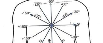
ECG is performed quite often: in 60%. At the same time, signs of cardiac injury such as large-focal injuries with changes in the T wave, a decrease in the RST interval were detected in 41.1%, rhythm disturbances – in 52%.
The diagnosis of cardiac injury before surgery was established in 75.3%.
According to the authors, progress in diagnosis is obvious, but mainly due to the “classical” clinical approach. This opinion is also shared by KKNagy et al., (1995), they classify clinical signs of damage and active surgical intervention as the most reliable diagnostic methods.
The following triad of symptoms should be considered characteristic signs of heart injury:
1) localization of the wound in the projection of the heart;
2) signs of acute blood loss;
3) signs of acute cardiac tamponade.
When the wound is located within the following boundaries: above - the level of the second rib, below - the epigastric region, on the left - the anterior axillary line and on the right - the parasternal line, there is always a real danger of injury to the heart.
When the wound is localized in the epigastric region and the blow is directed from bottom to top, the wound canal, penetrating into the abdominal cavity, goes further through the tendon center of the diagram into the cavity of the cardiac sac and reaches the apex of the heart.
The classic clinical picture of cardiac tamponade was described by K. Beck (1926): dullness of heart sounds; low blood pressure with a low rapid pulse (and low pulse pressure); high venous pressure with swelling of the neck veins.
If the patient's condition is stable, the diagnosis of cardiac injury can be confirmed by X-ray examination.
Currently, the most accurate and fastest non-invasive diagnostic method is echocardiography. In this case, within 2-3 minutes, the divergence of the pericardial layers (by more than 4 mm), the presence of fluid and echo-negative formations (blood clots) in the cavity of the heart lining, zones of akinesia in the area of the myocardial wound, as well as a decrease in myocardial contractility are clearly revealed.
Recently, surgeons have sometimes begun to use a minimally invasive method such as thoracoscopy to diagnose cardiac injuries. It is worth noting that indications for this method arise quite rarely, for example, in clinically unclear cases when it is impossible to diagnose a heart injury with echocardiography, when, on the one hand, it is dangerous to continue observation and examination over time, and on the other hand, it is dangerous to perform classical thoracotomy (for example, in patients with decompensated diabetes mellitus).
When the heart or pericardium is injured, after opening the pleural cavity, it is clearly visible how blood shines through the walls of the tense pericardium. Further manipulations of the surgeon and his assistants, the entire team on duty, including the anesthesiologist, must be clearly coordinated. The surgeon places two suture holders on the pericardium and opens it wide, parallel and in front of the phrenic nerve.
The holding assistant widely spreads the pericardial wound, and, at the same time, frees the pericardial cavity from liquid blood and clots, and the surgeon, guided by the pulsating stream of blood, immediately plugs a small heart wound with the second finger of his left hand, or, if the wound size exceeds 1 cm, with the first finger, bringing your palm under the back wall of the heart.
In cases of more extensive wounds, a Foley catheter can be used to achieve temporary hemostasis. Inserting a catheter into the heart chamber and inflating the balloon with gentle tension can temporarily stop bleeding. This task can also be accomplished by inserting a finger into the myocardial wound. We successfully used the last technique in four observations. When suturing a heart wound, exclusively non-absorbable suture material is used, preferably with an atraumatic needle. It should be remembered that thin threads are easily cut through when suturing a flabby wall, especially in the area of the atria.
In these cases, it is better to use thicker threads and place patches under them, cut in the form of strips from the pericardium. In cases of injury to the appendage of the heart, instead of applying sutures, it is better to simply bandage the appendage at the base, after first applying a windowed Luer clamp to it.
In order to avoid myocardial infarction when the branches of the coronary arteries are dangerously close to the wound, vertical interrupted sutures should be applied, bypassing the coronary artery.
Of no small importance for the postoperative course is careful sanitation and proper drainage of the cavity of the cardiac membrane. If this is not done, then postoperative pericarditis inevitably develops, leading to an increase in the duration of hospital treatment, and, in some cases, to a decrease in the patient’s ability to work.
Therefore, the cavity of the cardiac membrane is thoroughly washed with a warm isotonic solution, a section of about 2-2.5 cm in diameter is excised in the posterior wall of the pericardium, making a so-called “window” that opens into the free pleural cavity, and rare interrupted sutures are placed on the anterior wall of the pericardium to preventing dislocation of the heart and “strangulation” of it in a wide wound of the pericardium.
In cases of abdominal-thoracic wounds with damage to the heart from bottom to top, it is more convenient to suture the heart wound through the diaphragmatic-pericardial approach, without performing a lateral thoracotomy.
Noteworthy is the proposed Trinkle J.K. (1979) subxiphoid fenestration of the pericardium. It consists of dissecting the soft tissue in the area of the xiphoid process, resection of the latter, reaching the pericardium, applying holders to it, opening and evacuating blood clots in an open manner. This operation can be performed under local anesthesia and is life-saving in cases where it is necessary to gain time, but it is not possible to perform a thoracotomy.
We studied the results of using subxiphoid partial pericardiectomy in 10 patients with cardiac injury. The operation ended with the installation of a silicone drainage tube with a diameter of 5 mm into the cavity of the cardiac membrane. To improve the outflow from the pericardial cavity, the distal end of the drainage was connected to the aspiration system.
So, depending on the conditions of care, there may be different solutions to tactical problems for cardiac injuries.
Diagnostics
A preliminary diagnosis of “heart injury” is usually made by emergency medical services teams.
For the initial diagnosis of heart injury, two signs are important:
- presence of a wound in the left side of the chest or upper left abdomen;
- a progressive decrease in pressure, pronounced pallor of the skin, cold sweat, agitation, which gives way to apathy and loss of consciousness, frequent weak pulse, swollen veins in the neck (signs of internal bleeding and tamponade).
In cases where a heart injury is suspected, but the victim’s condition is stable, additional diagnostic tests can be used - echocardiography, chest x-ray, sometimes pericardial puncture (does not reveal cardiac injury in a third of victims), ECG (changes may be absent).
Types of injuries
Some traumatic situations lead to a combination of different types of open and closed heart injuries.
Depending on the nature of the damage, blunt cardiac injuries are as follows:
- tear or rupture of the pericardium;
- myocardial rupture;
- heart bruise;
- damage to the valve apparatus;
- damage to the coronary arteries;
- concussion;
- aortic damage.
Electrical shock is a separate group of blunt cardiac injuries.
Heart injuries can be:
- single;
- multiple.
Some severe traumatic situations can result in a combination of open and buried cardiac injuries. In addition, the victim’s condition may be aggravated by damage to other organs.
Spasm of coronary vessels and organ bruise - differences
The clinical picture of the bruise resembles a heart spasm (suddenly occurring coronary insufficiency). Treatment of pathologies is different, so differential diagnosis is necessary. Reliable results are provided by diagnosis, but before it is carried out, a drug test can be carried out - nitroglycerin reduces or completely relieves pain during spasm of the coronary vessels, whereas in case of a bruise, the painful sensations do not go away.
It is also necessary to find out from the patient the entire history of the disease. If in the days preceding the deterioration of health there were blows to the chest, then only a doctor can rule out a bruise.