Blockage of arteries in the vertebrobasilar region leads to the development of ischemic stroke with localization of the infarction zone in various parts of the brain stem, thalamus, occipital lobes and cerebellum. Individual characteristics of the location of the arteries and the variety of pathogenetic mechanisms very often determine the individual characteristics of the neurological clinic in acute ischemic strokes in this area. Along with the presence of typical neurological syndromes, doctors at the Yusupov Hospital often note atypical symptoms of acute cerebrovascular accident. In this clinical situation, they use brain neuroimaging methods that help confirm the diagnosis (computed tomography and magnetic resonance imaging).
Neurologists at the Yusupov Hospital assess the degree of impairment of neurological functions during patient hospitalization, during treatment and at the end of therapy. All patients admitted to the neurology clinic undergo the following examinations:
- Doppler ultrasound of the great vessels of the head in the extracranial region;
- transcranial Doppler sonography;
- duplex scanning.
A 12-electrode ECG is also performed, blood pressure is monitored, and the volumetric maximum blood flow through the internal carotid and vertebral arteries is determined. Spiral computed tomography of the brain at the Yusupov Hospital is performed in all cases immediately upon admission of patients to the hospital. In the presence of several foci of cerebral infarction, neurologists use a more sensitive neuroimaging technique - diffusion-weighted magnetic resonance imaging.
A modern sensitive technique for neuroimaging of the brain - perfusion-weighted magnetic resonance imaging - allows doctors at the Yusupov Hospital to obtain information about the state of blood supply to brain tissue, and identifies blood supply disturbances both in the ischemic core zone and in surrounding areas.
Types of ischemic strokes in the vertebrobasilar region
The following ischemic cerebral infarctions in the vertebrobasilar region are distinguished:
- lacunar strokes due to damage to small perforating arteries, caused by microangiopathies against the background of arterial hypertension and diabetes mellitus;
- non-lacunar strokes that developed due to damage to the short or long circumflex branches of the vertebral and basilar arteries in the presence of sources of cardioembolism and the absence of narrowing of the large vertebrobasilar arteries;
- non-lacunar strokes due to blockage of the vertebral and basilar arteries in the intracranial and extracranial parts, caused by their damage.
They have different symptoms and require differentiated therapy.
Stages
The development of CNMK goes through 3 stages. At the initial stage, tissue damage is insignificant, the lesions are small in size. Properly selected treatment will correct the resulting pathology. Violations are found mainly in the emotional sphere and are usually attributed to overwork and excessive nervous tension.
A person quickly gets tired, becomes apathetic, irritable, absent-minded, whiny, impulsive, and forgetful. There is a decrease in performance, difficulties with the perception and processing of new information. Headache occurs periodically. After a good rest, all these signs disappear.
At the second stage, the symptoms worsen and become more vivid. The patient loses interest in work, in what previously fascinated him. A decrease in motivation leads to unproductive, monotonous useless work, the purpose of which cannot be explained by the patient himself. Memory and intelligence decrease. Bouts of inexplicable aggression appear. The patient experiences uncontrolled mouth movements, problems with fine motor skills, and slows down movements.
Headaches become more frequent and intense; they are localized mainly in the forehead and crown area. The examination reveals signs of anatomical damage.
At the third stage, the resulting changes become irreversible. Clear signs of dementia appear. The patient often becomes aggressive and cannot control himself. Doesn't understand where he is, unable to tell the time. Problems with vision and hearing arise. He loses the ability to take care of himself, does not understand the meaning and consequences of simple actions. Incontinence of urination and defecation occurs.
Symptoms of ischemic stroke in the vertebrobasilar region
Lacunar strokes in the vertebrobasilar region occur as a result of damage to a separate paramedian branch of the vertebral artery, common artery or branch of the posterior cerebral artery against the background of arterial hypertension, which is often combined with high levels of lipids in the blood or diabetes mellitus. The disease begins suddenly and is accompanied by dizziness, nausea, and vomiting. There are disturbances in motor function caused by damage to the motor pathways in the area of the base of the bridge, which are supplied with blood by small arteries branching from the main artery:
- incomplete paralysis of facial muscles;
- arm paralysis;
- impaired movement of the arm and leg on one side of the body.
Lacunar infarctions in the thalamus cause the development of a purely sensory syndrome, the cause of which is damage to the lateral parts of the thalamus due to blockage of the thalamogenicular artery. Complete hemisensory syndrome is manifested by a decrease in superficial or deep sensitivity, or numbness of the skin of one half of the body. Some patients have unilateral decrease in sensitivity of the corner of the mouth, palm and foot.
When ischemia spreads towards the internal capsule, a sensorimotor stroke develops. It is manifested by motor impairments, which are preceded by sensory disorders. If the lacunae are located in the area of the bridge, doctors at the Yusupov Hospital determine the following signs of ischemic stroke:
- impaired coordination of movements on one half of the body;
- moderate leg weakness;
- Mild paresis of the arm.
Nonlacunar ischemic infarction in the vertebrobasilar region develops as a result of damage to the short or long circumflex branches of the vertebral or basilar arteries and is manifested by the following symptoms:
- systemic dizziness;
- headache;
- hearing loss with noise in the same ear;
- motor and cerebellar disorders;
- sensory disturbances in one or both limbs of one side of the body.
Blockage of the posterior inferior cerebellar artery is manifested by the following symptoms:
- systemic dizziness;
- nausea;
- vomit;
- swallowing disorder;
- speech and hearing impairment;
- sensitivity disorders on the face of a segmental type;
- cerebellar ataxia (impaired stability) on the side of the ischemic lesion;
- movement disorders, decreased pain and temperature sensitivity on the limbs and torso on the opposite side.
When the branches of the main artery supplying the midbrain are blocked, paresis of the muscles innervated by the oculomotor nerve occurs on the side of the lesion and paralysis of the limbs on the opposite side. With a heart attack in the quadrigeminal artery basin, upward gaze paralysis and convergence insufficiency develop, which is combined with involuntary high-frequency oscillatory eye movements.
Cerebellar infarction in most cases occurs due to cardiac or arterial embolism of the anterior inferior cerebellar artery or superior cerebellar artery.
Blockage of the vertebral artery can occur both inside and outside the skull. When the extracranial section is blocked, the following symptoms are noted:
- short-term loss of consciousness;
- systemic dizziness;
- visual impairment;
- oculomotor and vestibular disorders;
- violations of statics and coordination of movements.
Often, patients suddenly fall, their muscle tone is impaired, autonomic disorders develop, breathing and cardiac activity are impaired.
Treatment of cerebral venous outflow dysfunction
Allopathic medicine traditionally treats diseases. Venotonics and other vascular drugs are widely used in the treatment of venous pathology.
Yes, they can improve the quality of the venous flow and general well-being, but without systemic regular treatment, the problem of venous outflow of the brain will again make itself felt.
To get rid of discomfort, brain activity drugs, diuretics, and anticoagulants are also used. The home first aid kit and knowledge in the field of pharmaceuticals are replenished, but this does not make you feel healthier, except for periods.
Treatment with osteopathic techniques is aimed at long-term effects. This is achieved by soft and gradual removal of compensation and the revival of natural functions
It is important not only to establish venous outflow, but also to eliminate the root causes of the disorder
Next, work is carried out on the changes caused by venous insufficiency.
At the first appointment, the osteopath removes the disorders visible to him:
- Muscle tone that compresses and squeezes the veins of the head and cervical area;
- Displacements of the cranial and bones;
- Deformations in the spinal column.
Since the basis for impaired venous outflow is often osteochondrosis, the osteopath interacts with the spine along its entire length, paying attention to the pelvic area. How is the pelvis connected to the venous system of the head? Directly
Osteoscience perceives the body as a single structure in which there is nothing separate. Even an old injury to the coccyx can, over a period of time, cause disruption in the functioning of the blood vessels of the brain, affecting the spine, which in turn puts pressure on the cranial vault and causes tension in the muscles and veins of the neck
How is the pelvis connected to the venous system of the head? Directly. Osteoscience perceives the body as a single structure in which there is nothing separate. Even an old injury to the coccyx can, over a period of time, cause a disruption in the functioning of the cerebral vessels, affecting the spine, which in turn puts pressure on the cranial vault and causes tension in the muscles and veins of the neck.
There is another connection. Since cerebrospinal fluid continuously circulates in the spinal canal, it is necessary that its rhythm correspond to the micromovements of the cranial plates. In osteopathy, their mobility is usually compared to breathing, the disruption of which provokes multiple diseases. Treatment includes restoring bone mobility in the head.
During treatment, the osteopath may stand motionless for several seconds or minutes with his eyes closed, placing his hands on one part of the body, then on another. From the outside it seems that nothing is happening, and the patient does not feel any pain or pressure. This is how doctors find flaws and initiate self-regulation.
Most of our clients noted positive changes after starting treatment - ease of breathing, a feeling of vigor after sleep, improved memory and motivation.
But in some cases, so-called side effects occur. They are short-term and are explained by the body ridding itself of acquired compensations. He strives to preserve himself and, in response to damage, includes all kinds of protection to the detriment of full functionality. The structures of vital activity adapt to function in less than healthy conditions, and after eliminating the problem, it is difficult for them to return to their original state. The osteopath’s task is to do this as gently as possible.
Any alarming symptoms that arise should be discussed with your doctor. If it is moderate pain, insomnia or too much sleep, weakness, nausea - most likely everything is within normal limits.
These symptoms will quickly pass, but the treatment cannot be considered complete, since one session (regardless of how you feel after it) is not enough to work through all the problems. Subsequent visits to the clinic are necessary to assess the intermediate result and determine further tactics.
Treatment of ischemic stroke in the vertebrobasilar region
Neurologists at the Yusupov Hospital take an individual approach to the treatment of each patient diagnosed with ischemic stroke of the basilar artery. In the presence of high blood pressure, antihypertensive therapy is carried out. To stimulate spontaneous formation of channels in a blocked artery and prevent re-embolization in atherothrombotic and cardioembolic subtypes of nonlacunar ischemic infarction, direct anticoagulants, as well as antiplatelet agents, are used.
Complex therapy of acute ischemic strokes in the vertebrobasilar region also involves the prior use of neuroprotectors. In order to determine the feasibility of neuroprotective therapy, doctors at the Yusupov Hospital use diffusion-perfusion MRI studies, which help identify viable areas of the ischemic penumbra. After this, neuroprotective drugs are prescribed.
The Neurology Clinic of the Yusupov Hospital is equipped with the necessary equipment to diagnose complex locations of cerebral infarction. Neurologists treat patients with modern medications that have a pronounced effect on blockage of the vertebrobasilar artery. Call and they will make an appointment with a neurologist.
Classification of cerebrovascular pathology
CVD is a brain disease associated with progressive tissue damage due to impaired circulation in the organ. It can arise due to pathologies of cerebral vessels, which cause changes in the blood circulation of brain tissue and hypoxia. It is mainly diagnosed in older people, but in rare cases, the disease can also occur in a child.
Cerebrovascular disease develops gradually. At the very beginning, the brain begins to experience oxygen deficiency due to vascular dysfunction.
With chronic hypoxia, all brain functions begin to suffer. First, transient and then persistent changes in brain tissue occur.
Cerebrovascular disease contributes to the development of grade 2 DEP, which causes brain damage.
Based on the classification, CVD can be divided into transient, acute and chronic.
Acute cerebrovascular insufficiency includes:
- Encephalopathy of the hypertensive type.
- Ischemic attacks.
- Stroke (multilacunar form, ischemic, hemorrhagic, unspecified).
Chronic cerebrovascular insufficiency (CVI) is divided into:
- Embolism, which results in blockage of blood vessels by blood clots. Which arise in the large arteries of the body and can enter small ones with the blood.
- Bleeding resulting from rupture of the vascular walls. This condition provokes the development of hemorrhagic stroke.
- Thrombosis is a pathology when the lumen of blood vessels narrows and becomes clogged with plaques.
Neurologist Mikhail Moiseevich Shperling will tell you more about the ischemic form of stroke:
The transient form of cerebrovascular disease includes:
- Cerebral hypertensive crisis.
- Transient ischemic attacks.
List of sources
- Kamchatnov P.R., Gordeeva.T.N., Kabanov A.A. and others. Clinical and pathogenetic features of vertebrobasilar insufficiency syndrome. J. Stroke, 2001; 1; 55-57.
- Diseases of the nervous system. Guide for doctors. T.1/ Yakhno N.N., Shtulman D.R., Melnichuk P.V. and others. Ed. Yakhno N.N. - M. Medicine 1995.
- Losev R.Z., Nikolenko V.N., Sholomov I.I. et al. Diagnosis and treatment of patients with circulatory insufficiency in the vertebrobasilar region // Saratov Medical Scientific Journal. – 2009. – Volume 5. – No. 4. – P. 629-634.
- Samsonova I.V., Solodkov A.P., Burak G.G., Novikova O.V. Vertebro-basilar insufficiency: problems and prospects for solutions // Bulletin of Vitebsk State Medical University. – 2006. – Volume 5. – No. 4. – P. 1-15.
- Yakovlev N.A. Vertebro-basilar insufficiency (vertebrobasilar arterial system syndrome). – M.: Provincial Medicine, 2001. – 400 p.
Basic principles of vascular health
The vertebrobasilar basin is not the only vascular conglomerate in which certain disorders can be observed that can lead to serious consequences. Any blood vessel plays its role in the body, and its blockage and narrowing of the lumen can cause pathology. That is why the prevention of vascular disorders should become one of the principles of maintaining one’s own health for every person. Unfortunately, many pathologies develop in the womb, and it is often very difficult to compensate for them with the real efforts of doctors, the patient and his relatives. But acquired vascular problems are the fault of the person himself, who neglects his health and does not listen to his body. A correct daily routine, a change in activities, a rational, nutritious diet, regular medical examinations, and careful attention to your health will help maintain the functionality of the cardiovascular system.
The vertebrobasilar basin is a vascular structure that supplies blood to the brain. It carries a huge load, providing oxygen and nutrients to many parts of the main organ. Disturbances in the blood flow of this system can cause pathological processes in many systems of the human body, deterioration of its vital functions and even death. To prevent this from happening, you need to listen to the signals that your body gives about impending problems.
What is the job of the vascular complex?
Blood supply to the body is a complex task, solved by the cardiovascular system depending on the functionality of the endocrine and immune systems. Providing oxygen and nutrients is a load that must be performed regularly, without serious disruptions that can lead to pathological changes in all structures of the body. Poor circulation of the vertebrobasilar system leads to many problems, because the vessels included in this conglomerate supply blood to the posterior regions of the brain, as well as the pons, the structure responsible for transmitting information from the spinal cord to the brain.
Any problems with blood supply to areas localized in the access area of this vascular system will certainly affect the health status and deterioration of a person’s life.
Prevention
To prevent the occurrence of manifestations of vertebrobasilar syndrome, a person predisposed to the development of such pathologies should adhere to certain rules, namely:
- practice proper nutrition, primarily limiting salt intake;
- do as much physical exercise as possible;
- spend more time outdoors;
- avoid stress and injury ;
- give up all bad habits;
- control blood pressure ;
- create comfortable conditions for work and sleep (in terms of body position);
- be periodically examined by a neurologist.
In order to prevent relapse after an attack of VBI, the patient must follow all the recommendations described above, as well as follow preventive therapy (antihypertensive, anticoagulant, antiplatelet, etc.) prescribed during the hospital stay.
Methods for diagnosing the disease
This type of stroke, as well as vertebrobasilar insufficiency itself, is very difficult to diagnose. This is due to the fact that the disease manifests itself differently in different people. In addition, some patients cannot distinguish specific manifestations of the disease from subjective discomfort. As a result, when collecting anamnesis, the doctor cannot understand what specific disease to look for. In addition, the general symptoms of brain diseases are similar. The following diagnostic methods are used:
- MRI or CT. Magnetic resonance imaging can provide a more detailed picture of brain structures, but it cannot be done if the patient has implants in the mouth. For such cases, there is computed tomography. Thanks to it, you can see bleeding and all the brain changes that appeared immediately after the attack.
- Angiography. Contrast is injected into the vessels, and then pictures are taken. This diagnostic method allows you to obtain expanded information about the state of the vascular system and the pool in question as a whole. Any narrowing of the diameter of the vessels will be shown on the images.
- X-ray of the spine. Necessary for assessing the general condition of the vertebrae.
- Infrared thermography. Allows you to obtain information about the thermal characteristics of a specific part of the body.
- Functional tests. They will help determine if any area of the brain is severely affected after a circulatory disorder.
- Blood research in the laboratory.
Structure of the vascular conglomerate
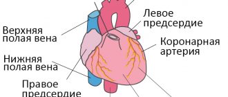
Blood vessels are peculiar channels that transport blood from the heart and lungs to every cell of the body. They differ not only in their size, but also in their purpose. Two arteries - vertebral and basilar - form the basis of this vascular complex, which is named after their name.
The vertebral artery is a rather complex formation - the right and left vertebral arteries originate from the subclavian artery. They both consist of the following parts:
- the first section passes through the transverse foramen of the sixth cervical vertebra;
- the second section passes upward through the transverse openings of the cervical vertebrae, it is intertwined with veins and sympathetic fibers;
- the third section passes into the so-called fissure of the atlas and enters the foramen magnum;
- the fourth section penetrates the dura mater, then, connecting with the opposite vertebral artery, forms a single whole.
The supply of blood to the brain is carried out by such an extensive structure as the vertebrobasilar basin. The arteries combined in it must work together, fully carrying blood to the brain.
The second part of this vascular association is the basilar artery. It is also a large blood vessel that is formed by the fusion of the left and right vertebral arteries. This artery is located along the basilar groove. In fact, the vertebrobasilar basin of the brain is a single structure, which is only conditionally divided into several components.
Two complexes - one job
The brain is the director of all processes on which not only all life support processes depend, but also human life itself. If the blood supply to this organ is difficult or disrupted for any reason, then the entire body suffers, even to the point of death. The blood supply system to the brain, including the supply of blood to it, is complex, but experts distinguish two conglomerates in it, which have received characteristic names - the carotid system and the vertebrobasilar system. They are the two main systems involved in delivering blood to the brain.
The carotid basin is the union of the two carotid arteries and adjacent small vessels. It begins in the chest area - the left one branches off from the aorta, and the right one originates from the brachiocephalic trunk. The work of this system is to provide both hemispheres of the brain, visual organs, and soft tissues of the head with oxygenated blood. The features of the vertebrobasilar basin will be discussed below. Both systems are important for the proper functioning of the most important organ of the human body; impaired vascular patency leads to a lot of problems that can end very badly.
Part of the vascular system
The cardiovascular system is the main transport route, supplying all cells with oxygen and useful substances carried in the blood, as well as removing waste products for metabolism, utilization and excretion. It consists of three main types of vessels - arteries, veins and capillaries, and in some organs, specialists combine them into certain structures in order to characterize the pathology as accurately as possible, if necessary. One of these structures is the vertebrobasilar basin of the brain.