The heart begins to beat long before we are born. Surprising and even magical, right? However, this means that this organ works longer and harder than others. And not only the quality of our life, but also life itself depends on how carefully we pay attention to his condition!
More than 17 million people die every year from heart and vascular diseases. It has been proven that 80% of premature heart attacks and strokes could be prevented!
Fortunately, most cardiovascular diseases are successfully diagnosed thanks to modern innovative equipment and the professionalism of doctors. And as you know, establishing an accurate diagnosis means taking an important step towards healing.
One of the most informative and safe studies is cardiac echocardiography (EchoCG) or, in other words, ultrasound of the heart.
1 ECHO-KG in "MedicCity"
2 Echocardiography at MedicCity
3 Ultrasound of the heart at MedicCity
The essence of echocardiography
Ultrasound of the heart is the process of studying all the main parameters and structures of this organ using ultrasound.
When exposed to electrical energy, the echocardiograph transducer emits high-frequency sound that travels through the structures of the heart, is reflected from them, captured by the same transducer and transmitted to the computer. He, in turn, analyzes the received data and displays it on the monitor in the form of a two- or three-dimensional image.
In recent years, echocardiography has been increasingly used for preventive purposes, which makes it possible to identify cardiac abnormalities at an early stage.
What does an ultrasound of the heart show:
- heart size;
- integrity, structure and thickness of its walls;
- sizes of the cavities of the atria and ventricles;
- contractility of the heart muscle;
- operation and structure of valves;
- condition of the pulmonary artery and aorta;
- pulmonary artery pressure level (to diagnose pulmonary hypertension, which can occur with pulmonary embolism, for example, when blood clots from the veins of the legs enter the pulmonary artery);
- direction and speed of cardiac blood flow;
- condition of the outer shell, pericardium.
1 ECHO-KG in "MedicCity"
2 Echocardiography at MedicCity
3 Ultrasound of the heart at MedicCity
Echocardiography with Doppler analysis
Doppler echocardiography is a variant of ultrasound examination that allows one to evaluate blood flow (velocity and turbulence) in the cavities and large vessels of the heart. The method is based on the Doppler effect: the speed of an object affects the frequency of the supplied signal. This version of echocardiography is highly informative and safe, so it is mandatory for pregnant women and children.
Advantages of the method:
- non-invasive;
- fast processing of information in real time;
- accuracy.
The examination helps to identify:
- pathological blood flows;
- mitral valve prolapse;
- blood clots and cardiac aneurysms;
- defects, congenital and acquired.
Three main modes of echocardiography with Doppler analysis:
- Pulse-wave . Diagnostics of low-speed flows.
- Constant wave . Assessment of blood flow velocity in the aorta (diagnosis of aortic stenosis).
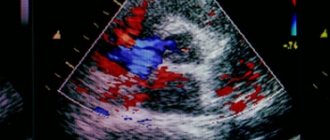
- Color mapping . Visualization of valve and turbulent flows at the site of blockage. Diagnosis of blood clots, aneurysms, atherosclerotic plaques, determination of the nature of the neoplasm.
Who is prescribed echocardiography?
The following are routinely examined:
- infants - if congenital defects are suspected;
- teenagers are a time of intensive growth;
- pregnant women with existing chronic diseases - to decide on the method of childbirth;
- professional athletes - to monitor the state of the cardiovascular system.
An echocardiogram is required if:
- anomalies of the endocardium and valve apparatus:
- heart tumors;
- arrhythmias;
- threat of a heart attack or a heart attack;
- IHD;
- failures of cardiac activity due to various intoxications;
- attacks of angina pectoris;
- pericarditis of various origins;
- hypertension;
- heart failure.
And also in the process of treating heart diseases, before and after cardiac surgery.
What does Echo KG show?
The procedure is carried out by an ultrasound diagnostic specialist, and the duration of the examination is 15-30 minutes. Echocardiography is prescribed for frequent or recurrent pain in the heart, irregular heartbeat, severe shortness of breath, high or low blood pressure, suspected heart disease, or a previous heart attack. The main indicators of cardiac echocardiography are presented below:
- ventricular myocardium mass (right and left);
- condition and contractility of the valve apparatus;
- the speed of blood flow through the heart valves and chambers;
- the thickness of the partitions between the chambers of the heart.
There is no cause for concern if your cardiac ultrasound results are normal. However, the decoding results may differ from the standard ones - the data obtained will tell about anomalies in the functioning of the cardiovascular system. Based on what the Echocardiogram of the heart shows in adults or children, the doctor will be able to make an accurate diagnosis:
- pericarditis, myocarditis, cardiomyopathy;
- pre-infarction condition/myocardial infarction;
- heart failure (left-sided, right-sided);
- coronary heart disease (myocardial damage).
A comprehensive ultrasound of the heart with Doppler sonography also displays indicators that allow you to assess the condition of the blood vessels. The procedure makes it possible to determine the speed of blood flow, as well as identify pathological changes in the walls of blood vessels. Thanks to the study, the doctor can timely detect atherosclerotic processes, aortic aneurysm and heart defects.
When is a cardiac ultrasound performed?
Indications for cardiac ultrasound are:
- alarming changes in health (increased or interrupted heartbeat, shortness of breath, swelling, weakness, prolonged fever, chest pain, cases of loss of consciousness);
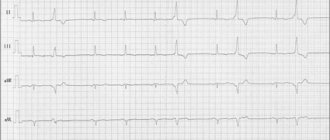
- changes detected in the last ECG;
- increased blood pressure;
- heart murmurs;
- cardiomyopathy;
- manifestations of coronary heart disease;
- heart defects (congenital, acquired);
- pericardial diseases;
- lung diseases.
Indications for echocardiography (ultrasound of the heart)
Echocardiography is performed in the case of:
- shortness of breath, complaints of dizziness, weakness, loss of consciousness;
- pain in the heart and behind the sternum;
- heart rhythm disturbances (arrhythmias);
- detected heart murmurs;
- high blood pressure;
- presence of changes on the ECG;
- suspicion of diseases leading to heart damage (for example, rheumatism);
- chest injuries;
- suspected heart failure (enlarged liver, edema);
- chronic tonsillitis, as well as previous tonsillitis or flu in severe forms (for timely detection of possible complications on the heart).
For children, echocardiography is indicated in cases of suspected congenital heart disease.
Echocardiography is also done to monitor the condition of the heart:
- in case of treatment with antibiotics for cancer;
- after myocardial infarction;
- after operations on blood vessels and the heart;
- pregnant women;
- athletes (for professional sports, the examination should be carried out at least once a year).
Preparation for cardiac echocardiography
Ultrasound of the heart does not require special preparations. On the eve of the procedure, the patient is free to eat as usual and perform normal activities. The only thing that will be asked of him is to give up alcohol, caffeine-containing drinks, and strong tea.
If the patient is constantly taking medications, this must be warned in advance so that the results of the study are not distorted.
For each subsequent ultrasound of the heart, you should take a transcript of the previous one. This will help the doctor see the process over time and draw the right conclusions about your condition.
The examination itself takes from 15 to 30 minutes.
The patient, undressed to the waist, is lying on his back or side. A special gel is applied to his chest, ensuring easier sliding of the sensor over the area under study (the patient does not experience discomfort).
The specialist performing echocardiography has access to any areas of the heart muscle - this is achieved by changing the angle of the sensor.
Sometimes standard ultrasound of the heart does not provide complete information about the functioning of the heart, so other types of echocardiography are used. For example, fat on the chest of an obese person can interfere with the passage of ultrasound waves. In this case, transesophageal echocardiography is indicated. As the name suggests, the ultrasound probe is inserted directly into the esophagus, as close to the left atrium as possible.
And to screen cardiac function under stress, the patient may be prescribed stress echocardiography. This study differs from the usual one in that it is performed with a load on the heart, achieved by physical exercise, special medications or under the influence of electrical impulses. It is used primarily to identify myocardial ischemia and the risk of complications of coronary artery disease, as well as for certain heart defects to confirm the need for surgery.
Ultrasound of the heart: norm and interpretation of results
In various fields of medicine, ultrasound examination of internal organs is the main diagnostic method.
Ultrasound of the heart in cardiology is called echocardiography . The study allows us to identify changes, anomalies, disorders and malformations in the functioning of the heart. The procedure is painless, non-invasive, and can be prescribed to people of all ages and even pregnant women. Ultrasound examination of the heart is carried out already at the stage of intrauterine development of the fetus.
For the examination, special equipment is used - an ultrasound machine or scanner. A special gel is applied to the patient’s skin, which promotes better penetration of ultrasound into the heart muscles and other structures, and a sensor is attached. The data is displayed on the monitor and automatically recorded.
The procedure lasts from 30 to 60 minutes. Echocardiography is carried out by a cardiologist, as well as in the direction of a pulmonologist, neurologist, endocrinologist, and gynecologist.
The doctor prescribes an examination if the patient has complaints of dizziness, shortness of breath, chest pain, weakness, arrhythmia, tachycardia, high blood pressure, signs of heart failure, and fainting. People who have had cardiac surgery or a heart attack need to undergo the procedure once a year.
Echocardiography is used for:
- determining murmurs, - diagnosing the condition of valves, - detecting changes in structures, - assessing the functioning of parts of the heart in people with chronic diseases, - detecting fluid accumulation, - assessing and monitoring birth defects, blood flow, the condition of blood vessels, - detecting blood clots in the chambers
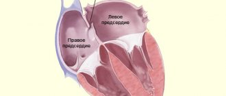
Ultrasound of the heart shows the condition of the pericardium, myocardium, blood vessels, mitral valve, and ventricular walls.
During the procedure, the cardiologist records the readings obtained. Decoding the data makes it possible to identify diseases, abnormalities, pathologies, and anomalies in the functioning of the heart. Based on the information received, the doctor makes a diagnosis and prescribes treatment. Often, in addition to echocardiography, Doppler ultrasound is prescribed. This procedure allows you to see the direction of movement, determine the speed and turbulence of blood flow in the chambers of the heart.
The doctor makes a conclusion in the diagnostic results report, which displays the data obtained from the ultrasound machine.
Echocardiography is used to diagnose:
— pre-infarction state; - heart failure; - ischemic disease; - myocardial infarction; - hypertension, hypotension; - heart defects; - rhythm disturbances; - cardiomyopathy; - myocarditis, pericarditis; — vegetative-vascular dystonia; - rheumatism.
In order to obtain data on the work of the heart during physical activity, stress echocardiography is performed. The patient is given a certain physical activity or the heart muscle is stimulated to work harder with the help of drugs, and readings are taken.
Quite rarely in practice there are cases when it is not possible to carry out a standard ultrasound procedure of the heart: deformation of the chest, the presence of prosthetic valves, a layer of subcutaneous fat, abundant hair growth. In this case, transesophageal (transesophageal) echocardiography is performed.
Types of echocardiography:
- One-dimensional in M-mode. The sensor produces waves along one selected axis. An image is displayed on the monitor - a top view. Makes it possible to see the aorta, atrium, and ventricles.
- Two-dimensional. Makes it possible to obtain an image in two planes and analyze the movement of structures.
Only a cardiologist can conduct a complete and accurate analysis of the cardiogram transcript and draw a conclusion.
Heart ultrasound standards:
Left ventricle
- myocardial mass (men: 135-182 g; women: 95-141 g)
— myocardial mass index (men: 71-94 g/m2; women: 71-80 g/m2)
- volume at rest (men: 65-193 ml; women: 59-136 ml)
- resting size (men: 5.7 cm; women: 4.6 cm)
- size during contraction (men: 4.3 cm; women: 3.1 cm)
— wall thickness outside of heart contractions during work: 1.1 cm. An indicator of 1.6 cm indicates hypertrophy
— ejection fraction of at least 55-60%. The indicator indicates the volume of blood that the heart ejects with each contraction. A lower value indicates heart failure
— stroke volume: 60-100 ml (the amount of blood ejected per contraction)
Right ventricle
— wall thickness 5 mm
— size index 0.75 — 1.25 cm/m2
- size at rest 0.75 - 1.1 cm
Valves
— A decrease in the diameter of the valve opening, difficulty pumping blood, indicates stenosis
— Heart failure is diagnosed if the valve flaps prevent the reverse flow of blood and do not perform their intended function
Pericardium
— Liquid norm is 10-30 ml. When the reading is over 500, normal heart function is difficult. The onset of an inflammatory process is possible - pericarditis, accumulation of fluid, formation of adhesions of the heart and pericardial sac.
Carrying out a cardiac ultrasound procedure helps to detect diseases of the cardiovascular system at an early stage and take the necessary measures in time.
Decoding EchoCG
After the examination, the doctor draws up a conclusion. First, a visual picture with the presumed diagnosis is described. The second part of the study protocol indicates the patient’s individual indicators and their compliance with standards.
Decoding the data obtained is not a final diagnosis, since the study can be done not by a cardiologist, but by an ultrasound diagnostic specialist.
It is the cardiologist, based on the collected medical history, examination results, interpretation of tests and data from all prescribed studies, who can draw accurate conclusions about your condition and prescribe the necessary treatment!
Carrying out echocardiography (ultrasound of the heart) at the “Family Doctor”
You can get an echocardiography (ultrasound of the heart) in Moscow at JSC “Family Doctor”. In the Family Doctor clinics, you can undergo an examination (including Doppler analysis) quickly and in comfortable conditions. Doppler echocardiography allows you to evaluate the speed and direction of blood movement in each of the chambers of the heart, which makes it possible to use the diagnostic capabilities of cardiac ultrasound to the fullest extent possible.
No special preparation is required for echocardiography (ultrasound of the heart). However, it is advisable that echocardiography be performed no earlier than three hours after the last meal, as a high position of the diaphragm may interfere with some aspects of the study. The procedure is performed in a lying position or on the left side. The average duration of the study is no more than half an hour.
Sign up for diagnostics Do not self-medicate. Contact our specialists who will correctly diagnose and prescribe treatment.
EchoCG norm: what some parameters indicate
There is a range of normal values for one or another cardiac ultrasound indicator for adults (in children the norms are different and directly depend on age).
Thus, along with other important parameters, echocardiography helps to obtain information about the ejection fraction of the heart - this indicator determines the efficiency of the work performed by the heart with each beat.
Ejection fraction (EF) is the percentage of blood volume ejected into the vessels from the heart ventricle during each contraction. If there were 100 ml of blood in the ventricle, and after the heart contracted, 55 ml entered the aorta, it is considered that the ejection fraction was 55%.
When the term ejection fraction is heard, we are usually talking about the left ventricular (LV) ejection fraction, since it is the left ventricle that ejects blood into the systemic circulation.
A healthy heart, even at rest, pumps more than half of the blood from the left ventricle into the vessels with each beat. As the ejection fraction decreases, heart failure develops.
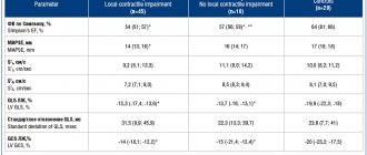
The normal left ventricular ejection fraction for an adult is 55-70%. A value of 40-55% indicates that EF is below normal. An ejection fraction of less than 40% and even lower ejection fractions indicate heart failure in the patient.
Is cardiac echocardiography safe?
During this study, there is no radiation or other load on the organ. Therefore, if necessary, it can be prescribed even several times a week.
This study is characterized by the absence of complications and side effects.
EchoCG does not harm either the expectant mother or the fetus during pregnancy.
Limitations for the procedure may include inflammatory diseases of the skin of the chest, chest deformities and some other reasons.
How to do an ultrasound of the heart at the MedicCity clinic?
It is advisable for every person to have an echocardiogram at least once in their life. The fairly low cost of cardiac ultrasound is another reason to prefer this particular diagnostic method.
At MedicCity you can undergo all types of cardiac diagnostics - ECG, EchoCG, bicycle ergometry, HOLTER, ABPM, etc.
Just type in a search engine: “Ultrasound of the heart, Savelovskaya metro station, Dynamo metro station.” Or call us at: +7 (495) 604-12-12.
The doctors of our multidisciplinary clinic will do everything possible to relieve your heart pain!
How is an ultrasound of the heart performed?
First of all, we should consider the classic diagnostic method through the surface of the skin in the chest area (transthoracic). The procedure can be carried out in a hospital or at home - the person undresses to the waist and lies down on a flat surface. After applying the gel to the patient’s chest, the specialist begins the examination, which lasts 15-20 minutes. To decipher an ultrasound of the heart in an adult and draw up a conclusion, it will take an additional 10-15 minutes.
A much less common and more complex method of cardiac ultrasound, which is performed only in a hospital setting, is transesophageal cardiography: the sensor is inserted into the oropharynx and lowered down the esophagus to the required depth. The main advantage of this method is high detail. It is possible to obtain more accurate data than with transthoracic ultrasound of the heart. Transesophageal echocardiography is indicated before surgery to repair heart valves, ruptured aortas, repair heart defects, treat bacterial infections of the heart or valves, or endocarditis.
