Congenital heart defects (CHD) are developmental anomalies that lead to disruption of the morphological structure of the heart, including the valve apparatus and great vessels.
CHDs occur during the period of intrauterine development (usually at 2-8 weeks) as a result of disruption of embryogenesis processes. These anomalies can occur either alone or in combination with each other.
The overall prevalence of this group of diseases is up to 5-8 cases per 1000 births. Congenital defects can be associated with chromosomal abnormalities, however, non-chromosomal congenital heart defects are often diagnosed.
The overall incidence of non-chromosomal congenital heart disease is up to 7 cases per 1000 births, of which up to 3.5% are perinatal losses, 20% are diagnosed prenatally, 5.6% of pregnancies are terminated due to a detected fetal anomaly. Complex non-chromosomal heart defects are less common, approximately 2 cases per 1000 births. The outcome in 8% of cases is perinatal death, 40% are diagnosed in utero, and 14% cause termination of pregnancy.
Causes of congenital heart disease
The main leading causes in the formation of defects are most often structural and quantitative chromosomal abnormalities and mutations, i.e.
primary genetic factors. It is also necessary to pay attention to potentially teratogenic environmental factors: various intrauterine infections (rubella viruses, cytomegalovirus, coxsackie, infectious diseases in the mother in the first trimester), medications (vitamin A, antiepileptic drugs, sulfasalazine, trimethoprim), constant contact with toxic substances ( paints, varnishes). In addition, it must be remembered that maternal factors have a negative impact on intrauterine development: reproductive problems preceding a given pregnancy, the presence of diabetes mellitus, phenylketonuria, alcoholism, smoking, age, but also factors from the father - age, drug use ( cocaine, marijuana).
The leading role belongs to the multifactorial theory of the development of congenital heart defects (up to 90%).
Types of congenital heart defects
- Atrial septal defect (ASD) or patent foramen ovale is diagnosed when one or more holes are identified in the interatrial septum. One of the most common congenital heart defects. Depending on the location of the defect, its size, and the strength of blood flow, more or less pronounced clinical signs are determined. ASD is often combined with other cardiac anomalies and is associated with Down syndrome.
- Ventricular septal defect (VSD) is diagnosed when the interventricular septum is underdeveloped at various levels with the formation of a pathological communication between the left and right ventricles. It can occur either alone or together with other developmental anomalies. With a small defect, there is often no pronounced lag in physical development. VSD is dangerous because it can lead to the development of pulmonary hypertension, and therefore must be promptly corrected surgically.
- Coartation of the aorta is a segmental narrowing of the aortic lumen with disruption of normal blood flow from the left ventricle to the systemic circulation. Up to 8% of all cases of congenital heart disease are detected, more often in boys, and are often combined with other anomalies.
- Patent ductus arteriosus is diagnosed when the duct of Batallus is not closed, which is detected in newborns and becomes overgrown in the future. As a result, a partial discharge of arterial blood from the aorta into the pulmonary artery occurs. With this congenital heart disease there are often no severe clinical manifestations, however, the pathology requires surgical correction, since it is associated with a high risk of sudden cardiac death.
- Pulmonary atresia - underdevelopment (full or partial) of the pulmonary valve leaflets with the development of backflow of blood from the pulmonary artery into the cavity of the right ventricle is diagnosed. Subsequently leads to insufficient blood supply to the lungs.
- Pulmonary valve stenosis is an anomaly in which narrowing of the pulmonary valve opening is diagnosed. As a result of pathology, most often of the valve leaflets, normal blood flow from the right ventricle to the pulmonary trunk is disrupted.
- Tetralogy of Fallot is a complex combined congenital heart disorder. Combines ventricular septal defect, pulmonary stenosis, right ventricular hypertrophy, and aortic dextraposition. With this pathology, a mixture of arterial and venous blood occurs.
- Transposition of the great vessels is also a complex congenital disorder. With this pathology, the aorta originates from the right ventricle and carries venous blood, and the pulmonary trunk originates from the left ventricle and carries arterial blood, respectively. Paroxysm is severe and is associated with high mortality in newborns.
- Dextrocardia is an anomaly of intrauterine development characterized by the right-sided placement of the heart. Often, there is a “mirror” arrangement of other unpaired internal organs.
- Ebstein's anomaly is a rare congenital heart defect, diagnosed when the location of the tricuspid valve leaflets changes. Normally - from the atrioventricular fibrous ring, with an anomaly - from the walls of the right ventricle. The right ventricle is smaller, and the right atrium is elongated, including abnormal valves.
Mitral valve prolapse
Among ultrasound methods for diagnosing primary mitral valve prolapse, preference is given to two-dimensional echocardiography, since one-dimensional echocardiography results in a large number of false-positive and false-negative results. With two-dimensional echocardiography, it is necessary to measure the area of the atrioventricular valve and the circumference of the atrioventricular ring, which are significantly larger in children with mitral valve prolapse than in healthy ones. Sometimes it is possible to identify a violation of the architectonics of the valves, elongation, atypical attachment and incorrect distribution of the chords. The end-diastolic diameter of the left ventricle in children with primary mitral valve prolapse is reduced: in one third it corresponds to the 5th and in half to the 25th percentile. The combination of a reduced left ventricular cavity with an increase in the area of the mitral valve contributes to the occurrence of valvular-ventricular disproportion, which is one of the pathogenetic aspects of leaflet prolapse.
Methods for diagnosing congenital heart disease
Diagnosis of congenital heart disease is based on collecting medical history data (presence of developmental defects, including congenital heart defects, genetic diseases in close relatives; information about pregnancy and the presence of etiological factors in parents).
When collecting complaints, attention is paid to the child’s developmental delay, poor weight gain, poor appetite, sluggish sucking from the breast or bottle, breast refusal, cyanosis, and frequent respiratory infections.
Physical examination
During a physical examination, pay attention to the color of the skin, determine pulse and blood pressure (on the right arm and either leg), perform auscultation of the heart and lungs, pay attention to the presence of peripheral edema, perform pulse oximetry, and determine diuresis.
Instrumental diagnostics
However, the leading role in the diagnosis and confirmation, differential diagnosis of congenital heart disease is played by instrumental examination methods: x-ray examination of the chest organs, electrocardiography, echocardiography, MRI, CT, catheterization of the cardiac cavities.
Sign up for diagnostics To accurately diagnose the disease, make an appointment with specialists from the Family Doctor network.
Treatment of minor cardiac anomalies
If, nevertheless, the child is bothered by any of the above syndromes, then care must be taken to ensure that the daily routine is followed. Limiting exercise is not necessary; on the contrary, you need to spend more time in the fresh air and play active games. At the same time, we must not forget about the benefits of proper nutrition, this will help you quickly restore your strength and support your immune system. Surgery is required in very rare cases. In order to remove the anomaly, the surgeon will insert an instrument through large vessels and correct the defect. The operation is not considered difficult, complications are usually not observed and the person quickly returns to their previous lifestyle. The doctor may suggest a course of magnesium supplements in order to improve the functioning and structure of heart tissue.
Microanomalies of the right atrium
The Chiari network is a fibrous structure extending from the crest border or tubercle inferiori to the eustachian or basal valve. The frequency among congenital anomalies is 4%.
Prolapse of the pectineal muscles in the cavity of the right atrium - in the form of thin thread-like and mobile structures in the area of the wall of the right atrium or appendage. Frequency among congenital anomalies - 9.7%
Eustachian valve
The Eustachian valve (valvula venae cavae inferioris) is located at the level of the anterior arch of the inferior vena cava and, usually after the neonatal period, does not exceed one centimeter in length or is completely rudimentary. The valve is a fold of the endocardium with an average width of 1 cm. In the embryo, the valve directs a stream of blood from the vein to the foramen ovale. After birth, in the absence of communication between the atria, this valve function loses its significance. According to sectional data, the Eustachian valve is found in 86% of children.
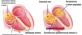
In population studies using echocardiography, an unusually long Eustachian valve (more than 1 cm) is detected in 0.20% of the population and is considered a stigma (Fig. 161). According to our data, this small anomaly predisposes to supraventricular arrhythmias, probably reflexively due to irritation of the pacemaker formations of the atrium.
Fig. 161
Enlarged Eustachian valve of the inferior vena cava.
EchoCG criteria.
Two-dimensional echocardiography:
Visualization of a valve-like structure in the right atrium at the junction of the inferior vena cava.
Microanomalies of the papillary muscles
This anomaly includes changes:
- forms
- quantities
- location
According to the autopsy:
- Two papillary muscles - 65.2%
- Three papillary muscles - 13%
- Four papillary muscles - 4%
- Five papillary muscles - 4.4%
- Six papillary muscles - 4.4%
Tags: abnormal trabecula, Abnormally located trabeculae, heart disease, true chord, false chords, macroanomalies of the heart, minor cardiac anomalies, MAS, Microanomalies of the papillary muscles, LLC, Patent foramen ovale, right atrium, mitral valve prolapse, pulmonary artery dilatation, heart, Chiari network, Chiari network, trabecula, Elongated Eustachian valve
Aneurysm of the interatrial septum
It occurs in connective tissue dysplasia significantly more often than in the general population; its combination with mitral valve prolapse is noted. The aneurysm is most often located in the fossa ovale area. Probably, the occurrence of aneurysmal protrusion of the septum may be associated with spontaneous closure of the defect in children under the age of 5–6 years. The absence of hemodynamic disturbances, in turn, gives the right to classify a small aneurysm of the interatrial septum as a minor anomaly of cardiac development. Clinically, an aneurysm can be suspected by the presence of clicks in the heart, similar to those with mitral valve prolapse.
EchoCG criteria
Two-dimensional echocardiography
- Aneurysmal protrusion towards the right atrium in the area of the oval window, intensifying in systole (Fig. 159 - 160).
- Absence of volume overload of the right departments.
Fig. 159
Aneurysmal protrusion of the interatrial septum towards the right atrium in the area of the oval window.
Fig. 160
Aneurysmal protrusion of the interatrial septum towards the right atrium in the area of the oval window.
Idiopathic dilatation of the pulmonary artery
It is characterized by an expansion of the trunk in the absence of heart disease and pulmonary pathology. Dilatation of the pulmonary artery trunk occurs against the background of hereditarily determined connective tissue pathology, which is confirmed by the simultaneous detection of other markers of connective tissue dysplasia of the heart and the frequent detection of dilation in hereditarily determined syndromes, for example, Marfan syndrome. Auscultation can detect a systolic murmur of moderate intensity at the base of the heart, which decreases with an upright position. With idiopathic dilatation of the pulmonary artery, dynamic observation and examination is necessary.