How does the left ventricle work?
The left ventricle is a cavity formation.
Its volume depends on the person’s age and type of activity, since the myocardial muscles are amenable to “training”. The thickness of the LV wall is 10-14 mm and consists of three layers in which the fibers have different directions (circular, straight and oblique). The inside of the chamber is lined with endocardium, a connective tissue membrane that forms valves near the atrioventricular openings. The shape of the left ventricle is oblong-oval, cone-shaped. There are two divisions of the structure:
- apex - a narrow part, which in a healthy person is located in the 4-5 intercostal space on the left;
- the outer edge is a rounded contour that forms the lateral (pulmonary) surface - the most massive part of the heart muscle.
Inside, the LV consists of two parts: the posterior and anterior sections, through which blood gradually moves from the chamber cavity to the aortic cone.
In the area of the atrioventricular opening there is a valve consisting of two leaflets made of endocardial tissue. The main task of this structure is to regulate blood flow and prevent regurgitation (backflow). Attached to the ventricular myocardium by papillary (papillary) muscles, the tone of which determines the amplitude of movement of the valves.
The aortic valve is represented by three semilunar valves, which are attached around the circumference of the opening between the conus arteriosus and the aorta. The coordinated operation of the valves ensures the movement of fluid into the systemic circulation during contraction of the LV myocardium and prevents regurgitation when the chamber is filled during diastole.
The excitation wave that causes the ventricle to contract passes from the atrioventricular junction along the left bundle branch and its two branches. Cardiomyocytes located at the apex of the heart receive impulses through Purkinje fibers.
Volume of the ventricular system
Normal values of the volume of the ventricular system (in Table 3) [8].
| Ventricular system (cm3) | Meaning | RMS deviation |
| Intracranial volume | 1444.6 | 108.1 |
| Right lateral ventricle | 5608.3 | 1310.8 |
| Left lateral ventricle | 6047.2 | 1214.5 |
| Both lateral ventricles | 11655.4 | 2368.1 |
| Third ventricle | 910.5 | 151.3 |
| Fourth ventricle | 1793.6 | 356.9 |
Table 3
Main functions of the department
The left ventricle of the heart is responsible for the following functions:
- systolic - synchronous contraction of cardiomyocyte fibers ensures adequate release into the systemic circulation to supply all organs and tissues with oxygen;
- diastolic - relaxation of the walls of the ventricle, opening of the atrioventricular valve and filling the cavity with blood from the atrium.
The normal pumping function involves the coordinated work of all departments. The cardiac cycle consists of alternating states: systole and diastole, which are characterized by contraction and stretching of the muscle fibers of cardiomyocytes.
Periods of the systolic phase:
- tension with the stage of asynchronous and isometric contraction;
- expulsion (two periods - fast and slow).
The asynchronous period is characterized by an uneven distribution of the excitation impulse, so individual fibers do not contract simultaneously. This phase is necessary for complete closure of the mitral valve leaflets, after which intraventricular pressure increases (due to the ongoing contraction of cardiomyocytes). Having reached a level of 70-80 mm Hg. Art., the semilunar valves open and blood enters the aorta (at first quickly, and then more slowly). The closure of the valve is caused by high pressure inside the vessel.
Relaxation of the myocardium of the LV walls opens the mitral valve, which starts the flow of a new portion of blood. To ensure adequate ejection, it is necessary that all parts of the ventricle, leaflets, papillary muscles, trabeculae and chords are in good functional condition.
Diagnosis of diseases: what methods will determine the problem?
There are two types of left ventricular dysfunction (dysfunction):
- Systolic, which is characterized by a decrease in the ejective capacity of the LV. Most often, pathology develops when contractile cardiomyocytes are damaged during myocardial infarction, inflammatory or autoimmune disorders.
- Diastolic. It is characterized by the inability of muscle fibers to completely relax and create the required volume of the chamber (the condition is most often diagnosed with pathologies of the pericardium or neighboring structures).
If you have complaints of shortness of breath during exercise or at rest, swelling in the legs, cough, pain in the heart area, you should contact a cardiologist and undergo a comprehensive examination to find out the cause.
ECG
Using electrocardiography (ECG), it is possible to identify not only the correct rhythm and heart rate, but also disturbances in the functioning and structure of the left ventricle. Signs of ECG changes in various LV pathologies:
- Hypertrophic changes in the walls of the left ventricle, regardless of the cause:
- displacement of the electrical axis of the heart to the left (angle α ranges from -90 to 0°);
- intraventricular conduction disorders (complete or incomplete blockade of the left bundle branch);
- repolarization disorders – deformation of the T wave in I, aVL, V5, V6.
- Scarring in the area of the left ventricle:
- altered Q wave in leads I, II, aVL, as well as in V1-6;
- location of ST on an isoline;
- low and flattened T wave.
Ultrasound
Ultrasound or echocardiography (echocardiography) helps determine the size of the heart chambers and the thickness of their walls, assess the functional state of the valves, and the efficiency of myocardial contraction. Each department has its own standard indicators.
The main ultrasound parameters of the LV are presented in the table.
| Criterion | Standards | Changes |
| Muscle mass |
| Enlargement – with hypertrophy (symmetric and asymmetric growth are distinguished) |
| Myocardial mass index (LVMI) |
| |
| Interventricular septal thickness (IVS) |
|
|
| End-diastolic volume (EDV) |
| |
| Final size |
| |
| Ejection fraction (EF) | 55-60% | A decrease in the indicator is characteristic of left ventricular systolic dysfunction |
| Stroke volume (SV) | 60-100 ml |
|
Scintigraphy
Perfusion scintigraphy is a method for assessing the blood supply to the muscular layer of the left ventricle using radiopharmaceuticals that are administered intravenously and accumulate in the myocardium. The advantage of the study is the accurate detection of ischemia even in small vessels, which is not always possible to detect with angiography. Scintigraphy can be static (performed at rest) and dynamic (during dosed physical activity on a bicycle ergometer).
Indications:
- myocardial ischemia of unknown origin;
- assessment of the effectiveness of operations to restore coronary blood flow (angioplasty, bypass surgery, etc.);
- studies of myocardial blood flow in non-coronary diseases (mainly inflammatory in nature);
- with a questionable ECG (to confirm the diagnosis of acute myocardial infarction).
MRI and CT
Recently, X-ray methods for studying the heart have become in demand, which make it possible to obtain three-dimensional images of the organ and identify changes in the structure.
Computed tomography (CT) takes a series of X-rays of the heart from different angles in separate sections of the organ. Modern devices are capable of providing three-dimensional visualization of the internal structure.
Magnetic resonance imaging (MRI) has a similar principle. However, the study does not use ionizing radiation, but waves of a magnetic nature. Tissues “respond” to this influence in different ways, which is what the device records.
Both methods can provide the following information:
- LV wall thickness;
- changes due to myocardial infarction;
- condition of blood vessels, presence of atherosclerotic plaques;
- hemodynamic adequacy;
- condition of the pulmonary arteries.
Bicaudal index
The bicaudal index (BCI) is a commonly used linear measure of the lateral ventricles (Fig. 3). To account for natural changes in ventricular size with aging, (BCI) is then divided by the upper limits of “normal” age to calculate the relative bicaudal index [6–7].
BKI = A/B, where A = width of the frontal horns at the level of the caudate nuclei; B = brain diameter at one level [6-7].
| Age | up to 46 years old | 46-55 years old | 56-65 years old | Over 66 years |
| Meaning | 0.11 | 0.13 | 0.15 | 0.15 |
| Range | 0.10-0.12 | 0.11-0.14 | 0.13-0.16 | 0.14-0.17 |
Table 2 Normal BCI values stratified by age group.
The diagnosis of hydrocephalus is established when the BCI is greater than 1. Normative values determined in subjects without neurological disease in the mid-to-late 1970s [6-7]. Divide the width of the frontal horns of the lateral ventricles, at the level of the caudate nuclei, by the corresponding diameter of the brain. Take the measurement on a section that includes the foramen Monro [6-7].
Fig. 3 Measurement of the bicaudal index (left) and asymmetry of the lateral ventricles in the absence of an organic cause (right).
What pathologies most often affect the function and structure of the LV?
There are many pathological conditions and provoking factors that lead to changes in the anatomical structure and physiological dysfunction of the left ventricle. In some cases, LV hypertrophy is a normal variant, for example, in athletes or pregnant women.
Pathologies that are accompanied by dysfunction of the left ventricle:
- congenital defects: coarctation of the aorta, anomaly of the interventricular septum, combined disorders - tetralogy or pentade of Fallot;
- inflammatory diseases: endo-, myo-, pericarditis (children and adolescents are more often affected);
- acquired heart defects (more often diagnosed in adults as a consequence of rheumatism);
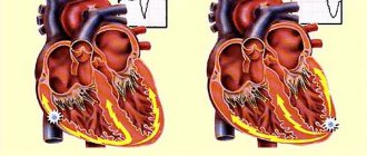
- IHD: angina pectoris, post-infarction cardiosclerosis, necrosis of myocardial walls;
- chronic lung pathology;
- arterial hypertension;
- atherosclerosis of the aorta;
- consequences of metabolic syndrome in combination with obesity and diabetes;
- pathologies of the kidneys, liver, thyroid gland.
Without adequate treatment and hemodynamic correction, the patient’s condition worsens: heart failure progresses, the brain and all internal organs experience oxygen starvation.
At the first manifestations of left ventricular dysfunction, it is recommended to visit a general practitioner (cardiologist) and undergo examinations to prevent further development of the pathology. Patient management depends on the underlying disease and involves the use of pharmacological drugs or cardiac surgery.
Additional therapy includes lifestyle modification and compensation for associated disorders.
conclusions
Of all the parts of the heart, the left ventricle plays the greatest role in ensuring adequate blood circulation. The large chamber volume, thick muscular wall and opening into the aorta are key characteristics that determine the importance of the structure. Damage to the left ventricle by ischemic, traumatic, inflammatory processes is accompanied by severe hemodynamic defects with symptoms of heart failure. Shortness of breath, swelling, chest pain, a feeling of interruptions in the functioning of the organ are signs of serious disorders that require comprehensive diagnosis and adequate therapy.
Source
- Methodological manual for students of medical institutes. Compiled by: Gubsky L.; Borkina P.; Abovich Y.; Evstifeeva N.; Timakov V. link
- Orlov Yu.A. Hydrocephalus. – Kyiv, 1995. – 75 p.
- Cerebral lateral ventricular asymmetry on CT: how much asymmetry is representing pathology? Kiroğlu Y1, Karabulut N, Oncel C, Yagci B, Sabir N, Ozdemir B.Surg Radiol Anat. 2008 May;30(3):249-55. doi:10.1007/s00276-008-0314-9. Epub 2008 Feb 6. link
- Grosman H, Stein M, Perrin RC, Gray R, St Louis EL. Computed tomography and lateral ventricular asymmetry: clinical and brain structural correlates. (1990) Canadian Association of Radiologists journal = Journal l'Association canadienne des radiologists. 41 (6): 342-6. Pubmed
- Shapiro R, Galloway SJ, Shapiro MD. Minimal asymmetry of the brain: a normal variant. (1986) A.J.R. American journal of roentgenology. 147(4):753-6. doi:10.2214/ajr.147.4.753 - Pubmed
- Gijn, Jan van et al. “Acute Hydrocephalus After Aneurysmal Subarachnoid Hemorrhage.” Journal of Neurosurgery 63.3 (1985): 355-362. link
- Dupont, Stefan and Alejandro A Rabinstein. “CT Evaluation Of Lateral Ventricular Dilation After Subarachnoid Hemorrhage: Baseline Bicaudate Index Balues.” Neurological Research 35.2 (2013): 103-106. link
- Morphometric Changes in Lateral Ventricles of Patients with Recent-Onset Type 2 Diabetes Mellitus. Junghyun H. Lee, Sujung Yoon, Perry F. Renshaw, Tae-Suk Kim, Jiyoung J. Jung, Yera Choi, Binna N. Kim, Alan M. Jacobson, In Kyoon Lyoo. Published: April 4, 2013 “PLOS one”.link
- "Third and Fourth Cerebral Ventricular Sizes among Normal Adults in Zaria-Nigeria." Ahmed Umdagas Hamidu, Solomon Ekott David, Sefiya Adebanke Olarinoye-Akorede, Barnabas Danborno, Abdullahi Jimoh, Olaniyan Fatai. Year:2015, Volume:2, Issue:2, Page:89-92 link