The four-chambered human heart allows blood to pass in one direction, this is facilitated by the valve apparatus, consisting of bicuspid, tricuspid and mitral valves.
The mitral valve consists of soft valves, the movement of which is regulated by papillary muscles connected by chordae.
When the anatomical structure of the valve apparatus changes (insufficiency or stenosis), the hemodynamics of the human body are disrupted due to prolapse, which allows the reverse flow of blood.
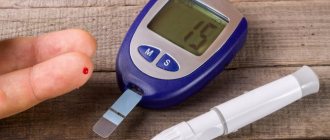
Prolapse without regurgitation has a name: prolapse of the anterior leaflet of the mitral valve, grade 1, hemodynamically insignificant. This is a fairly common phenomenon, mainly detected on a cardiogram during routine examinations.
What is hemodynamically insignificant MVP? How dangerous is this condition? The initial stage of MVP develops with pathological changes in the connective tissue, causing the valves to bend into the atrial cavity during the contraction stage.
The degree of valvular insufficiency depends on the volume of the thrown blood flow; the degree of prolapse is determined by the size of the valve cusp. The first degree has a deviation of no more than six millimeters.
The cause of the defect, if it is a congenital defect, is considered to be an abnormal structure of the connective tissue that changes the valve leaflets and chords.
Minor prolapse is often detected during routine medical examinations, as it does not produce pronounced symptoms, and parents do not consult a doctor about this.
Main causes of violation:
- rheumatic valve disease caused by streptococcus, which also affects the joints.
- ischemia;
- mechanical heart injuries.
Basic treatment methods
In the absence of symptoms, treatment is not necessary, but the patient needs medical supervision and an annual hardware examination. The cardiologist advises strengthening the heart muscle by running, race walking, and swimming.
Admission to competitive sports is issued by a doctor. Sports involving weight lifting are prohibited.
If the disease manifests itself with symptoms, the cardiologist selects drugs to relieve them.
Main drugs:
- sedatives;
- beta blockers;
- drugs to improve myocardial nutrition: riboxin; panangin; Magnerot.
If there is a threat of thrombosis, a serious complication, anticoagulants are prescribed.
For minor prolapse, herbal medicine is indicated. But before using herbs, you should consult a doctor.
Discussion
In world practice, hybrid operations on the carotid arteries are described, the essence of which is the reconstruction of the ICA and the use of carotid access for stenting the proximal part of the common carotid artery or brachycephalic trunk [6]. Combined treatment of tandem stenosis of the internal carotid artery was first described by T. Terada et al. [7] in 1998, who reported 3 cases of simultaneous carotid endarterectomy and angioplasty of the intracranial ICA for multilevel lesions in asymptomatic patients with good postoperative and mid-term results within 13 months. They also developed a temporary Y-shunt with a side outlet to perform balloon angioplasty of the distal ICA. In 1999, D. Widenka et al. [8] reported a successful case of hybrid treatment of a patient with retinal transient ischemic attacks and a good long-term period. S. Gock et al. [9] in 2001 described 8 clinical cases of hybrid treatment of asymptomatic tandem stenosis. Only 1 of 8 patients experienced a complication of ipsilateral stroke within 24 hours after surgery. O. Hartung et al. [10] in 2004 reported one patient aged 71 years with 60 and 90% left bifurcation stenosis and ipsilateral petrosal stenosis who complained of persistent tinnitus and dizziness. After carotid endarterectomy and stenting of the petrous part of the ICA, the symptoms regressed. The patient remained asymptomatic for 19 months. Similar results were described by K. Uda et al. [11] in 2003
In our case, the patient had a bilateral hemodynamically significant lesion, which manifested itself in the form of frequently recurrent transient ischemic attacks in the territory of the left middle cerebral artery. The procedure was performed on the natural blood flow without clamping the carotid arteries through the inflow of an autovenous patch of the “proboscis” type (see Fig. 3). No additional puncture was required.
Currently, a technique called “intravascular near-infrared spectroscopy” is being studied in detail, which makes it possible to quantify the amount of lipids in the vascular wall, i.e. identify unstable atherosclerotic plaques rich in lipids.
The technique is automated: an intravascular sensor reads information, the device automatically calculates a color index characterizing the amount of lipids in each consecutive segment of a coronary artery 4 mm long (maxLCBI4mm). The normal wall of the coronary artery is colored red, lipids are colored yellow. In the device used today, the sensor is combined with an intravascular ultrasound probe. It is technically possible to combine it with a sensor for optical coherence tomography.
In September 2021 The results of the Lipid-Rich Plaque (LRP) study were reported. The study included 1563 patients with suspected coronary artery disease who underwent diagnostic coronary angiography and, if necessary, PCI. In half of the cases, coronary angiography was performed in connection with ACS. On average, about 2 of the coronary arteries in each patient were also analyzed using IVUS and intravascular spectroscopy.
The incidence of major adverse events (cardiovascular death, cardiac arrest, ACS, readmissions, repeat myocardial revascularization procedures) not related to stenoses undergoing PCI at 24 months was significantly associated with the maxLCBI4mm value and increased by 18% for every 100 conventional units of the value of this index. The incidence of adverse events in patients with a maxLCBI4mm index of more than 400 was 12.6%, less than 400 - 6.3%.
According to the authors, the data obtained make it possible to plan prospective randomized studies designed to evaluate the feasibility of any additional measures aimed at reducing the risk of ischemic complications. This can be either aggressive medical techniques (high doses of lipid-lowering drugs, anti-inflammatory therapy), or, possibly, a modification of the approach to invasive treatment of coronary artery disease.
Based on materials:
Waksman R. Assessment of coronary near-infrared spectroscopy imaging to detect vulnerable plaques and vulnerable patients. Presented at: TCT 2021. September 24, 2021. San Diego, CA.
NIRS-IVUS Imaging Spots Vulnerable Plaques, At-Risk Patients - Medscape - Oct 05, 2021.
Text: Ph.D. Shakhmatova O.O.
Hemodynamically significant tortuosity of the common carotid arteries in adult patients
Bakhmetyev A.S., Dvoenko O.G., Makarova Ya.I., Sukhoruchkin V.A. State Budgetary Educational Institution of Higher Professional Education “Saratov State Medical University named after. IN AND. Razumovsky” of the Ministry of Health of Russia, Saratov
Purpose of the study
To identify the incidence of pathological tortuosity (PI) of the common carotid arteries (CCA) in adult patients who complained of headaches.
Material and methods
The study included 2150 patients (4300 CSA in 1455 women and 700 men, average age 50.7 ± 12.3 years) who underwent routine triplex scanning of the brachiocephalic arteries (TS BCA) at the Department of Ultrasound and Functional Diagnostics of the Clinical Hospital named after. S.R. Mirotvortsev SSMU in the period from June 2012 to July 2016. The study was conducted on an expert-class device Philips HD11 XE.
Hemodynamically significant PI of the CCA was considered in the presence of turbulent blood flow in it, an increase in peak systolic velocity (PSV) of more than 150 cm/s at the inflection point of the CCA, and also with a PSV gradient of more than 2.5 (the ratio of PSVmax to PSV in the proximal part of the CCA).
results
PI OSA was detected in 850 (39.5%) patients (1120 OSA in 600 women and 250 men, mean age 57.4 ± 8.7 years). 135 (6.3%) of those who applied had PI OSA on both sides. The structure was dominated (720 patients – 33.5%; 930 arteries) by hemodynamically insignificant C-shaped PI CCA. S-shaped PI CCA at an obtuse or right angle – in 80 (3.7%) cases (125 arteries).
In 2 (0.1%) patients there was a hemodynamically significant PI at a right angle (PSC gradient 2.65). Kinky tortuosity of the CCA - in 48 (2.2%; 63 arteries) patients (in all cases - hemodynamically significant; average PSC gradient - 2.69 ± 0.22; average PSC in the inflection - 154.35 ± 31.5 cm/s ). In 2 (0.1%) patients there was a loop-shaped hemodynamically significant tortuosity of the CCA (2 arteries; average PSC - 2.73; average linear systolic velocity at the inflection - 179.4 cm/s).
Note that out of 52 (2.4%; 67 arteries) patients with hemodynamically significant PI CCA, 48 (2.2%) suffered from uncontrolled arterial hypertension for at least 5 years. There was no correlation between hemodynamically significant PI CCA and gender and age.
conclusions
Hemodynamically significant PI OSA in the population of adult patients occurs only in 2.4% of cases. The main share of significant PIs is due to kinking and loop-like tortuosity of the CCA. TS BCA is the leading non-invasive method for identifying hemodynamically significant PI CCA.
- Views: 1938
- Comments:
Did you like the post? Do you find it useful or interesting? Support the author!
You must log in to the site
To perform this action, you must log in to the site or register. Interesting 5
S and C shaped tortuosity of the internal carotid artery
There are several types of pathology.
More common is the s-shaped tortuosity of the ICA with a smooth transition from one dislocation site to another. Usually with this type there is a deviation at two points. Most often, s-tortuosity of both ICAs occurs with deformities of the cervical spine. Straightening the cervical lordosis and static overstrain of the neck muscles entails a change in the position of the bed of this cerebral vessel. S-shaped tortuosity of the internal carotid artery in most cases does not give pronounced clinical manifestations. It is discovered during a study for another disease. If left untreated for 2-3 years, the disease will progress. In this case, the clinical picture worsens and emergency treatment of vascular pathology is required.
C-shaped tortuosity of the internal carotid artery develops in people suffering from high blood pressure. This type is typical for elderly patients suffering from atherosclerosis of cerebral blood vessels.
Children and adolescents are characterized by this type of pathology called kinking. This is a bend of the cerebral blood vessel at an angle of 45 degrees. It is a congenital vascular pathology. Gives attacks of acute cerebrovascular accident during excessive physical exertion. Such children and adolescents often complain of attacks of dizziness and fainting during physical education lessons. Parents should pay attention to the appearance of such complaints and conduct an examination.
Pathological and hemodynamic tortuosity
Congenital pathological forms of vascular structure are characterized by:
- detection of signs of cerebral circulation disorders at a young age and in children;
- the lesion is bilateral;
- at the same time there are connective tissue diseases (Marfan syndrome, Ehlers-Danlos syndrome), vascular underdevelopment, coarctation of the aorta;
- bending shape - loop (coiling), C- and S-shaped.
Coarctation of the aorta as a result of vascular tortuosity.
Elastic and muscle fibers are enlarged in the walls of the arteries, and there is swelling. As they grow, some of the vessels straighten. Due to the good ability of collateral circulation, strokes and transient attacks occur rarely. In the clinic, convulsive syndrome predominates. The prognosis is usually favorable.
Hemodynamic forms of tortuosity occur against the background of significant circulatory impairment, and are more common in older people. Atherosclerosis and hypertension in such patients have a long-term and continuous effect on the walls of blood vessels , causing irreversible changes - hardening, sclerosis, and degenerative processes.
Therefore, the formed deformities cannot disappear on their own, and weakening of cerebral hemodynamics is manifested by dyscirculatory encephalopathy, acute cerebral ischemia in the form of a brief attack or stroke.
Moderate loop-like tortuosity of the ICA
The loop-like tortuosity of the ICA is called coiling. This pathology has a very significant impact on hemodynamics. The loop can close at a certain moment, this provokes a sharp cessation of blood supply to a large part of the brain.
- Blockage and stenosis of the carotid artery
Even a moderate form of coiling-type tortuosity of the ICA requires constant monitoring by a doctor. When the first signs of insufficiency of cerebral blood supply appear, it is imperative to begin treatment.
Pay attention to the following clinical symptoms of this vascular pathology:
- regularly occurring attacks of dizziness with nausea;
- tension headache (occurs at the end of a working day);
- orthostatic dizziness;
- disruption of the vestibular apparatus, which is expressed in discoordination of movements of the arms and legs;
- nystagmus of the pupils of the eyes without other signs of brain injury;
- whistling, rustling, pulsation and other types of extraneous sounds in the ears;
- fainting and confusion during severe physical exertion;
- constant fatigue, drowsiness and decreased mental performance.
To diagnose this type of vascular pathology, you can use Doppler ultrasound and ecoscanning, spectral analysis of the structure of the carotid artery, computer and magnetic resonance tomography, angiography using an X-ray machine with preliminary administration of a contrast agent.
Coronary angiography does not always reveal stenosis of the coronary vessels of the heart
Coronary angiography does not always reveal stenosis of the coronary vessels of the heart
Stenosis of the coronary vessels of the heart is a fairly common disease - it occurs in 30-40% of cases when visiting a cardiologist. The consequences of stenosis are heart failure, the development of coronary heart disease, myocardial infarction and thrombosis. In advanced cases, the disease increases the likelihood of developing a dissecting aneurysm, which can lead to internal bleeding. In the absence of adequate drug therapy, death occurs.
The “gold standard” for detecting coronary artery stenosis is still coronary angiography (CAG), but this method also has its limitations.
As an example, we can cite the medical history of a 58-year-old patient with a history of hypertension, who went to the Medservice clinic. Prior to this, she was hospitalized in the hospital with pain in the chest.
Based on the results of the electrocardiogram, clinical and laboratory data, a diagnosis of myocardial infarction (without q wave) was made. Coronary angiography was performed, where no hemodynamically significant stenoses were detected.
A month after hospitalization, during dynamic observation at the Medservice clinic, the patient underwent echocardiography (EchoCG - ultrasound examination of the heart) to assess the contractility of the myocardium and coronary arteries. In the absence of obvious visual contractility disorders, when assessing myocardial deformation using the left ventricular function analysis (LVAF) method, a zone of segmental disorders in the anterior septal wall of the heart was revealed, as well as a slight acceleration of blood flow in the middle segment of the anterior interventricular branch (LAD). The patient was undergoing treatment.
Repeated coronary angiography revealed no hemodynamically significant narrowings. But intravascular ultrasound revealed extensive hemodynamically significant stenosis of the LAD (75%, length 25 mm). The patient underwent stenting of the affected area.
Currently, the patient is still regularly monitored at the Medsrvis clinic. Angina pectoris is not about LVAF; there is an area of impaired contractility, but its area has decreased. Ultrasound examinations of the coronary arteries showed that blood flow in the anterior interventricular branch was normal.
Thus, the use of all available methods in the diagnosis and treatment of patients with coronary artery disease usually gives a complete picture of the patient and helps in correct treatment.