directions
An electrocardiogram is a common non-invasive test to obtain data on the condition of the heart. The reason is general accessibility, simplicity, low cost of use and high information content. An ECG of the heart is a mandatory part of the examination at an appointment with a cardiologist, and is also used by therapists.
An ECG records changes in the heart and determines whether each of its major parts is functioning correctly. The study reveals arrhythmias, heart blocks, pathologies and disruptions in the functions of the heart muscle.
Prices for services
- Interpretation, description and interpretation of data from electrocardiographic studies (ECG) 220a
- Carrying out electrocardiographic studies (ECG) 880a
- Carrying out electrocardiographic studies (ECG) with a load of 1455a
- Appointment (examination, consultation) with a cardiologist + ECG 2070a
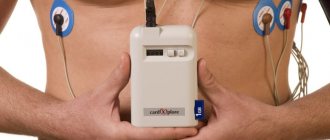
- Holter heart rate monitoring (Holter ECG) 2900a
The information and prices presented on the website are for reference only and do not constitute a public offer.
Heart Echo - evaluates the anatomy, functions and changes in the structures of the heart!
Another test that cardiologists order when they need more information is an echocardiogram, more commonly known as a cardiac echo, simply an ultrasound of the heart. The specialist uses an ultrasound scanner to examine the heart, most often using the transthoracic method. It is non-invasive and does not require special preparation. Usually lasts from 15 to 30 minutes. During the examination, the doctor can evaluate the anatomical and functional aspects of the heart, i.e. confirm or rule out the presence of birth defects, leaks, and structural defects of the heart. He can also evaluate the condition and function of the heart valves, that is, recognize valve defects. Cardiac echo helps evaluate contractility and function of the heart muscle, which are associated with myocardial infarction, coronary artery disease, and ischemia.
IMPORTANT! An echocardiogram is a repeatable test, but its diagnostic value depends on the experience and skill of the treating physician. In case of diagnostic doubt, the diagnosis should be expanded to include, for example, magnetic resonance imaging (MRI) or computed tomography (CT). Transthoracic echocardiography allows you to determine the state of the disease, the degree of its development and the effectiveness of the treatment. An indication for its implementation is also a suspicion of pulmonary arterial hypertension. After this, if in doubt, transesophageal echocardiography can be prescribed.
Our clinics in St. Petersburg
Structural subdivision of Polikarpov Alley Polikarpov 6k2 Primorsky district
- Pionerskaya
- Specific
- Commandant's
Structural subdivision of Zhukov Marshal Zhukov Ave. 28k2 Kirovsky district
- Avtovo
- Avenue of Veterans
- Leninsky Prospekt
Structural subdivision Devyatkino Okhtinskaya alley 18 Vsevolozhsk district
- Devyatkino
- Civil Prospect
- Academic
For detailed information and to make an appointment, you can call +7 (812) 640-55-25
Make an appointment
Method of measurement
This method is based on tracking the electrical potentials that occur with each heartbeat. Electrodes installed on the patient's body receive signals, conduct it to an electrocardiograph, which records the fluctuations and displays them on a display or paper.
The Nobel Prize was awarded in 1924 for the design of the first device for measuring the ECG of the heart, the details of decoding the electrocardiogram and the description of disturbances in the functioning of the heart. The basis of Willem Einthoven’s developments is also used in modern medicine.
Study frequency
When conducting an ECG, sensors that are attached to the chest do not have any harmful effects on the body. The sole task of these sensors is to capture and transmit cardiac impulses. Thus, an ECG does not cause any harm to the human body, even if the study is performed daily.
Doctors believe that the following frequency of ECG is justified:
- healthy population under 40 years of age can conduct the study once a year;
- people with chronic diseases and patients suffering from cardiovascular diseases - the ECG schedule is determined by the attending physician, as a rule, once a quarter;
- workers associated with risk, for example, the Ministry of Emergency Situations, etc. doctors advise having an ECG every six months;
- people who have crossed the forty-year mark should undergo a heart examination every 3-6 months, the frequency depends on the state of health and will be determined only by a doctor.
Technique for taking an ECG of the heart
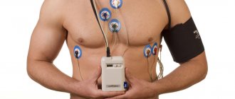
To take an ECG, the patient is positioned in a horizontal or any other position convenient for him. His breathing should be calm, the electrode locations (wrists, ankles, chest) should be exposed. The skin of the subject is degreased at the places where the electrodes are applied; in some cases, electrode paste is used for better contact with the skin. From each sensor there is a marked wire that transmits information to the electrocardiograph.
According to the standard scheme, electrodes are placed on the intercostal spaces at the right and left edges of the sternum; along the left parasternal line; in the intercostal spaces along the left midclavicular line; along the left anterior and middle axillary lines. The doctor is required to correctly determine the location of the sensors and securely attach them to the patient’s body in order to avoid signal distortion.
The doctor sets the same amplification of the electrical signal, the speed mode for ECG recording, and other parameters in accordance with the patient’s age and weight.
The whole procedure takes 5-10 minutes . The short duration of the conduction is both an advantage and a disadvantage of the ECG. A 20-second recording is not always enough if a person suffers, for example, from arrhythmia. When data on the work of the heart is needed not at rest, but during the patient’s usual activities, they resort to Holter ECG monitoring.
To assess changes in the myocardium characteristic of coronary heart disease, an ECG test is performed with dosed physical activity (bicycle ergometer, treadmill). According to the doctor’s recommendations, tests are carried out with medications and hyperventilation.
How does electrical wiring work in the heart?
So, an electrocardiogram, according to its name, records electrical processes occurring in the heart. Let's figure out what and how is happening there. In the depths of the heart muscle there are special groups of cells that make up the so-called conduction system of the heart. For simplicity, you can imagine it in the form of electrical wiring embedded in the wall, although in reality everything is a little more complicated.
The “power source” of a healthy heart is the sinus node , which is located in the right atrium. For those who are familiar with electricians, it can be compared to a capacitor. The sinus node accumulates a charge and then emits electrical impulses at a certain frequency that cause the heart to contract. Consequently, if “the battery is good,” then the first line of the cardiogram conclusion will read: sinus rhythm .
Sinus rhythm is the normal physiological rhythm of the heart.
The heart has four chambers - two atria and two ventricles. The atria contract first, then the ventricles. In order for this to happen in exactly this sequence, it is necessary that the electrical impulse first excites the atria and then switches to the ventricles. This switching occurs in the so-called atrioventricular node. More often it is called in Latin the atrioventricular node (atrium - atrium, ventriculum - ventricle), and even more often - simply AV node .
Two “wires” emerge from the AV node, which, after the author’s surname, are called bundle branches .
Through the right bundle branch, the electrical signal is mainly conducted to the right ventricle, and through the left bundle branch, of course, to the left ventricle. Since the left ventricle is the largest chamber of the heart, and it needs a lot of electrical supply, the left leg is also divided into anterior and posterior branches. This is how the complex conduction system of the heart turns out. If an accident occurs in a particular area of the power supply, we will call this a “conduction block” or a disturbance in the conduction of the heart.
Registration and interpretation of ECG
The doctor diagnoses organic and functional changes in the heart by looking at ECG data transferred to paper. The cardiologist analyzes the elements of the cardiogram: leads, waves, segments, intervals and their deviations from the norm.
The main indicator of the ECG is the heart rhythm. The heart rate of a healthy person is 60-80 beats per minute. Deviations from the norm to a lesser extent are diagnosed as bradycardia (this indicator also occurs in athletes); to a greater extent – tachycardia (occurs during physical activity, emotional experiences).
ECG is a universal test that helps diagnose diseases such as chronic heart failure, myocarditis, myocardiosclerosis, myocardial dystrophy, myocardial infarction, and the presence of post-infarction scars. It is worth highlighting the most common problem today - left ventricular hypertrophy; Using an ECG, the problem can be tracked and treatment can be started on time, whereas if not diagnosed on time, the disease often leads to the sudden death of the patient.
Rules for preparing for an ECG
Many people have no doubt that an ECG requires absolutely no preparation. Sometimes in such patients the result of the cardiogram may be false. There may even be elements indicating the development of heart pathology. To eliminate the risk of receiving false results, cardiologists recommend not ignoring the rules for preparing for an ECG.
You need to start preparing the day before the test.
- Maintaining complete calm - on the eve of the study, it is necessary, if possible, to completely remove yourself from stressful situations. Experiences and high anxiety can distort the results of the cardiogram.
- Get a good night's sleep - be sure to get a good night's sleep the night before the test. A tired body contributes to abnormalities in the functioning of the heart, as it functions in an enhanced mode.
- Drinking alcohol before the study is strictly prohibited. The fact is that alcohol causes blood to thicken, as a result of which the heart muscle begins to work much stronger and more active.
- Proper nutrition - do not overload the body with too much dinner or breakfast. On the eve of the study, it is advisable to exclude heavy foods from the diet and avoid fatty foods. Otherwise, the body will devote all its energy to digesting food and the heart will be forced to work harder.
- Refusal of physical activity - a cardiogram will show reliable results if you refuse to play sports the day before and do without jogging and exercise in the morning.
- Avoid thermal procedures - you cannot visit baths and saunas before taking a cardiogram. Doctors also advise against taking baths. Any thermal procedures can increase blood circulation and heart function, which will distort the result of the study.
- Taking medications - it is not recommended to take any medications the day before the examination, other than those prescribed by the doctor on a regular basis. In this case, you must inform your doctor about the medications you are taking.
On the day of the cardiogram, the following recommendations should be followed:
- give up coffee - it is advisable to avoid drinking coffee on the day of the study, this applies to all caffeine-containing drinks; it is strictly forbidden to take energy drinks;
- exclude the use of creams before the procedure - do not apply any greasy lotions or creams to the chest; these cosmetics can form a thin film on the surface of the body through which the electrodes will have poor contact;
- stop smoking 3 hours before the test - you must stop smoking, as nicotine can cause severe vasoconstriction, which leads to excessive heart activity;
- choose the right clothes - sensors that record heart impulses are installed in the chest area, as well as in the calves and wrists, these areas of the body will need to be exposed before the study, so choose clothes that will be easy to remove and will not create any interference for the installation of electrodes .
ECG during pregnancy
During pregnancy, a woman's heart is subject to increased stress, because... The volume of circulating blood increases and the concentration and composition of hormones changes. An ECG test is performed for every pregnant woman upon registration. When developing a labor management plan (at a later date), a repeat study is carried out. These measures help to identify disturbances in the functioning of the heart muscle and prevent the development of complications. If any changes are detected on the ECG, the woman is referred for consultation and, possibly, treatment to a cardiologist.
The absolute safety and simplicity of the study serves as a recommendation for its use in pregnant women and children of any age.
How is an ECG performed?
The examination is carried out in the supine position. To conduct the examination, you will be asked to remove clothing above the waist and expose your lower legs. To make the procedure more comfortable, wear comfortable clothing that can be quickly removed or unbuttoned at the chest. The places where the electrodes will be attached will be treated with alcohol and a special gel will be applied. Then, ten electrodes will be attached to the arms, legs and chest using cuffs and suction cups, which monitor the rhythm of contractions of the heart muscle. All you have to do is lie quietly for a couple of minutes and wait for permission from the doctor to get up.
Where to do an ECG of the heart
In the problem of competently taking and interpreting an ECG, the main role is played by the choice of a specialist and a clinic. This measurement is quite common - the device is available in all free public clinics, but there are many arguments in favor of private ones.
Reasons to have an electrocardiogram done at the Medicenter:
No queues . By making an appointment at a time convenient for you, you will quickly receive results and a professional transcript and go about your business.
Professional doctors. Medicenter employs highly qualified cardiologists with extensive experience. They have knowledge and specificity in taking cardiograms in children and adults.
Possibility to conduct research at home. Not many medical centers offer such services to patients. This offer is especially relevant for elderly patients and children.
The latest equipment. To take an ECG, the Medicenter uses a 3-channel ECG machine “Cardiomax” (Japan), which has the full range of capabilities of modern cardiac equipment.
Low price.
Matters of the heart: why and who needs to do an ECG?
Useful articles / December 14, 2019
- Cardiovascular diseases are rapidly becoming younger every year: coronary artery disease is often found in those over 40, and myocardial infarction is diagnosed in people in their early twenties. Stress, nerves, and eating according to the principle of “whatever comes to hand” do not improve the health of the heart muscle. What to do to prevent the “fiery engine” from stalling?
According to the World Health Organization, cardiovascular disease (CVD) is the leading cause of death worldwide. No other pathology, including cancer, claims as many lives as problems with the heart muscle. About 18 million people die from CVDs every year. Doctors say the main reasons for the development of diseases are a sedentary lifestyle, frequent stress, poor diet and bad habits. 80% of premature heart attacks and strokes are preventable. Therefore, prevention and timely diagnosis now occupy a special place in the fight against diseases.
What is electrocardiography?
ECG is the main method in diagnosing heart function. Muscle activity is measured using electrodes connected to the body. The received information is displayed on the device monitor in graphical form or printed on paper. A cardiogram allows you to determine:
- heart rate; - heart rate; - various types of arrhythmias; — various types of conduction blockades; - myocardial infarction; — ischemic and cardiodystrophic changes; — Wolff–Parkinson–White syndrome; - ventricular hypertrophy; — position of the electrical axis of the heart.
It often happens that sudden heart problems are not actually sudden, but were not diagnosed in time. The role of the therapist is important, who will refer you for an ECG in time, and the cardiogram will tell you a lot. How often should an ECG be done? Is there a need for a cardiogram for preventive purposes?
“The frequency of the study depends on the disease, the treatment being carried out, the need to monitor the heart rhythm and its frequency,” explains Lyubava Kazakova, therapist at the ELISA Medical Center . — An ECG is recommended for persons over 40 years of age annually, even in the absence of complaints. This is necessary to identify asymptomatic heart disease. People who suffer from cardiovascular diseases, diabetes, rheumatism, or have had a stroke should undergo an ECG at least once every three months. And if suddenly, during treatment and for no apparent reason, pain appears in the chest, pain in the heart area, the lower third of the sternum of a pressing and squeezing nature, interruptions in the work of the heart, palpitations, shortness of breath at rest or with minor physical activity - this is a reason to perform an ECG in urgently. Persons under 40 years of age undergo an ECG if they have complaints (pain in the heart, back, chest, abdomen, neck; swelling in the legs; shortness of breath; fainting; interruptions in heart function) or after examination by a therapist/cardiologist if pathology from the cardiovascular system is suspected. It is also necessary to undergo an ECG in preparation for surgical treatment, pregnancy, planned hospitalization in a hospital, for issuing a sanatorium-resort card, a GTO certificate, and undergoing a medical examination upon admission to a kindergarten, school or university.
Indications for ECG
There is a general list of symptoms for which doctors refer patients for an ECG: - Pain in the chest and under the shoulder blade (often the pain radiates to the left arm or jaw); - swelling of the limbs and face; - labored breathing; - increased blood pressure; - shortness of breath even at rest; — chronic diseases of the musculoskeletal system (with damage to the cardiovascular system).
However, abnormalities in the functioning of the heart can sometimes be asymptomatic, or a person does not pay attention to these symptoms.
“For example, Wolf-Parkinson-White syndrome (WPW syndrome) is a congenital anomaly of the structure of the heart; in some patients, clinical manifestations may not be detected and can be detected during an ECG or Holter (24-hour) cardiogram monitoring,” explains Lyubava Kazakova . —Also, left ventricular hypertrophy recorded on the ECG may indicate the presence of hypertension. Even if the patient does not complain about increased blood pressure, for example, he simply does not control his blood pressure or his blood pressure rises only at night.
ECG interpretation
A cardiogram is the basis for diagnosing cardiovascular pathologies. On a cardiogram, experts see 5 main “teeth”. They are designated by the letters P, Q, R, S, T. They record periods of contraction and recovery of various parts of the heart muscle. The changes in the line on the paper reflect the cells' response to the impulses. The ECG is deciphered by a functional diagnostics doctor, a cardiologist, and in the absence of doctors’ data, the ECG is deciphered by a therapist. At an ambulance, an emergency ECG must be interpreted by a paramedic.
“The nurse who conducts the research should also see changes (myocardial infarction, atrial fibrillation and flutter, overload of the right or left parts of the heart), requiring an urgent examination by a doctor followed by possible hospitalization,” says Lyubava Kazakova . — I had a real case in the clinic, when a patient ran into the office without an appointment and asked to write out a referral for an ECG, which was urgently required for a medical examination at work. Since the patient is working, he could not wait for a ticket to see his therapist and turned to the one who was working at that moment. The patient was given a referral for an ECG, and his blood pressure was measured - it was normal. After 15 minutes, a nurse from the ECG room came up to me and asked me to come and look at the film, as she suspected a myocardial infarction. When I came to the office, to my surprise, the same man was lying there, who had only come in for directions and nothing bothered him. The cardiogram revealed a slight depression of the ST segment and a negative T wave. With a diagnosis of myocardial infarction without ST segment elevation, the patient was provided with emergency treatment, and an ambulance team was called for transportation to the hospital. The diagnosis was confirmed in the hospital and the patient received specialized care in a timely manner.
How is the ECG procedure performed?
The procedure is absolutely painless and usually does not take more than five minutes. No special preparation is required. The classic study is carried out with the patient at rest. If more detailed data is needed, the doctor may order additional tests. The advantage of the classical ECG technique can also be considered the absence of contraindications.
How to prepare for an ECG: - Get enough sleep before the study; - do not smoke on the eve of the study and do not drink alcohol; - reduce physical activity; - be moderate in diet.
Prevention of cardiovascular diseases
The earlier a person begins to monitor their heart, the lower the risk of developing early diseases. To maintain the health of blood vessels and heart muscle into old age, you need to: - be less nervous; - monitor your diet and avoid foods rich in cholesterol; - eliminate bad habits such as alcohol and smoking; - maintain physical activity; - regularly undergo cardiovascular examinations.
Remember that day after day our lifestyle leaves an imprint on the work of the heart. Negative influences always accumulate like a snowball and sooner or later cause a malfunction in the body. But following the simple principles of a healthy lifestyle strengthens the heart. Moreover, if all the above factors and good heredity are met, problems with the heart muscle may not be experienced at all. At the ELISA Medical Center, patients have access to an ECG service, which you can sign up for by calling. The cost of an ECG with and without interpretation can be found on the research page.
Indications for ECG
Everyone needs to check their heart function from time to time. Data on the condition of the heart muscle is necessary in the following cases:
- arterial hypertension;
- age over 40 years;
- smoking;
- increased cholesterol levels in the blood;
- pregnancy;
- past infections;
- upcoming surgery;
- other diseases with suspected involvement of the heart in the pathology;
- deterioration of the condition of patients with “heart disease” (pain, arrhythmia, shortness of breath);
- occupational risk factors (shift work at different times of the day, nervous overstrain, occupational hazards);
- expert assessment of the working capacity of drivers, sailors, pilots.
Make an appointment for an ECG or consultation with a cardiologist.
Main indications for an ECG
Traditionally, ECG diagnosis is carried out:
- to record the electrical activity of the heart muscle;
- searching for the causes of unexplained pain in the chest area;
- Diagnose heart disease if symptoms are present (eg, dizziness, irregular or rapid heartbeat, fainting, difficulty breathing);
- Identifying side effects of taking medications, assessing the degree of their impact on heart health;
- checking the operation of implants (pacemakers, etc.);
- diagnostics of heart health (if there are signs and risk factors for the development of pathologies - hereditary predisposition, smoking, diabetes, high cholesterol, blood pressure, etc.);
- assessment of heart function (ECG of pregnant women) with increasing load on the body.