Aritel® Plus (Arital® Plus)
With the simultaneous use of the drug Aritel® Plus with phenytoin (with intravenous administration) and drugs for inhalation general anesthesia (hydrocarbon derivatives), the severity of the cardiodepressive effect and the likelihood of an excessive decrease in blood pressure (due to the content of bisoprolol in the drug) may increase.
The clearance of lidocaine and xanthines may decrease due to a possible increase in their concentrations in the blood plasma, especially in patients with an initially increased clearance of theophylline (due to the content of bisoprolol in the drug).
With the simultaneous use of calcium and/or vitamin D preparations in high doses, hypercalcemia may develop and the risk of metabolic acidosis increases (due to the content of hydrochlorothiazide in the preparation).
Aritel® Plus is contraindicated for use in combination with floctafenine, sultopride, MAO inhibitors (except for MAO type B inhibitors).
It is possible to enhance the hypotensive effect of the drug Aritel® Plus when used simultaneously with antidepressants, neuroleptics, blockers of “slow” calcium channels (SCBC) (amlodipine, felodipine, nifedipine, nicardipine, nimodipine, nitrendipine), angiotensin-converting enzyme inhibitors (incl. captopril, enalapril), irbesartan, diuretics, clonidine, sympatholytics, hydralazine and other antihypertensive drugs.
The hypotensive effect of Aritel® Plus may be weakened when administered simultaneously with glucocorticosteroids (for systemic use), estrogens, non-steroidal anti-inflammatory drugs (indomethacin, piroxicam, naproxen, phenylbutazone) and tetracosactide.
When used simultaneously with Aritel® Plus, the effect of non-depolarizing muscle relaxants and the anticoagulant effect of coumarin derivatives may be enhanced.
Cardiac glycosides, methyldopa, reserpine, guanfacine, BMCC (verapamil, diltiazem, amlodipine, felodipine, nifedipine, nicardipine, nimodipine, nitrendipine), antiarrhythmic drugs, as well as drugs that can initiate arrhythmias (astemizole, bepridil, erythromycin, halofantrine, pentamidine, sparfloxacin, terfenadine), increase the risk of developing and/or worsening bradycardia, atrioventricular block and chronic heart failure. When used simultaneously with sotalol, hypokalemia and the development of ventricular arrhythmias are possible.
With the simultaneous use of Aritel® Plus with lithium salts, the concentration of the latter in the blood may increase to a toxic level.
The effectiveness of insulin and oral hypoglycemic agents may be reduced.
Non-hydrogenated ergot alkaloids increase the risk of developing peripheral circulatory disorders.
When using the drug Aritel® Plus simultaneously with allergens used for immunotherapy, or with allergen extracts for skin tests, as well as with allopurinol or with iodine-containing X-ray diagnostic agents for intravenous administration, the risk of developing allergic reactions increases.
When Aritel® Plus is co-administered with mefloquine, bradycardia may develop; with carbamazepine - hyponatremia; with cyclosporine - an increase in serum creatinine is possible.
Sulfasalazine increases the concentration of bisoprolol in the blood plasma; rifampicin - shortens the half-life of bisoprolol.
Aritel®
Monitoring of patients taking Aritel should include measuring heart rate and blood pressure (at the beginning of treatment - daily, then once every 3-4 months), conducting an ECG, determining blood glucose levels in patients with diabetes mellitus (once every 4-5 months). months). In elderly patients, it is recommended to monitor renal function (once every 4-5 months).
The patient should be trained in the method of calculating heart rate and instructed about the need for medical consultation if heart rate is <50 beats/min.
Before starting treatment, it is recommended to conduct a study of external respiratory function in patients with a burdened bronchopulmonary history.
In approximately 20% of patients with angina, beta blockers are ineffective. The main causes are severe coronary atherosclerosis with a low ischemic threshold (heart rate <100 beats/min) and increased end-diastolic volume of the left ventricle, impairing subendocardial blood flow.
When smoking, the effectiveness of beta-blockers is lower.
Patients who use contact lenses should take into account that during treatment the production of tear fluid may decrease.
When used in patients with pheochromocytoma, there is a risk of developing paradoxical arterial hypertension (unless effective alpha-blockade is previously achieved).
In thyrotoxicosis, Aritel can mask certain clinical signs of thyrotoxicosis (for example, tachycardia). Abrupt withdrawal of the drug in patients with thyrotoxicosis is contraindicated, as it can increase symptoms.
In diabetes mellitus, Aritel can mask tachycardia caused by hypoglycemia. Unlike non-selective beta-blockers, it practically does not enhance insulin-induced hypoglycemia and does not delay the restoration of blood glucose concentrations to normal levels.
When taking clonidine concomitantly, it can be discontinued only a few days after Aritel is discontinued.
It is possible that the severity of the hypersensitivity reaction may increase and there will be no effect from usual doses of epinephrine against the background of a burdened allergic history.
If planned surgical treatment is necessary, the drug should be discontinued 48 hours before the start of general anesthesia. If the patient took the drug before surgery, he should select a drug for general anesthesia with minimal negative inotropic effects.
Reciprocal activation of the vagus nerve can be eliminated by intravenous atropine (1-2 mg).
Medicines that reduce the supply of catecholamines (including reserpine) can enhance the effect of beta-blockers, so patients taking such combinations of drugs should be under constant medical supervision to detect a pronounced decrease in blood pressure or bradycardia.
Patients with bronchospastic diseases can be prescribed cardioselective adrenergic blockers in case of intolerance and/or ineffectiveness of other antihypertensive drugs. An overdose is dangerous due to the development of bronchospasm.
If increasing bradycardia (heart rate <50 beats/min), a pronounced decrease in blood pressure (systolic blood pressure <100 mm Hg), or AV blockade is detected in elderly patients, it is necessary to reduce the dose or stop treatment.
It is recommended to discontinue therapy if depression develops.
Treatment should not be abruptly interrupted due to the risk of developing severe arrhythmias and myocardial infarction. Cancellation is carried out gradually, reducing the dose over 2 weeks or more (reduce the dose by 25% in 3-4 days). It should be discontinued before testing the content of catecholamines, normetanephrine, vanillylmandelic acid and antinuclear antibody titers in the blood and urine.
Impact on the ability to drive vehicles and operate machinery
During the treatment period, care must be taken when driving vehicles and engaging in other potentially hazardous activities that require increased concentration and speed of psychomotor reactions.
Aritel in Moscow
Patients should not interrupt treatment with the drug abruptly and change the recommended dose without first consulting a doctor, because this may lead to a temporary deterioration in heart function.
Treatment should not be interrupted suddenly, especially in patients with coronary artery disease. If discontinuation of treatment is necessary, the dose should be reduced gradually.
Monitoring of patients taking bisoprolol should include monitoring heart rate and blood pressure (at the beginning of treatment - daily, then once every 3-4 months), ECG, blood glucose concentration in patients with diabetes (once every 4-5 months). In elderly patients, it is recommended to monitor renal function (once every 4–5 months). The patient should be taught how to calculate heart rate and instructed about the need for medical consultation if the heart rate is less than 50 beats/min.
Before starting treatment, it is recommended to conduct a study of external respiratory function in patients with a burdened bronchopulmonary history.
In approximately 20% of patients with angina, β-blockers are ineffective. The main causes are severe coronary atherosclerosis with a low ischemic threshold (heart rate less than 100 beats/min) and increased end-diastolic volume of the left ventricle, impairing subendocardial blood flow.
In smokers, the effectiveness of β-blockers is lower.
Patients using contact lenses should take into account that during treatment the production of tear fluid may decrease.
When used in patients with pheochromocytoma, there is a risk of developing paradoxical arterial hypertension (if effective α-blockade is not previously achieved).
In hyperthyroidism, the drug may mask certain clinical signs of thyroid hyperfunction, such as tachycardia. Abrupt withdrawal of the drug in patients with hyperthyroidism is contraindicated, as it can increase symptoms.
In diabetes mellitus, it can mask tachycardia caused by hypoglycemia. Unlike non-selective beta-blockers, it practically does not enhance insulin-induced hypoglycemia and does not delay the restoration of blood glucose to normal levels.
When taking clonidine simultaneously, it can be discontinued only a few days after discontinuation of Aritel® and Aritel® Cor.
It is possible that the severity of hypersensitivity reactions and the lack of effect from usual doses of epinephrine (adrenaline) may increase against the background of a burdened allergological history.
If planned surgical treatment is necessary, the drug should be discontinued 48 hours before general anesthesia. If the patient took the drug before surgery, he should select a drug for general anesthesia with minimal negative inotropic effect. The anesthesiologist should be warned that the patient is taking Aritel® or Aritel® Cor.
Reciprocal activation of the vagus nerve can be eliminated by intravenous atropine (1–2 mg).
Drugs that reduce the reserves of catecholamines (including reserpine) can enhance the effect of beta-blockers, so patients taking such combinations of drugs should be under constant medical supervision to detect arterial hypotension or bradycardia. Patients with bronchospastic diseases can be prescribed cardioselective blockers in case of intolerance and/or ineffectiveness of other antihypertensive drugs, but the dosage should be strictly monitored. An overdose is dangerous due to the development of bronchospasm.
In case of increasing bradycardia (less than 50 beats/min), arterial hypotension (SBP below 100 mm Hg), AV blockade, bronchospasm, ventricular arrhythmias, severe liver and kidney dysfunction in elderly patients, it is necessary to reduce the dose or stop treatment.
It is recommended to discontinue therapy if depression caused by taking β-blockers develops.
Treatment should not be abruptly interrupted due to the risk of developing severe arrhythmias and myocardial infarction. Cancellation is carried out gradually, reducing the dose over 2 weeks or more (reduce the dose by 25% in 3-4 days).
Should be discontinued before testing the content of catecholamines, normetanephrine and vanillylmandelic acid in the blood and urine; antinuclear antibody titers.
Impact on the ability to drive vehicles or perform work that requires increased speed of physical and mental reactions.
During the treatment period, care must be taken when driving vehicles and engaging in other potentially hazardous activities that require increased concentration and speed of psychomotor reactions.
Guide to the treatment of chronic sinusitis
What are the diagnostic signs of acute and chronic sinusitis? Are antibiotics indicated, and if so, in what case? Which patients should be referred to specialists?
Reviews concerning the diagnosis and treatment of diseases of the paranasal sinuses (PSN) most often raise many new questions, since an accurate diagnosis is complicated by the non-specificity of non-invasive examination methods. Empirical treatment, especially with antibiotics, is generally considered successful, although many cases will resolve spontaneously without any treatment.
The purpose of this review is to highlight current understanding of the nature of inflammation of PPN and to provide logical and factual justification for medical or surgical treatment.
Anatomy and physiology. The nasal cavity and PPN are endowed with important physiological functions. Inhaled and exhaled air passes primarily through the nasal cavity, so the nose must have protective mechanisms that can protect the airways from inhaled pathogens and foreign bodies.
| Figure 1. Mucus flows back into the nasopharynx due to the movement of cilia. |
The glands of the nasal ciliated epithelium and the PPN produce the superficial mucous layer. It traps particles of substances, and the cilia, which are in constant motion, push them back into the nasopharynx (see Fig. 1).
Both the maxillary and frontal sinuses are ventilated through ducts that in turn pass through the anterior ethmoidal region. It is very important that these passages remain patent, since normal mucus flow is necessary to maintain air flow in the sinuses.
The important role of the anterior cells of the ethmoidal labyrinth and the middle meatus in the physiology of the PPN is confirmed by the fact that this region is called the “osteomeatal complex” (Fig. 2). It is believed that mild, localized inflammation in this area may lead to secondary infection of the maxillary and frontal sinuses. This is largely true, although the pathogenesis of sinusitis is more complex.
| Figure 2. Normal middle meatus - region of the “ostiomeatal complex” |
Microbiology. The nasal cavity and PPN are populated by normal bacterial flora; Normally, the same microorganisms are found there as in infected sinuses. Many infectious processes in the sinuses are viral in nature; bacteria are attached a second time.
In acute sinusitis, Streptococculs pneulmoniae, Heamophiluls influlenzae and Moraxella catarrhalis are most often isolated.
In chronic sinusitis, the same microorganisms are usually present, as well as anaerobes, such as strains of Fulsobacteriulm, Staphylococculs aulreuls, and occasionally gram-negative bacteria, such as strains of Pseuldomonas. In recent years, cases of diagnosis of sinusitis caused by fungi have become more frequent, usually in immunocompromised patients. Aspergilluls strains are most often detected, and the severity of clinical manifestations depends on the patient’s immune status.
| Figure 3. Pus in the middle meatus in acute sinusitis |
Allergic sinusitis is increasingly being diagnosed, often associated with nasal polyps.
Clinic. From the standpoint of otorhinolaryngological surgery, the concepts of the anatomy, physiology and pathology of PPN have radically changed with the advent of rigid endoscopy of the nasal cavity and the possibility of computer scanning (CT) of the sinuses.
However, none of these diagnostic methods are available to the general practitioner, who often has to diagnose and treat sinusitis based on clinical symptoms.
Often the complaints of patients with acute and chronic sinusitis are the same, so a timely approach suggests that when trying to distinguish between these conditions, the doctor relies on pathophysiology rather than on considerations of the duration of the disease.
| Figure 4. Computer scan of sinuses |
Sinusitis is considered acute when the infection resolves with drug therapy without leaving significant damage to the mucous membranes. Acute episodes may be recurrent in nature; Chronic sinusitis is a permanent disease that cannot be treated with medication alone. When distinguishing between these conditions, the problem is that there are always indications for surgical treatment, although in reality, long-term drug therapy is sufficient for many patients. In addition, surgery is not 100% successful.
In many patients with a history of acute sinusitis, the onset of the disease is preceded by a cold. Symptoms suggesting the development of acute sinusitis:
- purulent nasal discharge;
- nasal congestion;
- pain and tenderness during examination;
- fever and chills.
In some cases, there are local symptoms that suggest involvement of various sinuses. When diagnosing, the most reliable symptom is a complaint of purulent nasal discharge or its detection during examination (Fig. 3).
If a patient suffers from headaches or facial pain in the absence of purulent discharge, it is most likely not sinusitis.
When sinusitis is untreated, the infection sometimes spreads beyond the sinuses, leading to serious complications. More often this happens when the frontal and ethmoid sinuses are infected; Children are most susceptible to complications.
As the infection spreads forward from the frontal sinus, the soft tissues of the forehead become swollen and painful. Initially, cellulitis develops, then a subperiosteal abscess. Spread through the posterior wall of the frontal sinus leads to intracranial complications such as meningitis, subdural empyema, or anterior lobe abscess.
When the ethmoid sinus becomes inflamed, the infection spreads through the thin bone of the lamina paper, leading to damage to the orbit, accompanied by cellulitis and orbital abscess. Untreated eye socket infections almost always lead to blindness.
| Figure 5. CT scan of the sinuses demonstrating unilateral chronic sinusitis. |
If complicated sinusitis is suspected, especially if there is swelling of the soft tissues of the orbit in a child, an urgent consultation with an otolaryngologist and clarification of the diagnosis using a computer scan is necessary.
The clinical picture of chronic sinusitis is varied. As with an acute infection, nasal congestion and purulent discharge are constant symptoms. The temperature does not rise or rises moderately, and complaints of general malaise, headache and facial pain are typical. Additionally, many patients complain of a decreased sense of smell, and they feel a disgusting smell of pus in the nose.
A simple clinical examination of the nasal cavity using an otoscope can detect large polyps; small polyps are visible only during nasal endoscopy.
| Rinsing the maxillary sinus under local anesthesia is losing its former popularity, as it rarely brings long-term relief |
Over the past decade, cases of diagnosis of acute and chronic sinusitis in children have increased, especially in North America. Diagnosis and treatment of childhood sinusitis is complicated by many factors.
Recurrent symptoms of upper respiratory tract lesions in children appear quite often and, as a rule, indicate the presence of disease of the tonsils and adenoids, and not primary sinusitis. Computed tomography scans of children with upper respiratory tract symptoms often reveal abnormalities of the PPN, especially the maxillary one.
Clinical experience shows that the symptoms of sinusitis in children often go away on their own with age, but it has not yet been established whether “snotty” children grow into “snotty” adults.
There is no doubt that chronic sinusitis also occurs in children, especially if there is dysfunction of the ciliated epithelium. However, most British ENT surgeons believe that, as far as possible, conservative treatment methods for children should be followed.
Examination. In general practice, the diagnosis of sinusitis is usually made on the basis of clinical data.
| Figure 6. A “spur” of the nasal septum cutting into the middle turbinate is a possible cause of “contact pain” |
Plane radiography of the sinuses is extremely nonspecific and is not very informative for identifying pathological changes. Abnormalities on such x-rays are found in half the population. Thus, an x-ray may reveal thickening of the mucous membrane of the maxillary sinus, which does not coincide with the results of direct endoscopy. Despite this, planar films are used quite often, especially for chronic symptoms.
Guidance issued by the Royal College of Radiologists states that planar radiography is not a mandatory routine examination for PPN disease].
Review of planar films suggests that it is appropriate to prescribe a full course of topical steroids without radiography of the PPN in patients with chronic nonspecific sinusitis; If such treatment is ineffective or neoplasia is suspected, the patient should be referred for treatment to a specialist.
The most specific method for assessing the anatomy and pathology of the sinuses is computed tomography, usually in the projection of the coronal suture (Fig. 4).
Computer scanning of the sinuses provides accurate information about the patient's anatomy and the presence of pathological changes (Fig. 5). However, this study should be carried out only after a specialized examination, including nasal endoscopy.
- Treatment
Acute sinusitis. In acute sinusitis, there is no consensus on the choice of antibiotic and the duration of treatment. On the one hand, according to the recommendation of North American rhinologists, antibiotics should be taken for at least 14 days or another 7 days after the symptoms disappear. Some studies suggest that antibiotics have no benefit over placebo when it comes to treating sinusitis-like symptoms in general practice.
The presence of such opposing points of view often only confuses the general practitioner who is faced with acute sinusitis. The danger of prescribing a long course of antibiotics is the development of antibiotic resistance; in addition, patients often refuse long-term treatment. Inadequate treatment hides the risk of residual infection, and there is always a risk of complications, albeit small.
| Figure 7: Intranasal steroid therapy should be tried before referral |
Many patients presenting with sinusitis symptoms recover spontaneously without antibiotics; The doctor’s task is to determine in a timely manner whether such a recovery is possible.
It is assumed that CT scanning can help resolve this issue successfully. Patients with fluid levels or complete opacification of the maxillary sinuses require antibiotics, while patients whose scans show no abnormalities or only mucosal thickening are likely to recover spontaneously.
English GPs do not have immediate access to CT scans and are unlikely to be provided with one for the diagnosis of acute sinusitis, as it exposes the patient to significant radiation and is also quite expensive.
From a purely symptomatic standpoint, purulent nasal discharge and nasal congestion are more reliable signs of sinus infection than other symptoms such as headaches and facial pain. For patients with the first group of symptoms, the prescription of antibiotics is justified.
When choosing an antibiotic, it is necessary to take into account the possibility of the presence of penicillin-resistant strains.
First-line drugs are amoxiclav, erythromycin and cephalosporins, such as cefixime. The same antibiotics can be prescribed for chronic infections; in this case, quinolone derivatives such as ciprofloxacin are also useful.
Often, in acute sinusitis, decongestants, both local and systemic, are used as additional agents. Topical decongestants, such as xylometazoline, reduce mucosal swelling and improve air conductivity, which theoretically speeds recovery.
Steam inhalations, often with aromatic additives, such as menthol, bring relief to the patient, increasing the sensation of air flow in the nasal cavity, but do not objectively contribute to recovery.
Chronic sinusitis. The presence of a chronic PPN infection implies either a disease of the mucous membrane itself, or an anatomical obstacle to the aeration of the sinuses. In any case, chronic sinusitis does not respond to antibiotic therapy alone.
The cornerstone of treatment in this case is steroid therapy, usually via the nasal route. The point of prescribing steroids is to reduce inflammatory swelling and improve sinus ventilation.
Topical steroids are prescribed in drops or spray form. Topical betamethasone drops are often effective and should be administered with the correct position (head tilted down) (Fig. 7) and used for no more than six weeks to avoid systemic side effects. The advantage of new steroid sprays (triamcinolone, budesonide) is their single use during the day, which is more convenient for the patient.
Patients should be referred for specialist consultation if adequate medical treatment fails or if more serious diseases such as neoplasia or Wegener's granulomatosis are suspected. Often, a course of intranasal steroids alleviates the condition of patients with recurrent acute and chronic sinusitis. Such a course should be carried out before referral to an otolaryngologist.
There are a number of symptoms that raise suspicion of neoplasia and require early referral: unilateral nasal discharge, facial numbness, diplopia, deafness due to middle ear effusion, and identification of an intranasal mass on examination.
Surgical treatment is indicated for some patients, with endoscopic ethmoidectomy generally preferred by surgeons. Puncture of the maxillary sinus under local anesthesia is losing its former popularity, as it rarely brings long-term relief and is extremely disliked by patients.
New surgical and anesthetic techniques allow most centers to perform sinus surgery on a day hospital basis and avoid routine postoperative nasal packing.
Treatment of facial pain. A significant portion of a rhinologist’s working time is spent diagnosing patients with facial pain and headaches. With the advent of sinus surgery, impressive results have been achieved in the treatment of diseases accompanied by these symptoms.
Often the symptoms inherent in sinusitis and the complaints typical of migraines and cluster headaches are largely the same.
If a patient with facial pain does not have nasal congestion or purulent discharge, and the endoscopy and CT scan results are normal, then the problem is most likely not in the nose and sinuses, and sinus surgery is not effective, although the possibility of a placebo effect should not be discounted .
Recently there has been interest in so-called contact pain. In this condition, it is assumed that the nasal septum is in abnormal contact with the lateral wall of the nose. This usually occurs when a sharp spur extends from the septum and rests on the middle turbinate (Fig. 6). Typically, patients complain of pain around the central part of the face, extending to the forehead and eye sockets.
Note!
- Many PPN infections are caused by viruses; bacterial agents are secondary. Typically, Streptococculs pneulmoniae, Heamophiluls influlenzae and Moraxella catarrhalis are found in acute sinusitis
- In many patients with a history of acute sinusitis, the onset of the disease is preceded by a cold. Signs suggesting the development of acute sinusitis: purulent nasal discharge, nasal congestion, pain and tenderness during examination, fever and chills
- The most reliable symptom is a complaint of purulent nasal discharge or its detection during examination. If the patient suffers from headaches or facial pain in the absence of purulent discharge, it is most likely not sinusitis
- Plane radiography of PPN is extremely nonspecific and has little information for identifying pathological changes. Abnormalities on such x-rays are found in half the population
- Many general practitioners presenting with sinusitis symptoms recover spontaneously without antibiotics; The doctor’s task is to determine in a timely manner whether such a recovery is possible
- First-line drugs include amoxicillin/clavulanate, erythromycin, and cephalosporins such as cefixime. The same antibiotics can be prescribed for chronic sinusitis; in this case, quinolone derivatives such as ciprofloxacin are also useful
- Patients should be referred for consultation with an otolaryngologist if adequate medical treatment has failed or if more serious diseases such as neoplasia or Wegener's granulomatosis are suspected. Often, a course of intranasal steroids alleviates the condition of patients with recurrent acute and chronic sinusitis. This course should be completed before referring the patient to a specialist.
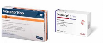
Aritel, 5 mg, film-coated tablets, 30 pcs.
Patients should not interrupt treatment with the drug abruptly and change the recommended dose without first consulting a doctor, because this may lead to a temporary deterioration in heart function.
Treatment should not be interrupted suddenly, especially in patients with coronary artery disease. If discontinuation of treatment is necessary, the dose should be reduced gradually.
Monitoring of patients taking bisoprolol should include monitoring heart rate and blood pressure (at the beginning of treatment - daily, then once every 3-4 months), ECG, blood glucose concentration in patients with diabetes (once every 4-5 months). In elderly patients, it is recommended to monitor renal function (once every 4–5 months). The patient should be taught how to calculate heart rate and instructed about the need for medical consultation if the heart rate is less than 50 beats/min.
Before starting treatment, it is recommended to conduct a study of external respiratory function in patients with a burdened bronchopulmonary history.
In approximately 20% of patients with angina, β-blockers are ineffective. The main causes are severe coronary atherosclerosis with a low ischemic threshold (heart rate less than 100 beats/min) and increased end-diastolic volume of the left ventricle, impairing subendocardial blood flow.
In smokers, the effectiveness of β-blockers is lower.
Patients using contact lenses should take into account that during treatment the production of tear fluid may decrease.
When used in patients with pheochromocytoma, there is a risk of developing paradoxical arterial hypertension (if effective α-blockade is not previously achieved).
In hyperthyroidism, the drug may mask certain clinical signs of thyroid hyperfunction, such as tachycardia. Abrupt withdrawal of the drug in patients with hyperthyroidism is contraindicated, as it can increase symptoms.
In diabetes mellitus, it can mask tachycardia caused by hypoglycemia. Unlike non-selective beta-blockers, it practically does not enhance insulin-induced hypoglycemia and does not delay the restoration of blood glucose to normal levels.
When taking clonidine simultaneously, it can be discontinued only a few days after discontinuation of Aritel® and Aritel® Cor.
It is possible that the severity of hypersensitivity reactions and the lack of effect from usual doses of epinephrine (adrenaline) may increase against the background of a burdened allergological history.
If planned surgical treatment is necessary, the drug should be discontinued 48 hours before general anesthesia. If the patient took the drug before surgery, he should select a drug for general anesthesia with minimal negative inotropic effect. The anesthesiologist should be warned that the patient is taking Aritel® or Aritel® Cor.
Reciprocal activation of the vagus nerve can be eliminated by intravenous atropine (1–2 mg).
Drugs that reduce the reserves of catecholamines (including reserpine) can enhance the effect of beta-blockers, so patients taking such combinations of drugs should be under constant medical supervision to detect arterial hypotension or bradycardia. Patients with bronchospastic diseases can be prescribed cardioselective blockers in case of intolerance and/or ineffectiveness of other antihypertensive drugs, but the dosage should be strictly monitored. An overdose is dangerous due to the development of bronchospasm.
In case of increasing bradycardia (less than 50 beats/min), arterial hypotension (SBP below 100 mm Hg), AV blockade, bronchospasm, ventricular arrhythmias, severe liver and kidney dysfunction in elderly patients, it is necessary to reduce the dose or stop treatment.
It is recommended to discontinue therapy if depression caused by taking β-blockers develops.
Treatment should not be abruptly interrupted due to the risk of developing severe arrhythmias and myocardial infarction. Cancellation is carried out gradually, reducing the dose over 2 weeks or more (reduce the dose by 25% in 3-4 days).
Should be discontinued before testing the content of catecholamines, normetanephrine and vanillylmandelic acid in the blood and urine; antinuclear antibody titers.
Impact on the ability to drive vehicles or perform work that requires increased speed of physical and mental reactions.
During the treatment period, care must be taken when driving vehicles and engaging in other potentially hazardous activities that require increased concentration and speed of psychomotor reactions.