Bleeding time is a laboratory test done to evaluate platelet function and the body's ability to clot. The test consists of puncturing a small area of skin and calculating the time required for the bleeding to stop (this moment is the appearance of dried crusts on the wound). The blood in a healthy person usually stops within 7-9 minutes, and in children within 10-13 minutes.
Note: determining the rate of blood clotting using the above method is almost never used in modern clinics. Modern equipment allows you to determine the rate of clotting literally from a milligram of blood taken from a finger or vein. But if the doctor does not have time to wait for laboratory test results, an incision may be made to the patient.
Basic test methods
The Ivey method is a test that is performed using a mechanical pressure measuring device. The cuff is placed on the elbow and inflated to 40 mmHg. The patient's forearm is wiped with alcohol and an incision is made 1 mm deep and 10 mm long with a sterile blade or scalpel. Since the Ivey method is designed to determine how long it will take for bleeding from the capillary vessels to stop, the incision is made from the inside.
This is what the Ivy method looks like:
Immediately after the blood begins to flow, the doctor starts the stopwatch. Then, every 30 seconds, a special paper filter is carefully applied to the wound. If the filter completely absorbs blood, this means that bleeding remains active. As soon as the bleeding has completely stopped, that is, there are no drops of blood left on the filter, the bleeding time is recorded and the air from the cuff is deflated. Thus, the duration of bleeding according to the Ivey method is defined as the time from the appearance of the first drops of blood until the blood completely stops being released.
The Duque method differs from the Ivey method in that it does not use a blood pressure machine. In addition, the Duque method is considered less aggressive, because the skin must be pierced in any place that does not contain large vessels, for example, the earlobe or finger. The puncture must be at least 3 mm deep. Then, as with the Ivey method, the manipulations with the filter paper are repeated every 30 seconds. Due to the fact that the Duque method is less invasive, it can be done independently at home.
Interpretation of test results
The norms in any analysis are the average values of many indicators of healthy people. To understand which norm will adequately assess the state of the body during a particular type of analysis, large-scale statistical studies are carried out.
At the same time, it is worth remembering that the average indicators are rather arbitrary, because people’s lifestyles may differ under the influence of non-pathological factors - individual norms sometimes go slightly beyond the boundaries of normal results.
This is one example of possible deviations from the norm. When analyzing the results of a hemostasiogram, a specialist must take into account specific factors in order to correctly assess the characteristics of the body’s functioning, identify possible pathology and prescribe treatment.
The ability to clot blood is determined using laboratory research methods, which use both capillary and venous blood.
A series of different tests may require a specific type of blood, which makes it possible to identify the characteristics of the course of hemostasis at different stages.
Diagnostic techniques for coagulation are studied:
- platelet content in the total blood volume - estimates the number of blood cells responsible for activating coagulation processes;
- fibrinogen content - determines the concentration of a special protein that plays an important role in strengthening blood clots;
- activated partial thromboplastin time (aPTT) - makes it possible to detect the activity of plasma factors independent of platelet activity;
- prothrombin index (PTI) - evaluates the ability of plasma blood factors to form clots under the influence of platelets. The analysis process reveals the ratio of prothrombin time indicators (in seconds) to normal prothrombin time;
- thrombosed time (TT);
- duration of bleeding according to Duke;
- clotting time – examines the ability of blood to form stable clots.
Capillary blood is used to determine platelet levels. The normal platelet count in adult women and men varies from 150 to 400 g/l, and in children - in the region of 150 - 350 g/l.
The amount of fibrinogen can be detected using venous blood. In adults, the norms vary around 2 - 4 g/l, and in newborns the values are slightly lower and range from 1.25 to 3.0 g/l.
To study APTT, material is taken from a vein, and the norms will be the same for everyone - from 35 to 50 seconds. The PTI of capillary blood (from a finger) should be in the region of 93–107%, and venous blood – 90–105%.
To study TV, blood is taken from a vein, and TV standards are kept in the range from 12 to 20 seconds. Duke capillary bleeding indicators should not exceed four minutes.
What processes occur during bleeding?
Since the time it takes for bleeding to stop is determined in laboratory conditions in most cases, it is necessary to know and understand the processes that accompany it. Indeed, the sheet of analysis results can indicate not only the time norm, but also hemostasis indicators.
The most accurate results can only be obtained in laboratory conditions
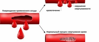
Hemostasis is a sequence of events that leads to the cessation of bleeding due to the formation of a special hemostatic plug of platelets and fibrin. A plug forms on the injured vascular wall from platelets and clotted blood. After platelets combine with endothelial cells and collagen in the damaged vascular wall, von Willebrand factor is released. Damaged cells also release tissue factor VII (blood clotting factor), and exposure to subendothelial collagen activates factor XII (beta globulin, which plays an important role in blood clotting).
This is what the process of platelet aggregation looks like under a microscope
After all factors are activated, platelet aggregation occurs. Soon after, additional platelets are taken from the bloodstream and aggregated, thanks to adenosine and thromboxane A2. Thanks to such complex chemical processes, the skin is restored after a cut or puncture. All of the above processes can be seen by the laboratory technician under a microscope, and if a failure occurs at any of these stages, the bleeding time increases.
Similar
| Brief results of the latest study on the topic “The Future of Quality” and about the book “Economic Benefits Study: International Case Studies” | Cuvette compartment, allowing the use of various types of cuvettes, including: round, dry, flow, etc. Allows you to study substrates, enzymes, electrolytes, hematological parameters. The photometer is an open system and can... |
| How American companies manage talent. Key Elements Half of American organizations cite talent management as a top priority. This is evidenced by research data... | Order No. 321 of the city of Nadym “On the organization and conduct of a district monitoring study of the readiness of first-graders to study at school in municipal... |
| The device is intended for binocular stereoscopic non-reflex examination of the fundus of the eye using the reverse ophthalmoscopy method when illuminated with white, blue and blue-green “red-free” light. The ability to operate the device from an autonomous power supply allows you to increase the productivity of medical personnel and carry out… | Documents1. /Basic principles of research/referat-online.at.ua.doc2. /Basic… |
| International scientific and practical conference “Social sciences: history, current state and research prospects” The organizer of the scientific and practical conference “Social sciences: history, current state and research prospects” is… | V. I. Tyupa narratology as an analyst of narrative discourse (“Bishop” by A. P. Chekhov) Tver, 2001 (Series “Literary text. Problems and research methods.” Appendix “Lectures” Series “Literary text. Problems and research methods.” Appendix “ Lectures in Tver" |
| Formation of the initial cost of a construction project when performing construction and installation work in an economic way; formulation of the relevance and goals of the study; Formulation of the relevance and goals of the study. One of the ways to receive fixed assets is new construction or expansion,… | Federal State Educational Institution of Higher Education "Voronezh State University" Voronezh Department of the Russian Geographical Society Faculty of Geography, Geoecology and Tourism Voronezh Department of Argo All-Russian Interdepartmental Scientific and Practical Conference "Municipal Entities of the Regions of Russia: Research Problems, All-Russian Interdepartmental Scientific and Practical Conference "Municipal Entities of the Regions of Russia: Research Problems ,… |
| Exploring a function using its derivative |
Documents
Documents
In what cases is a test prescribed?
Since platelets are the main factor in primary hemostasis, the results of the bleeding control test will directly indicate the functionality of the blood cells. Therefore, this test is prescribed to patients with suspected bleeding disorders or thrombocytopenia. A skin incision may also be done as a screening test in an outpatient setting. This is usually done in small clinics and outpatient clinics that do not have their own laboratory. This test is performed before invasive procedures in patients with suspected bleeding disorders to determine the likelihood that prolonged bleeding will occur.
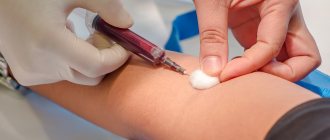
The test is quick and virtually painless
Laboratory assessment of clotting time may be ordered in patients suffering from prolonged or severe postoperative bleeding. Using a microscope, the doctor will be able to recognize dysfunctional platelets and choose the right treatment.
Why do you need to check blood clotting?
Blood clotting time is sometimes an indicator of serious deviations in the condition of the body. A blood test that determines clotting time is called a coagulogram.
Delayed or accelerated thrombus formation, to varying degrees, adversely affects the development and function of organs . The rate of blood clotting can change with age - in children this process occurs faster than in people of retirement age. Therefore, to prevent and timely correct possible hematopoietic pathologies, experts advise regularly undergoing preventive examinations and a full examination.
Causes of prolonged bleeding
The cause of abnormal bleeding time may be hereditary or acquired.
Among the hereditary causes of bleeding disorders are the following:
- von Willebrand disease;
- Glanzmann's thrombasthenia;
- Bernard-Soulier syndrome;
- connective tissue diseases (Ehlers-Danlos syndrome, Wiskott-Aldrich syndrome, Chediak-Higashi syndrome, hereditary hemorrhagic telangiectasia).
Among the acquired causes of abnormally long bleeding time, the most common are the following:
- long-term use of medications, especially aspirin, non-steroidal anti-inflammatory drugs, antibiotics, anticoagulants, tricyclic antidepressants, antipsychotics;
- vitamin C deficiency;
- frequent alcohol intoxication;
- uremia;
- liver failure;
- leukemia;
- myelodysplastic syndrome;
- amyloidosis.
This is why the patient should not take any blood thinning medications or alcohol for 7 days before the test as this may lead to false results.
A coagulogram is a study of the hemostatic system that allows you to evaluate the external and general pathways of blood coagulation and identify the risk of hypercoagulation (excessive clotting) or hypocoagulation (bleeding).
Synonyms Russian
Hemostasiogram: prothrombin index (PI), prothrombin time (PT); international normalized ratio (INR); factor I (first) of the plasma coagulation system.
English synonyms
Coagulation studies (coagulation profile, coag panel, coagulogram): Prothrombin time (Pro Time, PT, Prothrombin time ratio, P/C ratio); International Normalized Ratio (INR); Fibrinogen (FG, Factor I).
Units
% (percent), sec. (second), g/l (grams per liter).
What biomaterial can be used for research?
Venous blood.
How to properly prepare for research?
- Do not eat for 12 hours before the test.
- Avoid physical and emotional stress and do not smoke for 30 minutes before the test.
General information about the study
The hemostasis system consists of many biological substances and biochemical mechanisms that ensure the preservation of the liquid state of the blood and prevent and stop bleeding. It maintains the balance between blood clotting and anti-clotting factors. Significant disturbances in the compensatory mechanisms of hemostasis are manifested by processes of hypercoagulation (excessive thrombus formation) or hypocoagulation (bleeding), which can threaten the patient’s life.
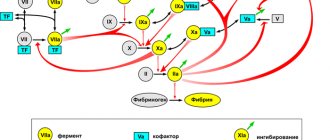
When tissues and blood vessels are damaged, plasma components (clotting factors) participate in a cascade of biochemical reactions, which results in the formation of a fibrin clot. There are internal and external pathways of blood coagulation, which differ in the mechanisms that trigger coagulation. The internal pathway is realized when blood components come into contact with the collagen of the subendothelium of the vessel wall. This process requires coagulation factors XII, XI, IX and VII. The extrinsic pathway is triggered by tissue thromboplastin (factor III) released from damaged tissues and the vascular wall. Both mechanisms are closely interrelated and from the moment of formation of active factor X they have common paths of implementation.
The study of indicators such as PTI (prothrombin index) and INR (international normalized ratio) allows us to assess the state of the external coagulation pathway. PTI is calculated as the ratio of the standard prothrombin time (the clotting time of control plasma after the addition of tissue thromboplastin) to the plasma clotting time, expressed as a percentage. INR is a prothrombin test standardized in accordance with international recommendations. It is calculated by the formula: INR = (patient prothrombin time / control prothrombin time) x MICH, where MICH (international sensitivity index) is the sensitivity coefficient of thromboplastin relative to the international standard. INR and PI are inversely proportional indicators, that is, an increase in INR corresponds to a decrease in the patient’s PTI and vice versa.
Reference PTI values depend on the set and characteristics of the reagents and differ in the activity of thromboplastin used in the test. The results of INR determination, thanks to standardization, make it possible to compare the results of different laboratories.
Tests for PTI (or a similar indicator - prothrombin according to Quick) and INR in a coagulogram help to identify disorders in the external and internal blood coagulation pathways associated with a deficiency or defect of fibrinogen (factor I), prothrombin (factor II), factors V (proaccelerin) , VII (proconvertin), X (Stewart-Prower factor). With a decrease in the concentration of these coagulation factors in the blood, the prothrombin time increases in relation to control laboratory parameters.
Plasma factors of the extrinsic coagulation pathway are synthesized in the liver. For the formation of prothrombin and some other coagulation factors, vitamin K is required, the deficiency of which leads to disturbances in the cascade of reactions and prevents the formation of a blood clot. This fact is used in the treatment of patients with an increased risk of thromboembolism and cardiovascular complications. Thanks to the prescription of the indirect anticoagulant warfarin, vitamin K-dependent protein synthesis is suppressed. PTI (or Quick prothrombin) and INR coagulation are used to monitor warfarin therapy in patients with factors that promote thrombosis (eg, deep vein thrombosis, presence of prosthetic valves, antiphospholipid syndrome).
Normally, a coagulogram in a healthy person has an INR in the range of 0.8-1.2; in patients being treated with indirect anticoagulants to prevent thromboembolic complications - 2.0-3.0, in patients with prosthetic valves and antiphospholipid syndrome - 2.5-3.5.
Simultaneous determination of fibrinogen in a coagulogram allows a comprehensive assessment of the state of the plasma hemostasis system.
Fibrinogen is blood clotting factor I, which is produced in the liver. Thanks to the action of the coagulation cascade and active plasma enzymes, it is converted into fibrin, which is involved in the formation of a blood clot and thrombus. Fibrinogen deficiency can be primary (due to genetic disorders) or secondary (due to excessive consumption in biochemical reactions), which is manifested by impaired formation of a stable blood clot and increased bleeding.
Fibrinogen is also an acute phase protein. Its concentration increases in the blood during diseases accompanied by tissue damage and inflammation. Determining the level of fibrinogen is important in the diagnosis of diseases with increased bleeding or thrombosis, as well as for assessing the synthetic function of the liver and the risk of cardiovascular diseases with complications.
What is the research used for?
- For a general assessment of the blood coagulation system.
- For the diagnosis of disorders of the external and general pathways of blood coagulation.
- To study the activity of coagulation factors I, II, V, VII, X.
- To monitor the patient's condition when prescribing anticoagulants.
- To assess the risk of cardiovascular complications.
- To assess the protein-synthesizing function of the liver (synthesis of blood clotting factors).
When is the study scheduled?
- With a comprehensive examination.
- When planning surgical interventions.
- When examining patients with nosebleeds, bleeding gums, blood in the stool or urine, hemorrhages under the skin and in large joints, chronic anemia, heavy menstrual flow, sudden loss of vision.
- When examining a patient with a history of episodes of thrombosis.
- With a hereditary predisposition to disorders of the hemostatic system.
- With a high risk of cardiovascular complications and thromboembolism.
- Before prescribing anticoagulants.
- When monitoring the hemostatic system while taking anticoagulants.
- For liver diseases.
What do the results mean?
Reference values (table of normal coagulogram indicators)
- Prothrombin according to Quick: 70 - 120%.
- INR
| Age | Reference values |
| Less than 3 days | 1,15 — 1,35 |
| 3 days – 1 month | 1,05 — 1,35 |
| 1 month – 1 year | 0,86 — 1,22 |
| 16 years | 0,92 — 1,14 |
| 6 – 11 years | 0,87 — 1,20 |
| 11 – 16 years | 0,97 — 1,30 |
| More than 16 years | 0,8 — 1,2 |
| Week of pregnancy | Reference values |
| 13-21st | 0,56 — 1,1 |
| 21st-29th | 0,5 — 1,13 |
| 29-35th | 0,58 — 1,17 |
| 35-42nd | 0,15 — 1,14 |
- Fibrinogen: 1.8 - 3.5 g/l.
| Week of pregnancy | Reference values |
| 1-13th | 2.12 - 4.33 g/l |
| 13-21st | 2.9 - 5.3 g/l |
| 21st-29th | 3 - 5.7 g/l |
| 29-35th | 3.2 - 5.7 g/l |
| 35-42nd | 3.5 - 6.5 g/l |
Reasons for an increase in INR and a decrease in the level of prothrombin according to Quick (indicates a possible deficiency of factors in the external hemostasis pathway and a tendency to increased bleeding):
- DIC syndrome (disseminated intravascular coagulation) during the period of hypocoagulation;
- hypofibrinogenemia (factor I deficiency);
- dysfibrinogenemia (synthesis of a defective protein that is unable to participate in the cascade of biochemical reactions);
- hereditary or acquired deficiency of factors II, V, VII;
- Factor X deficiency (eg, purpura due to amyloidosis);
- vitamin K deficiency;
- hemorrhagic disease of newborns;
- malabsorption with impaired fat absorption (due to celiac disease, chronic diarrhea);
- acute leukemia;
- antiphospholipid syndrome;
- congestive heart failure;
- liver pathology (hepatitis, cirrhosis, alcoholic liver disease);
- obstruction of the biliary tract, obstructive jaundice;
- pancreas cancer;
- Zollinger-Ellison syndrome (pancreatic adenoma);
- toxic shock syndrome;
- nephrotic syndrome (excessive urinary excretion of factors V and VII);
- oral anticoagulants (warfarin).
Causes of increased fibrinogen levels (indicates an increased risk of blood clots and cardiovascular complications):
- acute infection (eg pneumonia, tuberculosis);
- autoimmune diseases (rheumatoid arthritis, reactive arthritis);
- acute coronary syndrome, myocardial infarction;
- burns;
- cancer (breast, kidney, stomach);
- multiple myeloma;
- Hodgkin's disease (lymphogranulomatosis);
- glomerulonephritis, nephrotic syndrome, nephrosis;
- pregnancy;
- eclampsia;
- cerebrovascular disease, stroke;
- hepatitis;
- postoperative period;
- rheumatic fever;
- tissue damage.
Reasons for a decrease in INR and an increase in prothrombin according to Quick (indicates a tendency to form blood clots):
- DIC syndrome (period of hypercoagulation);
- deep vein thrombosis (initial stages);
- polycythemia;
- pregnancy (last months);
- increased activity of factor VII.
Reasons for decreased fibrinogen levels (may indicate an increased risk of bleeding):
- dysfibrinogenemia;
- hereditary afibrinogenemia;
- DIC syndrome;
- fibrinolysis;
- hemophilia A and B;
- liver pathology (hepatitis, cirrhosis);
- abortion;
- premature placental abruption;
- late stage of cancer;
- embolism (amniotic fluid, meconium, fat, tissue);
- anemia;
- eclampsia;
- leukemia;
- malabsorption;
- shock;
- sepsis;
- post-transfusion reactions.
What can influence the result?
- Factors that distort the result of the analysis: the presence of lupus anticoagulant in the blood (directly inhibits coagulation factors);
- transfusion of donor blood components in the last month (distorts the fibrinogen indicator).
- drinking alcohol, fatty foods;
- excess intake of vitamin K from food (found in beef or pork liver, green tea, broccoli, chickpeas, cabbage, turnips, soy, green leafy vegetables);
What do the test results indicate?
Depending on the duration of bleeding, health problems can be judged:
- 1-9 minutes is the norm;
- 10-15 minutes – platelet dysfunction;
- more than 15 minutes – obvious pathology.
Bleeding time is recorded to measure the primary phase of hemostasis (attachment of platelets to damaged capillaries, and then their activation and aggregation). If a patient has a low platelet count or dysfunctional platelets, the blood may not begin to clot even after 15 minutes.
Progress of the procedure
To carry out this type of laboratory test, a small amount of capillary blood is taken from the patient's ring finger.
After introducing a scarifier (puncture), the first drop of blood is removed, as it may contain elements of tissue fluid and pathogenic microorganisms. The next drop is the material for research, which is placed in the so-called Panchenkov apparatus. During the test, a sample of capillary blood is placed in a glass capillary, which is subsequently tilted left and right. In parallel with this action, the laboratory doctor records the time that will be spent on the formation of a blood clot. The onset of this process is not difficult to determine, since the blood in the glass capillary begins to coagulate and stops moving.
In simple terms, we can say that Sukharev’s technique is aimed at assessing the interval between taking a blood sample and the beginning of the formation of a fibrin clot.