Coronary angiography
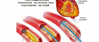
Coronary angiography is a radiopaque research method that evaluates the condition of the arteries of the heart. Coronary angiography is the most accurate and reliable way to diagnose coronary heart disease, which allows you to decide on the patient’s treatment tactics in each specific situation, for example, the possibility of continuing treatment with medications, the need for such treatment procedures as angioplasty and stenting or coronary artery bypass grafting. This procedure is invasive, which involves the introduction of a special catheter in an operating room and can be performed both for diagnostic purposes and to monitor the condition of blood vessels after operations already performed.
Is this examination dangerous?
Everyone who is about to undergo this procedure is interested in what the consequences and complications may be. This type of diagnosis is considered a safe manipulation; the process is controlled using an X-ray or computed tomograph. Nevertheless, the risk of complications is still present; the most dangerous consequence is damage to blood vessels by the catheter.
The likelihood of such a complication is very low; it can occur from sudden movements or carelessness of the doctor. If the patient has not undergone proper preparation, an allergic reaction or bleeding may occur (with poor clotting). All these risks can be reduced if you carefully prepare and relax before the procedure.
If you are overcome by fear and anxiety in the X-ray operating room, you can warn the doctor about this and he will prescribe sedatives.
Why is coronary angiography performed only in a hospital setting?
The main reason that made it mandatory for the patient to be in the hospital when performing this study was the need for puncture (puncture) of the peripheral artery (femoral) and associated complications. The femoral artery is a large vessel located at a depth of 2-4 cm from the skin surface in the groin area. To prevent bleeding, which can be dangerous, after the study the patient is prescribed a limited motor regimen under the supervision of medical personnel. But recently another access has appeared - through the small radial artery, which we palpate on the wrist. An approach called radial, which does not result in serious bleeding. The patient can get up immediately after the examination; bed rest is not necessary. Observation is limited to a few hours. In the CELT clinic, radial (radial) arterial access is used as the main approach. Experience with this technique indicates a lower incidence of local complications, which is 0-0.7%. The possibility of early rehabilitation of the patient after CAG with radial access and the almost complete absence of side effects make it possible to perform CAG on an outpatient basis.
Advantages and disadvantages of ultrasound and MRI of the heart
Today, high-quality cardiac MRI requires very expensive equipment - a tomograph with a magnetic field power of at least 3 Tesla. Research using such an ultra-high-field device will be expensive, so the main disadvantage of cardiac MRI is its high price. In addition, MRI of the coronary vessels and heart cannot measure the speed of blood flow and its actual volume. Cardiac ultrasound can better cope with this task.
Previous NextHow is outpatient coronary angiography performed?
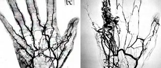
The algorithm for performing coronary angiography on an outpatient basis includes three stages. At the first stage, patients are selected for a diagnostic procedure, and the necessary additional examinations are carried out. Indications for coronary angiography : - identified or suspected ischemic heart disease; - pain in the chest, suspicious for angina pectoris; - myocardial infarction; - planned surgery for heart defects; - heart failure, ventricular arrhythmias. Indications for coronary angiography are determined by the attending physician in accordance with accepted criteria. In preparing the patient for coronary angiography , the necessary tests and studies are performed. In addition to these, additional studies may be prescribed. The second stage of outpatient CAG angiography procedure itself . The patient is admitted to the day hospital ward. After assessing the stability of his condition, premedication is administered and he is transported to the cath lab, where the coronary angiography . After anesthetizing the access area, research begins - a special catheter is passed through the artery of the forearm into the lumen of the coronary arteries . Using a catheter, a radiopaque substance is injected into the blood, thanks to which the lumen of the vessels becomes visible on a special device - an angiograph. During coronary angiography, the degree and size of damage to the coronary vessels is determined, which determines further treatment tactics. This procedure is low-traumatic, which allows it to be performed under local anesthesia without the use of general anesthesia. The duration of the procedure, as a rule, does not exceed 20 minutes. From the operating room, the patient, accompanied by medical personnel, is taken to the day hospital ward. The third stage of outpatient coronary angiography is observation of the patient in a day hospital ward for 4-5 hours after the examination. In the ward, the patient can drink water or juices without restrictions and have lunch. If there are no complications, the patient is sent home. On the day of the outpatient coronary angiography, the patient receives a conclusion with recommendations on further treatment tactics and a disk with the results of coronary angiography . If complications arise during coronary angiography or during the control period, patients are hospitalized in the intensive observation unit of the hospital.
Possible risks of coronary angioplasty
Although angioplasty is less invasive than bypass surgery, the procedure still carries some risks. Risks may be associated with the procedure itself and its long-term outcomes.
Early risks:
- Bleeding. There may be bleeding in the leg or arm where the vessel was accessed. Usually it just develops as a bruise, but sometimes there is serious bleeding that may require a blood transfusion or surgery.
- Heart attack. Coronary circulation disorders develop very rarely during surgery, but sometimes, due to technical reasons, a rupture or dissection of the coronary artery may develop. These complications may require emergency coronary artery bypass surgery.
- Kidney problems. The contrast agent used in angioplasty and stent placement can cause kidney damage, especially in people who already have kidney problems. If you are at higher risk, your doctor may take steps to protect your kidneys, such as limiting the amount of contrast material and increasing fluid intake to dilute the contrast.
- Ischemic stroke. During angioplasty, a stroke can occur if pieces of plaque break off as the catheter is passed through the aorta. Stroke is an extremely rare complication of coronary angioplasty. To prevent it, drugs that reduce blood clotting are prescribed.
- Heart arythmy. During the procedure, the heart may beat too fast or too slow. Such arrhythmias are usually short-lived, but sometimes a temporary pacemaker is required.
Late complications
- Repeated narrowing of the artery (restenosis). With angioplasty without stenting, the rate of restenosis is approximately 30% of cases. Stents have been developed to reduce restenosis. The use of plain metal stents reduces the risk of restenosis to 15%, and the use of drug-eluting stents reduces the risk to less than 10%.
- Stent thrombosis. Blood clots (thrombi) can block an artery, causing a heart attack. To reduce the risk of thrombosis, it is important to take aspirin, clopidogrel (Plavix), which helps reduce the risk of blood clots forming in the stent. Never stop taking these medications without talking to your doctor.
FAQ:
Question: I am 56 years old and have coronary heart disease . The cardiologist recommends a coronary angiography . I don't quite understand what this is? Answer: Coronary angiography is a study of the vessels of the heart, which, like a crown, surround the heart, supplying it with oxygen-rich blood. These are called coronary arteries. Narrowing of the coronary arteries leads to a decrease in blood supply to the heart, which leads to oxygen starvation, i.e. to cardiac ischemia. It is very important to know the condition of the arteries of the heart, because... the obstruction to blood flow can be eliminated, radically relieving the person of the symptoms of coronary heart disease . Question: How is coronary angiography performed? Answer: Coronary angiography is an x-ray examination in which a special contrast agent is injected into the vessels of the heart using a flexible thin probe. As the contrast agent passes through the vessels, short-term x-rays are taken with significant magnification using high-resolution equipment. The slightest changes in the coronary bed are visible “at a glance.” The results are recorded in digital format and are available for viewing on any personal computer. Question: Is coronary angiography performed under anesthesia? Answer: No general anesthesia is required. The pulse is determined in the groin area or on the wrist, and a probe or catheter is inserted into the lumen of the artery under local anesthesia. The subject does not feel how the catheter moves through the vessels, because There are no sensory nerve endings inside the arteries. There is no pain, the patient is fully conscious and, together with the operator, monitors the progress of the study on the monitor. The duration of the procedure is no more than 15-20 minutes. Further monitoring of the patient for several hours is necessary. At the CELT clinic, coronary angiography is performed exclusively through the radial artery (at the wrist). Immediately after the examination, the patient can get up and walk, the bandage on his arm does not limit him in any way (see ambulatory coronary angiography ). After 4 hours the patient is discharged home. If the examination is carried out through the groin, then bed rest is necessary and the period of stay in the clinic is extended to a day. Question: If coronary angiography does not reveal narrowing in the vessels of the heart, then there is no coronary heart disease? Answer: Yes, the absence of changes in the coronary arteries practically excludes the diagnosis of coronary heart disease . In rare cases, ischemia of the heart muscle can occur in the presence of “normal” coronary arteries, but the absence of damage to the arteries of the heart is the most reliable predictor of a good prognosis. This is very important information for choosing the right patient management tactics. Question: At what age is coronary angiography , what are the contraindications to coronary angiography? Answer: Coronary angiography is performed at any age, in all cases when there is a need for it, namely if the patient has angina pectoris after myocardial infarction. Coronary angiography, in the first hours of myocardial infarction , makes it possible to determine where the blockage of the vessel is causing the heart attack and immediately eliminate it. Coronary angiography is necessary for all adults with heart defects before surgery, before “major” vascular operations. Some patients at high risk for coronary heart disease , such as diabetes , may not have symptoms of the disease. In this case, coronary angiography is, in fact, the only reliable way to exclude or confirm coronary disease . Question: I need a coronary angiography . I contacted the Federal Center. They gave me a list of tests and put me on the waiting list. They said I needed hospitalization for 2-3 days. Answer: We coronary angiography on the day of admission! All tests and additional studies are carried out within 4-5 hours, including coronary angiography. Question: How can I get a coronary angiography at your center? Answer: You can come any day, appear at the clinic, at the cardiovascular surgery department in the morning at 08:30 on an empty stomach, PRIORLY AGREEING YOUR ADMISSION WITH US BY PHONE 305-34-04 or 788-33-88. Before admission, before performing coronary angiography , you should have on hand the results of the following tests and studies: - General blood test; — General urine analysis; - Biochemical analysis - protein, urea, creatinine, bilirubin and its fractions, lipid profile, potassium and sodium, glucose; — Coagulogram; — Blood type and Rh factor, Rh antibodies; — Blood test for HIV, RV, antibodies to hepatitis B and C; — Ultrasound of the heart, ECG. If you do not need tests and studies, then they can be performed with us on the day of admission. Question: coronary angiography cost you and what are the prices for coronary angiography in Moscow in other clinics? Answer: Coronary angiography including the stay costs 30,000 rubles. Today, if you analyze the prices for coronary angiography in Moscow, this is the minimum cost.
Contraindications for the procedure
Coronary angiography has only relative contraindications. There is a list of diseases that increase the risk of complications. But even against the background of such diseases, fluoroscopy of blood vessels can be performed if the patient’s life depends on it. Coronary angiography is performed with extreme caution when:
- impaired blood clotting;
- decompensated diabetes mellitus;
- allergies to contrast agent;
- arterial hypertension;
- severe renal failure - the contrast agent has a bad effect on diseased kidneys;
- internal bleeding - postponed until this problem is resolved.
All possible contraindications must be taken into account when prescribing coronary angiography. Absolute contraindications include only a severe allergic reaction to contrast. In most cases, the study is simply postponed.
Decoding the electrocardiogram
This procedure must be performed by a qualified medical professional - a clinical diagnostic doctor or a cardiologist, a therapist, who will subsequently prescribe effective treatment.
Some terms that are indicated in the cardiogram can be understood by patients themselves, including:
EOS - this indicator helps determine the location of the electrical axis, the heart muscle, and the functionality of its parts. The electrocardiogram may indicate a horizontal or vertical location with a shift to the right/left.
Heart rate is an indicator of the number of heartbeats. The norm is from 60 to 90 beats per minute. An increased heart rate is considered if it exceeds 91 beats per minute. An increased heart rate is called tachycardia, and a low heart rate (less than 59 beats/minute) is called bradycardia.
Non-sinus rhythm is an indicator of heart pathology in which non-essential electrical signals are produced outside the sinus node. Usually a sign of atrial fibrillation (old name: atrial fibrillation).
Regular sinus rhythm is an indicator of normal functioning of the heart muscle.
Atrial flutter is one of the types of arrhythmia and requires urgent medical intervention.
Ventricular hypertrophy – shows thickening of the walls of the ventricles or changes in their shape.
QT is an indicator of cardiac conductivity, and if abnormalities are visualized, frequent fainting can occur and can even lead to death.
Sinoatrial blockade - demonstrates disturbances in the conduction of impulses from the node to the atrium, often indicating the development of the following diseases: cardiosclerosis, cardiomyopathy, infarction, myocardium.
When is MRI better than cardiac ultrasound?
The level of anatomical detail of the heart area on MRI is higher than on ultrasound. During the scan, the doctor receives up to 1000 images of the mediastinal area. The scanning step of a 3 tesla device can be only 0.8 mm. This means that the doctor can look in detail at the condition of the heart itself and the neighboring coronary vessels. In this case, both planar and three-dimensional data will be provided. MRI best visualizes pathologies and diseases such as:
- inflammatory processes in tissues;
- tumor formations;
- blood clots, vascular malformations, occlusions, aneurysms;
- congenital anomalies of the heart structure;
- heart muscle defects;
- pericardial condition.
Author: Telegina Natalya Dmitrievna
Therapist with 25 years of experience
>
When is ultrasound better than cardiac MRI?
Cardiac ultrasound is well suited for diagnosing both children, from infancy, and adults. An ultrasound examination will clearly show the functional features of the heart:
- efficiency of the heart muscle;
- thickness of the heart walls;
- functional state of four chambers and valves;
- the size of the heart cavities and the pressure in them;
- speed of intracardiac blood flow.
Cardiac ultrasound is better suited than MRI for examining young children, since it does not require immobilization of the patient through anesthesia so that the baby can lie quietly. Ultrasound is an inexpensive way to initially screen the heart if you need to evaluate the condition of the organ due to some primary symptoms of illness or if you want to do an ultrasound of the heart as a preventive measure for cardiovascular diseases. Cardiac MRI is an expert diagnostic method. It is best used when a pathology is identified and it is necessary to verify or clarify the diagnosis.
Method of performing electrocardiography
Best materials of the month
- Coronaviruses: SARS-CoV-2 (COVID-19)
- Antibiotics for the prevention and treatment of COVID-19: how effective are they?
- The most common "office" diseases
- Does vodka kill coronavirus?
- How to stay alive on our roads?
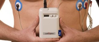
For the procedure, the patient is placed comfortably in a horizontal position on his back. Special electrodes are attached to the chest area, legs and arms. The device starts and the heart function is recorded. The duration of the procedure can vary from 1 to 10 minutes. The results obtained are sent to the doctor for interpretation and diagnosis.
