Electrocardiography is the simplest method for studying heart disease. Its result is an electrocardiogram - a graphic image obtained during the contraction and relaxation of the heart muscles. Ultrasound of the heart makes it possible to visually examine the internal organ, thanks to a special sensor that conducts rays and receives received impulses. Heart ultrasound (or ECHO CG) provides complete information about the structure of the organ, its size; with such an examination, it is possible to notice even the most minor deviations that affect the well-being of patients.
Difference between ECG and ultrasound (ECHO CG)
An electrocardiogram, carried out using an electric current that is invisible to the patient, makes it possible to find out the rhythm of the heart, determine the intervals of its relaxed state and contraction. An electrocardiogram is a mandatory examination during inpatient treatment of any disease; it is included in the list of medical preventive examination studies for children from 3 months of age.
An electrocardiogram, which is issued by a specialist after an examination, diagnoses normal heart function or various changes, without determining the cause of the disease.
Echocardiography (ECHO) is a modern method for studying heart disease, which not only detects the presence of abnormalities, but also determines the cause. During an ultrasound, the doctor’s monitor displays information about the size of the organ, its structure, the volume of cavities, it is possible to discern the presence or absence of scars, the condition of the myocardium and pericardium is assessed - all these processes are studied in real time. All information received is reflected in the protocol, which makes it possible to make the most accurate diagnosis and begin effective treatment.
Electrocardiogram
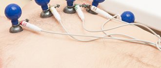
The heart is our “electric motor”. Electrical impulses are generated in a special group of cells in the atria. They cause parts of the heart to contract. An ECG is a recording of the electrical activity of the heart in the form of a waveform. Electrodes are placed on your body to take ECG readings. To increase the passage of electric current, the diagnostician lubricates the areas under the electrodes with a special gel. The procedure itself lasts several minutes. The study is usually carried out in a resting position, lying or sitting. There are types of ECG: stress testing, daily ECG monitoring according to Holper.
What does the ECG show?
- Heart rhythm disturbances: bradycardia and tachycardia, extrasystole, atrial fibrillation.
- Myocardial ischemia.
- Improper conduction of impulses or blockade.
The disadvantage of an ECG is that the study will show those disturbances in the functioning of the heart that affect its functioning at the time of recording. False-positive results are not uncommon for ECGs. If the doctor has doubts about the reliability of the ECG, he will additionally prescribe an ultrasound of the heart.
Indications for ECG
The cardiologist prescribes an electrocardiogram according to the following indications:
- heart rhythm disturbances;
- hypertension;
- infection, inflammation or other heart pathologies;
- alcohol and smoking abuse;
- pregnancy;
- increased blood cholesterol levels;
- previous sore throat;
- increased health requirements by profession for pilots, drivers, military personnel, athletes, etc.
Doctors prescribe an ECG for 80% of patients admitted for hospital treatment. There are no contraindications to the study. Now you can take an electrocardiogram even outside the hospital. For example, at the pharmacy or at the gym.
Remember! If you feel the slightest discomfort or pain in the heart area, then this is a reason to consult a doctor. Take care of your heart and be healthy!
How are ECGs and ultrasounds performed? Cost of heart ultrasound
ECHO KG:
- Ultrasound examination of the heart, or echocardiography, is performed using ultrasonic waves, which are completely safe for the patient.
- Unlike X-rays, which emit harmful compounds from the body, ultrasound can be performed several times a day. The patient simply lies on his back, and the doctor runs a sensor across his chest.
- To carry out an ECHO, the patient is not required to adhere to any regime - you just need to not strain during the examination.
- The affordable cost of cardiac ultrasound allows you to use this type of examination if you have symptoms of heart disease - this makes it possible to determine the cause of the disease at an early stage.
An electrocardiogram is performed on the patient in the supine position. The doctor attaches special vacuum suction cups to the surface of the chest and calves to obtain a clear cardiac impulse. There are no contraindications, as in the first option.
Features of echocardiography
How is echocardiography performed? The echo produced by the heart can show whether there are anatomical defects in the organ. Deviations often occur with cardiac malformations, myocardial infarction, cardiomyopathies of various origins, and coronary heart disease.
Impaired echogenicity may indicate the presence of thrombosis in blood vessels, benign and malignant tumors of the cardiac system, aneurysm, and infectious pathologies such as pericarditis, endocarditis and myocarditis.
How is echocardiography performed? During the examination, the doctor applies a special gel to the skin in the area of the heart. Then a sensor is used, which is passed over this area. The readings from the sensor are transmitted to a specialized monitor, which projects the detected changes in the heart.
When studying pathology based on analysis, the doctor uses a special attachment in his work, which is connected to an echo machine. After completing the diagnostic procedures, a clinical diagnostic specialist gives an opinion with which you can go for a consultation with a cardiologist.
It is worth saying that in cardiac diagnostics, ECG and EchoCG are not prescribed separately, but are used in combination, which in turn makes it possible to obtain a more accurate and reliable clinical picture of the disease.
Advantages of having an examination in our clinic
Among the main advantages of medical treatment are the following points:
- Affordable prices for all types of examinations and surgical procedures
- Wide range of modern equipment
- Highly qualified doctors
- City center location
Other services of our clinic:
- — Ultrasound of internal organs
- - Ultrasound of the uterus
- — Office hysteroscopy
- — Ultrasound of the heart
- — Ultrasound by week of pregnancy
- — Treatment of erosion
- — Treatment of urinary incontinence in women
- — Treatment for lack of orgasm
- — Androgyne in gynecology
So which study is better: ECG or ultrasound of the heart?
Ultrasound and ECG are two diagnostic methods that are used in cardiology to diagnose problems in the functioning of the heart muscle. Based on the results, the specialist assesses the degree of damage to the organ and selects the most appropriate treatment method.
The data obtained from both studies have equally high information content, but the results of the ultrasound examination provide a more meaningful picture. That is why, before conducting any examination, you should visit a cardiologist, who, based on the patient’s complaints, will be able to determine the most appropriate examination method. In most cases, an ECG is considered the primary procedure, because it allows you to get a general picture of the condition of the heart muscle. If the cardiologist does not like the resulting picture, then he can prescribe a more accurate procedure, namely an ultrasound.
Both methods are independent of each other and it is not possible to determine which one is better, because both of them must be completed periodically if there are complaints about the work of the body. Thus, timely detection of heart disease will help to avoid more serious consequences.
Features of diagnostics using ECG
The principle of the method is based on recording electronic heart impulses that occur during myocardial contraction. The resulting impulses are read by a cardiograph, displaying them in the form of a graphic curve on special paper with a scale.
Registration of cardiac impulses using ECG
Algorithm:
- Before starting the procedure, you must remove your jewelry.
- Expose the area of the hands, ankles and chest.
- Take a horizontal position and relax as much as possible.
- Before applying the electrodes, the nurse treats exposed skin with a cotton pad moistened with water for better conduction of impulses.
- Then he places 4 electrodes of different colors on the limbs in a certain order.
- Electrodes are fixed on the chest using 6 suction cups.
- Then the electrodes are connected to the device, after which they are registered.
A cardiogram is effective in identifying diseases of cardiac origin, is relevant for preventive examinations, and is mandatory before surgery.
Using an ECG, you can identify the following disturbances in the functioning of the cardiovascular system:
- Heart rhythm disturbances (arrhythmia, tachycardia, atrial fibrillation, extrasystole).
- Disturbances in myocardial nutrition (ischemia, myocardial infarction).
- Impulse conduction disorder (antiventricular block).
- Displacement of the heart axis (hypertrophy with different localization).
- Thickening of the myocardium (ventricular and atrium hypertrophy).
- Congenital and acquired defects (impairments in the structure of the valves, fibrous ring and notochord).
Persons over 40 years of age must undergo an electrocardiogram every year. This will help establish the functional state of the cardiovascular system and identify possible disorders at the initial stage.
The study is very effective and indicative, but despite this, to clarify the condition of the myocardium, it is necessary to resort to ultrasound diagnostics of the heart.
Why do a cardiac MRI?
We wrote about the principle of MRI in the material “CT or MRI: what to choose?” The method is used as often as Echo CG or Holter monitoring, but it gives particularly clear results in the study of heart tissue, and not its work.
- It is necessary to assess the extent of myocardial damage and impairment of its contractility after a heart attack.
- Consider whether atherosclerotic plaque or stenosis has accumulated in the blood vessels
- Check how recovery is going after treatment
All four types of research are absolutely safe. But even if you have thoroughly understood the information, only a doctor can select a diagnosis and analyze the results.
Find a cardiologist
Heart diagnostic methods
During the initial visit, the doctor will interview the patient and find out whether he has complaints of pain, shortness of breath, increased heart rate, high blood pressure and other symptoms.
In modern medicine, several types of diagnosing heart diseases are used:
- electrocardiogram (ECG);
- bicycle ergometry (ECG with exercise on an exercise bike);
- treadmill (ECG with exercise on a treadmill);
- computed tomography of the heart;
- coronary angiography of the heart;
- EchoCG (ultrasound of the heart);
- myocardial biopsy;
- 24-hour monitoring of heart and blood pressure according to Holter.
Holter: monitoring and analysis of heart function
A holter is attached to the patient, most often on a belt, in a special case and electrodes are applied. Doctors often use the phrase “hang up the Holter” or “make a Holter”, meaning that the device will be used for 24 hours, on an outpatient basis, in the patient’s usual living conditions - at home, at work, during night sleep. Patients are asked not to wet the device, but otherwise lead their usual lifestyle.
This process of taking readings is called Holter monitoring, named after the American research scientist Norman J. Holter, who first used this technique in 1961.
General information about procedures
There are no unnecessary techniques in cardiac examinations. Although many different research methods are used, each has its own advantages, disadvantages, indications and contraindications. Sometimes an accurate diagnosis is only possible with a comprehensive examination using several methods simultaneously.
- Echocardiography of the heart: principle of operation, capabilities and indications for the study
More about ECG
An ECG is performed using a special device - an electrocardiograph. The first device appeared back in 1903. The cardiograph records fluctuations caused by the work of the heart. Data enters the device through special electrodes, which are attached to the patient’s body before the procedure. ECG is safe and painless.
ECG results are a curved line that reflects the conduction of impulses. Typically the procedure is performed in 12 leads. An electrocardiogram helps to identify heart pathologies in the early stages and monitor the condition of the heart before surgical interventions. The procedure is also needed to determine how effective a particular therapy is.
More details about how an ECG is performed are shown in the video:
More about ultrasound
Although an electrocardiogram provides a detailed picture of the functioning of the heart muscle, the procedure cannot diagnose absolutely all pathologies. Until ultrasound equipment was invented, certain serious diseases could not be identified, which cost many their lives.
An ultrasound examination does not allow one to evaluate the work of the heart at the moment, but it makes it possible to see what condition the organ is in, how its valves work, what is the size of the muscle and the thickness of the walls of the heart.
An ultrasound of the heart allows you to see what the contractility of the heart is and whether the pressure inside the pulmonary artery is normal. This procedure is safe for the patient and does not cause pain.
Sometimes cardiologists use stress ultrasound. With this examination, the study is carried out first in a calm state, and then after exercise. Changing indicators helps to assess the condition of the heart and its reaction to the occurrence of irritating factors.
- Library of the State Autonomous Institution of Health Care Center - ECG is within the power of everyone
Diagnostics on the road
As part of my work, I spend a lot of time traveling on business in different regions. Tell me, is there a portable device that I could constantly carry with me and take “measurements” of pressure when I feel unwell?
Pavel, Surgut
– Yes, of course, you can purchase a portable blood pressure measuring device, which are sold in a large selection at every pharmacy. If the device shows a periodic increase in pressure (the maximum permissible for an adult is 140/90 mm Hg), contact a cardiologist for a more detailed examination. He will decide whether you need an in-depth examination (24-hour blood pressure monitoring and exercise testing).