About the operation
A pacemaker is a small device (artificial pacemaker) that creates electrical impulses and sends them to the chambers of the heart, forcing them to contract in a certain order, synchronizing the work of the atria and ventricles. A pacemaker eliminates the slow rhythm of the heart and prevents it from stopping.
The device consists of a generator and electrodes. The generator generates electrical impulses. The electrodes conduct impulses from the generator to the chambers of the heart that need to be influenced. Depending on the pacemaker, there can be from one to three electrodes. The device weighs 30-50 grams, is equipped with a battery and a microcircuit. It is installed by an arrhythmologist surgeon through a small incision in the upper chest. The case is made of titanium coating, so the risk of rejection is minimal. All information from the ECS is sent to the desktop computer of the medical institution where the device was installed. With its help, the doctor monitors the patient’s well-being, the work of the pacemaker, and, if necessary, makes changes in the device settings.
There are several types of pacemakers:
- Single-chamber – controls and stimulates only one chamber of the heart (atrium or ventricle). Not able to imitate the physiological contraction of the heart muscle;
- Dual-chamber – connects the generator through electrodes simultaneously to the atrium and ventricle. When the need for stimulation arises, the generator sequentially directs impulses from the atrium to the ventricles, restoring normal myocardial contraction;
- Three-chamber – designed to synchronize the ventricles of the heart. Electrodes are directed into the right atrium and both ventricles. Mainly used in the treatment of chronic heart failure.
According to the functionality of the EX, they are distinguished:
- Pacemakers – set the heart the correct rhythm;
- Defibrillators-cardioverters - in addition to imposing the correct rhythm, can stop attacks of arrhythmia and ventricular fibrillation.
Modern pacemakers operate on an on-demand basis. The device automatically monitors heart rate. If the heart rate is normal, the device does not send pulses. In case of a prolonged pause (above a threshold value), the device sends a signal to the myocardium.
There are also temporary pacemakers that are used for life-threatening conditions. In such a situation, the generator is not sewn under the skin, but is located outside the patient’s body. Also, a temporary pacemaker can be used until a permanent pacemaker is installed or to relieve an attack that may disappear (for example, with a drug overdose). The procedure is carried out only in a hospital under the constant supervision of specialists.
In some cases, installing an pacemaker is the only way to save the patient’s health and life.
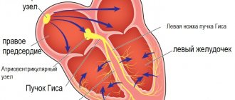
Normally, the regular contractions of our heart are supported by special cells that form the sinus node. This structure is located at the top of the right atrium. (Fig. 1)
Figure 1 Normal formation and propagation of electrical impulses
In some cases, the normal functioning of the heart may be disrupted; patients may experience an irregular, fast or slow pulse, pauses between contractions. All this is called heart rhythm disturbance or arrhythmia.
There are several types of arrhythmia:
Supraventricular rhythm disturbances:
- Atrial fibrillation and flutter
- Supraventricular paroxysmal tachycardia
- Atrial extrasystole
Ventricular arrhythmias:
- Ventricular extrasystole
- Ventricular tachycardia
What is paroxysmal tachycardia?
Under normal conditions, the normal heart rate is between 60 and 100 beats per minute. A heart rate greater than 100 beats per minute is called tachycardia.
With paroxysmal tachycardia, a sudden attack (paroxysm) of rapid heartbeat occurs, usually unrelated to physical activity.
Figure 2 Paroxysm of supraventricular tachycardia with a frequency of 180 bpm.
Paroxysm of tachycardia is a regular heartbeat with a frequency of usually 150 to 200 beats per minute, which is accompanied by weakness and shortness of breath. (Fig. 2) Sometimes patients may lose consciousness. In some patients, these episodes are short-lived and are not accompanied by significant symptoms.
How does extrasystole manifest?
An extrasystole is a premature contraction of the heart, which is felt by patients as a short-term interruption in the work of the heart, after which there may be a short pause followed by the restoration of a rhythmic heartbeat.
Treatment of arrhythmias
Both medicinal and surgical treatment methods are used to treat various types of arrhythmias. Among surgical treatment methods, the most common is catheter ablation.
What is ablation?
Catheter ablation is a minimally invasive operation that eliminates the “source” of arrhythmia using radiofrequency energy (RFA) or local cooling of heart tissue (cryoablation).
The emergence of catheter ablation techniques was the result of intensive development of science and the emergence of new medical technologies over the past 20 years.
Today, the catheter ablation method is widespread throughout the world (more than 1 million operations annually) and is the only treatment method that can radically eliminate a significant portion of cardiac arrhythmias in humans.
Types of catheter ablations
Most often in clinical practice, ultrahigh frequency (radio) frequency current is used - 500 kHz. This type of ablation is called radiofrequency catheter ablation (RFA). The effects of radiofrequency energy are carried out using special controlled catheters installed in the heart under X-ray control. (Fig. 3)
Figure 3 Placement of catheters in the heart during RFA.
Another type of impact on the source of arrhythmia is based on rapid, deep and local cooling of the heart tissue. This type of catheter ablation is called catheter cryoablation. Most often, catheter cryoablation is used to treat atrial fibrillation; a special cryoballoon catheter is used for this purpose. This type of surgery is called catheter balloon cryoablation. (Fig. 4)
Figure 4 Balloon placement in the left atrium during cryoablation.
What types of arrhythmias can be treated with catheter ablation?
All rhythm disturbances in humans are usually divided depending on the location of the “source” of arrhythmia in the human heart into “supraventricular” and “ventricular”. The cause (etiology) of arrhythmias in humans can be various pathological effects on the heart (inflammation, ischemia, etc.), or they can be caused by congenital features (anomalies) of heart development, incl. conduction system of the heart. Often it is not possible to detect the cause of arrhythmia. Such rhythm disturbances (in the absence of other heart diseases) are called “idiopathic”. The catheter ablation method allows in most cases to effectively eliminate arrhythmias that differ in both etiology and development mechanisms.
Benefits of Catheter Ablation
Treatment of heart rhythm disorders can be medication or interventional (surgical). It should be borne in mind that drug treatment involves long-term, often lifelong, antiarrhythmic drugs. Stopping medication or reducing the dose creates conditions for relapse of the arrhythmia. In addition, the use of medications is often impossible due to side effects or contraindicated due to the presence of concomitant heart diseases.
Currently, international and domestic recommendations for the treatment of cardiac arrhythmias consider catheter ablation as the main method of treating a significant part of arrhythmias. Such recommendations are based on the fact that catheter ablation can eliminate arrhythmia without further resorting to the use of antiarrhythmic drugs.
According to the guidelines, ablation is recommended in the following cases:
— as the main method of treatment for arrhythmias, where the use of catheter ablation is accompanied by high efficiency and safety (supraventricular tachycardia, atrial flutter)
-as an alternative treatment, usually when drug therapy is ineffective or side effects of antiarrhythmic drugs develop (atrial fibrillation, ventricular arrhythmias)
About catheter ablation
Preparation
Before performing RFA, the department performs the necessary examination (blood tests, instrumental diagnostic methods) for, as a rule, 2-3 days. On the day of surgery, the patient does not have breakfast; immediately before the operation, he puts on compression stockings (antithromboembolic stockings or elastic bandages).
RFA is performed by interventional arrhythmologists in a cath lab equipped with modern equipment for diagnostics and interventional treatment.
Ablation procedure
As the first stage of the operation, the so-called intracardiac electrophysiological study (ICE), the purpose of which is to clarify the type of tachyarrhythmia and search for the source of the arrhythmia. (Fig.3 and Fig.4)
To do this, a puncture of the vessels (femoral vein and, if necessary, artery) is performed, and special diagnostic electrodes are carried out under X-ray control. When performing VEPI, electrical stimulation of the heart of various parts of the heart is performed in accordance with diagnostic algorithms. This allows you to establish an accurate diagnosis and determine the location of the source of the arrhythmia.
In some cases (with atrial fibrillation), inhalation anesthesia is used as an anesthetic aid; in other cases, local anesthesia is used.
The duration of the operation is determined by its volume and takes from 1.5 to 3 hours.
After ablation
After RFA, a pressure bandage is applied to the puncture site of the vessel and the patient is transferred to the postoperative ward, where he is under the supervision of an anesthesiologist-resuscitator for at least 12 hours. All this time the patient is on strict bed rest.
To exclude possible complications after RFA, all patients undergo a comprehensive postoperative examination.
On average, the hospitalization period for RFA does not exceed 5 days.
Safety and effectiveness
The effectiveness of catheter ablation, depending on the type of arrhythmia, ranges from 70 to 99%. The risk of potential complications after surgery usually does not exceed 1%, but can reach 4-5% for the most complex heart rhythm disorders. The decision to carry out interventional treatment in each case is made by our specialists, taking into account the patient’s opinion, based on full information about the benefits and potential risks of surgical treatment.
Catheter ablation of atrial fibrillation
What is Atrial Fibrillation?
With atrial fibrillation (atrial fibrillation), instead of a regular heart rhythm, multiple electrical waves occur in the atria, leading to chaotic contractions of both atria at a very high frequency. [Rice. 5]
Figure 5. Atrial fibrillation
Atrial fibrillation (atrial fibrillation), as a rule, is manifested by a rapid, irregular heart rhythm, shortness of breath, and poor exercise tolerance. Often, atrial fibrillation is asymptomatic and is discovered accidentally during ECG recording.
Often, patients with atrial fibrillation (atrial fibrillation) are found to have another type of heart rhythm disorder - atrial flutter. [Fig.6]
Figure 6. Typical atrial flutter.
The symptoms of atrial flutter are not much different from atrial fibrillation. The exact diagnosis of these rhythm disturbances and treatment tactics should be determined by a cardiologist-arrhythmologist.
Why and how to treat atrial fibrillation?
Treatment of atrial fibrillation aims to: 1) eliminate the symptoms of arrhythmia, i.e. improving the quality of life of patients; 2) eliminating the threat of developing heart failure; 3) prevention of thromboembolic complications.
According to world medical statistics, atrial fibrillation (atrial fibrillation) is the most common (1-2% in the population) heart rhythm disorder. In a significant proportion of patients (up to 40%), AF is asymptomatic. In this category of patients, drug antiarrhythmic or non-drug treatment (catheter ablation) for AF is usually not carried out. Treatment of these patients consists of monitoring heart rate and administering anticoagulants to prevent thromboembolic complications.
Patients in whom atrial fibrillation is accompanied by the symptoms described above are prescribed continuous antiarrhythmic therapy aimed at preventing relapses of AF. In approximately one third of all patients with AF, it is possible to select an effective antiarrhythmic drug or their combination.
In 30% of patients with symptomatic, poorly tolerated atrial fibrillation, it is not possible to select effective antiarrhythmic therapy, or the use of antiarrhythmic drugs is contraindicated, is accompanied by the development of side effects, or patients do not want to adhere to long-term conservative drug treatment. In this category of patients, in accordance with modern international and Russian recommendations, catheter ablation is indicated.
It should be emphasized that the choice of treatment options in each specific case is the task of a cardiologist-arrhythmologist, taking into account the patient’s opinion and objective medical data.
Catheter and surgical ablation
Depending on the form of atrial fibrillation (paroxysmal, persistent or permanent), the presence of other pathologies of the cardiovascular system and concomitant diseases, 3 types of catheter (or surgical) ablation are used:
Intracardiac catheter ablation is the most widely used non-drug treatment for AF. Catheter ablation is performed in a cath lab using guided catheters moved into the chambers of the heart through vascular access (femoral and subclavian veins). The purpose of the operation is to radically eliminate the “sources” of arrhythmia in the left and (with atrial flutter) right atrium. Currently, two types of catheter ablation have found widespread clinical use: radiofrequency catheter ablation and balloon cryoablation.
· catheter ablation (destruction) of the AV node is a type of intracardiac catheter ablation, which is used in cases where AF is accompanied by a persistently high heart rate when drug control or radical elimination of AF is impossible. Ablation of the AV node is performed only after implantation of an artificial pacemaker (pacemaker).
· Operation “Labyrinth” – surgical ablation of AF. The “maze” operation (MAZE) is used in cases where a patient with AF is indicated for open-heart surgery due to the presence of an “underlying” heart disease: coronary artery bypass surgery, valve replacement, etc. As an independent intervention for AF, the “labyrinth” operation is used in the form of modified minimally invasive operations with thoracoscopic access (through a small incision in the chest) and only if previously performed attempts at catheter ablation are ineffective.
RFA or cryoablation?
According to modern concepts, the key role in the development of AF belongs to the so-called “arrhythmogenic” pulmonary veins (so-called AF triggers) - large vessels flowing into the left atrium. [Fig.7]
Figure 7. Left atrium and pulmonary veins. Multislice computed tomography
That is why most patients with paroxysmal and persistent forms of atrial fibrillation are indicated for catheter ablation (isolation) of the pulmonary veins.
How it works?
With radiofrequency catheter ablation, isolation of the pulmonary veins is achieved by applying a large number of targeted effects using high-frequency current. These effects should form a continuous chain of many sequential coagulative necrosis around each of the veins [Fig. 8A]. When using another technology - balloon cryoablation [video 1], a zone of necrosis around the veins is created due to the effect of low temperature (up to - 60ºC) in a cryoballoon located sequentially in each of the mouths of the pulmonary veins [Fig. 8B].
Rice. 8A - Radiofrequency catheter ablation (RFA); 1-circular diagnostic electrode of the “Lasso” type, 2-catheter for RFA
Rice. 8B — Catheter balloon cryoablation: 3- cryoballoon
Video 1. Balloon cryoablation.
In most cases, complete isolation is achieved with a single cryo-exposure for several minutes, which is an absolute advantage over radiofrequency ablation. Both types of catheter ablations are performed in a cath lab under general anesthesia or deep sedation. These interventions are high-tech types of medical care and should be performed by qualified specialists with sufficient experience in interventional interventions.
Efficiency and safety
The generally accepted definition of the effectiveness of catheter ablation for AF is the absence of any atrial arrhythmias after surgery without the use of antiarrhythmic drugs. Monitoring of effectiveness is carried out clinically (patient self-monitoring) or using long-term ECG recording systems (HM ECG or special implantable heart rate recorders).
One of the main factors determining the effectiveness of catheter ablation for AF is the duration of episodes of fibrillation. In cases where attacks of arrhythmia do not exceed several hours or days (the so-called paroxysmal form) and, as a rule, go away on their own, the operation is most effective. In comparative studies (the international Fire and Ice study), there were no relapses of AF during the first year in 65% of patients both after RFA and after balloon cryoablation. At the same time, there are observations that in individuals without concomitant cardiac pathology, the effectiveness of balloon cryoablation can reach 80-90%.
In patients with persistent AF, i.e. with arrhythmia lasting more than 7 days, and also requiring drug or electrical cardioversion to restore sinus rhythm, the expected effectiveness of catheter ablation is about 50-60%.
If, after catheter ablation, AF recurs with the same frequency and duration, re-intervention is justified.
Complications during catheter ablation of AF can manifest themselves in the form of vascular damage at the puncture site, perforation of the heart wall with the development of tamponade, the formation of blood clots in the heart cavity and thromboembolic complications, thermal damage to the esophagus, the development of phrenic nerve paresis and a number of others. The use of modern high-tech control methods during intracardiac interventions, sufficient experience and qualifications of the doctor allows these interventions to be performed effectively and without a significant risk of complications.
At the same time, it is necessary to clearly understand that the decision to carry out interventional treatment for AF should be made by a doctor with sufficient experience in treating this category of patients, objectively taking into account the pros and cons.
Catheter ablation of AF in the Department of Clinical Electrophysiology and X-ray Surgery of Rhythm Disorders
Interventional arrhythmology has been one of the main directions in the scientific and clinical work of the Department of Clinical Electrophysiology since its founding in 1990. It has almost 20 years of experience in treating various heart rhythm disorders using catheter ablation technology.
Since 2012, the department has introduced a catheter ablation method for AF. Today, the priority method used in the clinical practice of the Department of Clinical Electrophysiology for non-drug treatment of AF is the method of balloon cryoablation. This choice is based on the fact that cryoablation for AF is not inferior in effectiveness to radiofrequency, while being the safest method of interventional treatment of AF, which was proven by analyzing the many years of experience of the world's leading centers in the treatment of AF.
Specialists from the Department of Clinical Electrophysiology conduct a full preoperative examination of patients, perform interventional interventions and provide outpatient monitoring of all patients for at least 1 year after catheter cryoablation of AF. In cases where patients have complex concomitant heart rhythm disorders, complex (one-stage) interventional treatment, or the so-called. “hybrid therapy”, combining catheter intervention and subsequent drug treatment. If AF recurs, repeat balloon cryoablation or radiofrequency catheter ablation may be performed.
Radiofrequency ablation of paroxysmal supraventricular tachycardias
The most common types of paroxysmal supraventricular tachycardias are:
- Paroxysmal atrioventricular nodal reentrant tachycardia or AV nodal tachycardia. It is characterized by the presence of a congenital anomaly - an additional pathway in the AV node, which normally conducts impulses from the atria to the ventricles. This anomaly is the cause of tachycardia (Fig. 9);
Rice. 9. Paroxysm of AV nodal tachycardia on the ECG.
-Paroxysmal tachycardia in Wolff-Parkinson-White syndrome (WPW syndrome) also occurs due to a congenital anomaly - an additional pathway between the atria and ventricles (Kent's point). (Fig. 10);
Fig. 10 Kent bundle of left lateral localization
-Atrial tachycardia can have a source in both the left and right atria (Fig. 11), and can be a consequence of concomitant heart diseases (coronary heart disease, hypertension or valve defects), or have the so-called. idiopathic (without concomitant pathology) nature.
Fig. 11 Sources of atrial tachycardia in the left atrium (indicated by arrows)
An ECG recorded during a paroxysm of tachycardia often does not allow an accurate diagnosis to be made.
It can be clarified by such examination methods as transesophageal pacing (TEPS) or intracardiac electrophysiological study (ECES), which are standard methods for diagnosing arrhythmias.
Surgical treatment of the above types of tachycardias using radiofrequency ablation (RFA) allows you to completely get rid of arrhythmia; the effectiveness of the method is 97-99%. At the same time, the risk of potential complications of RFA, as a rule, does not exceed 1%.
Ventricular tachycardia
With ventricular tachycardia, a pathological impulse circulates in the myocardial tissues of the right or left ventricles.
This type of arrhythmia can occur in healthy individuals without any serious heart pathology. Often these heart rhythm disturbances have a benign course and do not always require treatment.
In other cases, ventricular tachycardia may be the result of serious illnesses such as myocardial infarction, which can cause sudden cardiac death. (Fig.12)
Figure 12 Ventricular tachycardia
Currently, non-drug treatment methods, such as radiofrequency ablation (RFA) or implantation of a cardioverter-defibrillator, are usually used to treat ventricular tachycardia.
To select the optimal treatment method, a comprehensive examination is necessary.
Indications for implantation
When drug treatment is not enough, the cardiologist recommends implantation of a pacemaker. The operation is intended to treat diseases:
- bradycardia, accompanied by constant fainting, loss of consciousness, dizziness, low pulse during physical exertion;
- atrial fibrillation;
- AV heart block with clinical manifestations;
- sick sinus syndrome;
- cardiomyopathy with impaired myocardial contractile function;
- chronic heart failure.
Relative indications include:
- AV heart block without clinical manifestations;
- fainting states are not associated with complete transverse block or ventricular tachycardia, but another cause of the condition cannot be established.
In this case, the decision on implantation is made by the doctor individually for each patient, taking into account physiological characteristics and the presence of concomitant pathologies.
In fact, there are no contraindications to installing an pacemaker. Surgery may be postponed due to viral or infectious diseases. After treatment of the concomitant pathology, the operation is carried out as planned after examinations.
Heart rhythm disturbances
Heart rhythm disturbances are based on a change in the conditions for the formation of excitation of the heart muscle or an anomaly in the paths of its propagation. Arrhythmias can be caused by both functional disorders and severe organic lesions of the heart. In some cases, the cause of heart rhythm disturbances is the congenital characteristics of the conduction system of the heart. The state of the nervous system plays a certain role in the occurrence of arrhythmia. For example, mental and emotional stress causes changes in the pace, and often in the rhythm, of heart contractions, including in healthy people. Arrhythmia often occurs in people with diseases of the central and autonomic nervous system.
Various diseases, accompanied by a violation of the anatomical structure of the heart or the metabolic processes occurring in it, cause types of arrhythmia of different duration and nature, and only a doctor can make a diagnosis, whose conclusions are based on clinical and electrocardiographic data.
Symptoms of arrhythmias
Quite often, patients do not feel the presence of arrhythmias. In these cases, arrhythmia is detected during a routine clinical examination. However, many patients experience a variety of sensations in the chest, which most often include:
- feeling of palpitations and chest pains;
- very rapid heartbeat;
- extremely slow heartbeat (“fading” of the heart);
- chest pain;
- dizziness;
- loss of consciousness or a feeling close to fainting.
Classification of arrhythmias
1. Automatic disorders:
a) nomotopic (pacemaker - in the sinus node):
- sinus tachycardia;
- sinus bradycardia;
- sinus arrhythmia;
- sick sinus syndrome (SSNS).
b) heterotopic (pacemaker - outside the sinus node):
- lower atrial rhythm;
- atrioventricular rhythm;
- idioventricular rhythm.
2. Excitability disorders:
a) extrasystoles:
- by source: atrial, atrioventricular, ventricular;
- by number of sources: monotopic, polytopic;
- by time of occurrence: early, interpolated, late;
- by frequency: single (up to 5 per minute), multiple (more than 5 per minute), paired, group;
- according to order: disordered, allorhythms (bigeminy, trigeminy, quadrigeminy).
b) paroxysmal tachycardia (atrial, AV, ventricular)
3. Conduction disorders
:
a) increased conductivity (WPW syndrome); b) decreased conductivity (blockades: sinoauricular, intraatrial, AV, bundle branch block).
4. Mixed (atrial flutter/fibrillation, ventricular fibrillation)
Diagnostics
ECG is the main method for diagnosing arrhythmias. However, it is not always possible to detect arrhythmia when taking a resting ECG. In such cases, the K+31 clinic performs 24-hour ECG monitoring (Holter monitoring). Some arrhythmias are provoked by physical activity; in such cases, stress tests (treadmill) are used to diagnose them.
Complications
A number of rhythm disturbances can lead to the development of severe complications. If the rhythm of contractions in the heart is abnormal, blood flow slows down, which leads to the formation of blood clots in the atria. In the absence of adequate therapy, these blood clots can spread through the bloodstream throughout the body, leading to blockage of the arteries of the brain (stroke), intestinal arteries, and lower extremities. Long-term existence of uncorrectable arrhythmia often leads to the development of heart failure.
Treatment of arrhythmias
- if arrhythmia is a consequence of any disease (for example, thyrotoxicosis), our doctors treat the underlying disease;
- antiarrhythmic drugs are prescribed (affecting the conduction system of the heart, as well as various ion channels).
To prevent the development of complications, dynamic monitoring of the patient by a cardiologist is recommended, even during the period of remission.
Preparing for surgery
Before the operation, the patient must undergo a full examination and pass a list of tests. Our cardiology center of the Federal Scientific and Clinical Center FMBA offers its patients planned hospitalization. During your stay in the cardiovascular surgery department, you will undergo all the necessary tests. The list of examinations includes:
- clinical and biochemical blood test (total protein, creatinine, urea, total bilirubin, direct bilirubin, glucose, pancreatic amylase);
- determination of blood group and Rh factor;
- detailed coagulogram;
- platelet aggregation indicator ADP;
- markers of infections: HIV, hepatitis B, hepatitis C, syphilis;
- general urine analysis;
- chest x-ray;
- ECG;
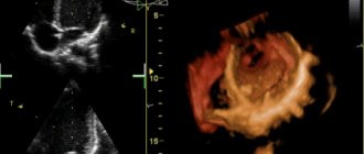
- EchoCG;
- EGDS;
- Ultrasound scanning of brachiocephalic arteries;
- Ultrasound scanning of the arteries of the lower extremities and veins of the lower extremities;
- FVD;
- Ultrasound of internal organs;
- TEE;
- coronary angiography.
If during the examination the doctor discovers a concomitant pathology, you may be prescribed additional tests and consultation with related specialists (neurologist, ophthalmologist, endocrinologist, etc.).
Our center is equipped with high-precision modern equipment. Thanks to their extensive experience and many years of work experience, doctors guarantee the most accurate diagnosis and interpretation of indicators. Our own clinical diagnostic center is responsible for the accuracy of test results. By undergoing an examination with us before implantation of an pacemaker, you can be confident in the quality of the services provided and the speed of their provision. Collecting all the necessary research can take months, while we will prepare you for surgery in just 1 day.
Before the operation, the patient needs to prepare; to do this, just follow simple recommendations:
- A few days before surgery, the doctor may stop taking medications. Alcohol must be eliminated (preferably a week in advance);
- on the day before pacemaker implantation, the patient has breakfast and lunch as usual. In the evening, a light dinner is brought, the last meal should be no later than 6-8 hours before the operation. You can drink water in unlimited quantities;
- at night the patient is given a cleansing enema;
- In the morning, the patient takes a shower and prepares the surgical field. It is necessary to shave the hair in the chest area.
Before the patient is taken to the operating room, he is examined by a surgeon and an anesthesiologist. A mild sedative may be administered to calm and relieve anxiety.
Implantation of a pacemaker
The operation to implant a pacemaker is performed under local anesthesia. Depending on the type of pacemaker, the procedure can take from half an hour when installing a single-chamber pacemaker, to 2.5 when installing a three-chamber pacemaker. If the patient is right-handed, the pacemaker is installed in the left subclavian region, if left-handed - in the right. This allows you to reduce the impact of the pacemaker on the working arm.
The cardiac surgeon and assisting staff perform the following actions:
- the surgical field is treated with antiseptics and anesthetized;
- the surgeon makes a small incision about 5 cm under the collarbone and creates a pocket for implantation of the pacemaker. The pocket may be located under or above the pectoralis major muscle. The location is determined by the surgeon;
- From the incision, the subclavian vein is punctured (pierced) with a special needle;
- A conductor is inserted into the vein through a needle into the cavity of the heart. The doctor controls the movement of the conductor using an X-ray unit;
- the needle is removed and an introducer (plastic tube) is inserted into the vein along the guidewire. Depending on the type of pacemaker, if it is necessary to introduce 2 or three electrodes, an additional introducer is inserted along the same path or through a puncture of another vein;
- the conductor is removed;
- 1 or several electrodes are inserted into the heart cavity through the introducer. The electrodes are attached to the inner lining of the heart using hooks at the end of the electrode or using a special fastener that resembles a corkscrew (the electrode seems to be screwed into the inner lining);
- when the electrodes are installed in the desired location, the introducer is removed;
- the doctor connects the other end of the electrodes to the pacemaker;
- the pacemaker is implanted into a previously formed pocket under the collarbone;
- the wound is sutured;
- an aseptic dressing is applied;
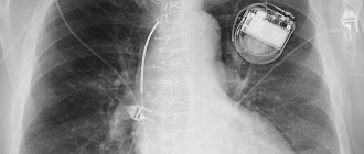
Pacemaker implantation
Pacemaker implantation
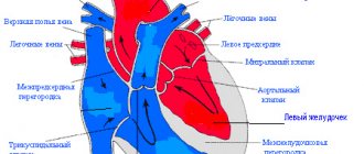
Electrical system of the heart
Your heart has its own electrical system that makes it beat and controls its rate. It includes two nodes and many pathways, organic or functional. Special tissues generate electrical signals that travel along the heart's "electrical wires" with each beat. The pacemaker in a healthy person is the sinus node, located at the posterior wall of the right atrium near the confluence of the superior vena cava. After the impulse is born here, it is carried out to the atria, causing them to contract and push blood into the ventricles. The impulse is then conducted to the ventricles. However, on the way to them, it lingers in the second node - the atrioventricular one, and only then spreads through the conduction system of the ventricles: the branches of the His bundle and the Purkinje fibers.
The complexity and sensitive response of this system make it sensitive to the slightest changes in the body. Breakage can occur at every stage of the pulse's movement.
This explains the fact that not only cardiological, but essentially any pathology can cause cardiac arrhythmias. A pacemaker (pacemaker) is for people whose hearts beat too slowly or irregularly. It is an excellent substitute for the natural pacemaker of the heart and restores one of the most essential rhythms of life - the rhythm of the human heart. Since the first pacemaker was successfully implanted, more than two million people have benefited from this remarkable invention. The pacemaker sends tiny electrical impulses to the heart muscle. Electrical impulses are individually set and precisely dosed, forcing the heart to work within its natural rhythm range.
Pacemaker device
To fully work, the electrical stimulation system must consist of two parts - an ECS unit and an electrode: The ECS unit (also called a pulse generator) is a small device weighing 22 - 45 g and measuring no more than 55 mm. It contains a battery and a microcircuit enclosed in a titanium case, which allows the pacemaker to remain in the human body for a long time without showing a negative reaction from the body.
The ECS block includes three main components:
- Battery - provides electrical energy for the pacemaker. This is a small, sealed lithium iodine battery that typically lasts for many years.
- The electronic circuit is similar to a mini-computer inside the ECS. It transforms energy from the battery into tiny electrical signals that are not felt by the patient. The circuit controls the timing of impulses directed to the heart. The characteristics of the quartz oscillator are so stable that they practically do not change during the entire service life of the ECS.
- The connecting block is a plastic connector located in the upper part of the metal casing of the pacemaker, where the electrode is in contact with the device.
- An electrode is an insulated wire connected to the pacemaker that carries an electrical impulse from the machine to the heart and transmits information regarding the natural activity of the heart back to the pacemaker. The electrodes are extremely flexible to resist twisting and bending caused by contractions of the heart itself or body movements. One end of the electrode is connected to the pacemaker in the connector block. The other end is installed in the right ventricle or right atrium of the heart. Depending on the type of pacemaker prescribed by your doctor, one or two electrodes are used.
The pacemaker manufacturer checks each component individually and the entire system as a whole at each stage of production. The pacemaker monitors information regarding the natural activity of the heart. The myocardium's own electrical signals are captured by the electrode and transmitted to the pacemaker sensor circuit, where extraneous noise and interference are filtered. If, when analyzing received signals and programmed parameters, the frequency of heart potentials is less than programmed, the electrical pulse generator will begin electrical stimulation of the heart.
Types of EX
In 1952, the first report of the successful use of pacemaker in the United States appeared. But the method used had a significant drawback: the pacemaker was located outside the body and the electric current it generated was conducted to the heart through wires through the skin. As a result, dangerous infections spread along the wires to the heart, putting patients' lives at great risk. In 1958, Swedish doctors successfully implanted a pacemaker under a patient's skin. This was a huge step forward, even though the battery had to be recharged outside every week. In the early 70s, pacemakers weighed 120 g, and patients were re-operated every 3 years. But the technology became more and more reliable, and the devices themselves became smaller and lighter. Now the pacemaker has become a common thing in the treatment of patients with heart rhythm disturbances. This fact is the best characteristic of such devices; it means that people have accepted this amazing remedy as reliable and trustworthy.
Not all patients have the same pathology of the cardiac conduction system. Taking this into account, various types of pacemakers have been designed:
- single-chamber and multi-chamber;
- with and without frequency adaptation;
- with telemetry and anti-tachy functions.
Currently, more than 100 ECS models are known. The type of pacemaker that will be implanted depends on the disease, concomitant pathology, and physical properties of the heart. You can be absolutely sure that the doctor is familiar with all existing options and will be able to choose the most suitable model.
Indications for pacemaker implantation:
- Sick sinus syndrome. This is a disease in which the sinus node, the heart's natural driver, generates electrical impulses too slowly. Sometimes, he simply “forgets” about his work. And then there is a pause during which the heart stops. The first organ to respond to a lack of blood is the brain. Due to inadequate oxygen supply, severe weakness, dizziness, darkening of the eyes and loss of consciousness for several seconds occur. These episodes are called Morgagni-Adams-Stokes attacks, named after the doctors who described them 200 years ago.
- AV block. In this disease, electrical impulses are blocked on the way from the atria to the ventricles. However, the heart is protected from such an unexpected error of nature; otherwise, it would stop beating. In this case, another part of the ventricle takes on the function of a pacemaker and makes the heart work, but with less efficiency. Thus, the chambers of the heart have different pacemakers and contract independently of each other: the atria - often, the ventricles - rarely (30-60 per minute).
- A combination of both forms of bradycardia. Bradycardia is a consequence of heart disease, myocardial infarction, cardiac surgery or the aging process. The exact cause may not be known.
- Atrial fibrillation with long pauses between heartbeats.
- Condition after some arrhythmological operations. In some cases, tachycardia requires surgery to eliminate this arrhythmogenic focus. To do this, the surgeon inserts several electrodes into the heart cavity through the great vessels and treats the area of the atrioventricular node with radiofrequency energy, stopping conduction from the atria to the ventricles. After this operation, the ventricles begin to contract rhythmically, but slowly, and this requires the installation of an pacemaker.
- Chronotropic competence of the myocardium. Insufficient increase in heart rate during exercise and sufficient myocardial contraction at rest.
Preparing for surgery
Once the patient has been admitted to the hospital, a brief preoperative assessment will be performed. Before the operation, the doctor will give special instructions. Some of them will be as follows:
1. Avoid eating any food or drinking any liquids after midnight the night before surgery
2. The evening before your implantation procedure and 30 minutes before you leave your room and go to the operating room, you will receive some sedatives and anesthetics (pain-relieving medications).
Progress of the operation
The pacemaker can be implanted in the endocardial or myocardial version. The first method is most often used. Endocardial implantation is performed under local anesthesia. The operating surgeon will treat the surgical field and numb it with several injections with an anesthetic - novocaine. The doctor will inject as much novocaine as needed to prevent pain. During the operation, the patient may not experience any sensations or feel that the doctor is performing some actions in the subclavian area. During surgery, the patient may doze or feel sleepy.
The vast majority of operations have the following sequence: A small incision 5-7 cm long is made under the collarbone (parallel to it). The location of the area where the pacemaker is placed (right or left) depends on the patient’s disease, age and lifestyle. The surgeon will then find and catheterize the subclavian vein and pass the endocardial lead through it into the heart. The electrode will be advanced under the control of an X-ray machine until it reaches the desired chamber of the heart.
Implantation of a single-chamber pacemaker requires only one electrode. The patient's disease determines which chamber the electrode is placed in (atrium or ventricle). When implanting a dual-chamber pacemaker, two electrodes are used. In this case, one electrode is passed into the right ventricle, and then the other electrode is passed into the right atrium appendage. After inserting the electrode into the appropriate chamber of the heart, the doctor will measure the electrical parameters of several areas of the heart muscle and place the tip of the electrode at the point with the lowest electrical resistance.
Then the outer end of the electrode will be connected to the pacemaker block, which is immersed in the subcutaneous fatty tissue here in the subclavian region. The operation ends with suturing the incision. The total duration of the operation, as a rule, does not exceed 1.5 - 2 hours.
The period after pacemaker implantation
The postoperative rehabilitation period after pacemaker installation is quite short. During this time, the patient may experience some pain around the pacemaker implantation site, in which case the doctor will prescribe a pain reliever. Of course, it will take some time to get used to the pacemaker, but the feeling of discomfort will pass very soon. At the first post-operative examination, the doctor will check the electrical activity of the heart and the functioning of the pacemaker. If necessary, the parameters of the pacemaker program will be adjusted to the individual needs of the patient. On average, the examination schedule includes visits to the doctor every 6 months. Almost immediately after the operation, the patient can continue to engage in his usual activities - working in the garden or around the house, driving a car. The attending physician will recommend what kind of physical activity the patient can handle, from what time it will be possible to resume sports, favorite activities, and resume sexual activity.
Stimulator operating life
As a rule, the operating life of a modern pacemaker is from 5 to 10 years.
How is a pacemaker replaced?
Pacemaker replacement is a safe and simple procedure. During the process of replacing the pacemaker, the properly functioning electrodes are preserved and the doctor only connects them to the new pacemaker.
Recommendations for patients with an implanted pacemaker
From time to time, a person with a pacemaker needs to take their pulse. Assessing the rhythm of the pulse and the difference in heart rate at rest and during movements of different tempos allows us to judge the serviceability of the device. The patient should periodically measure the pulse for a minute while sitting or lying down. Along with the pulse, it is important to monitor the appearance of shortness of breath during movements, since its intensification indicates a discrepancy between the performance of the heart and the required physical effort. It will be very useful to keep a diary in which you can record your heart rate, date and time of counting. If necessary, the patient can be helped by his family members.
If you suspect a malfunction of the device and any changes in the patient’s condition, you must inform your doctor. Using special equipment, the doctor can check the operation of the pacemaker and compare it with the condition of the heart, echocardiography data and the patient’s lifestyle. Only a specialist can adjust the frequency of the pacemaker pulses, recommend the permissible level of stress and, if necessary, prescribe the necessary medications.
The opinion that a person with a stimulator is deeply disabled and must constantly protect himself from unnecessary movements is fundamentally wrong. If the heart has not been seriously damaged by the disease, then acceptable physical activity will only be beneficial for it. Provided that the operation was successful and the device is working normally, the patient can begin his work within two to three weeks. At the same time, the permissible severity of work is determined by the condition of his heart and physical fitness. A person with a modern pacemaker can feel practically healthy if he does not have significant myocardial damage, valve defects or blockage of the coronary arteries.
Precautions to be observed by a patient with an installed pacemaker
Appliances
Most often, household electrical appliances are absolutely safe and should not affect the operation of the pacemaker. In addition, the pacemaker device provides built-in protection against interference from other electrical equipment. If the patient suspects that an electrical device is interfering with the operation of his pacemaker (causing dizziness and increased heart rate), simply move away from the device or turn it off. There are no permanent changes in the stimulator itself, so it will resume its normal mode of operation after the end of the external influence. If the patient is sure that interference in the operation of the device is associated with the functioning of household appliances, it would be advisable to consult a doctor so that he can check the pacemaker settings.
To reduce the likelihood of interference from household appliances, it is recommended to keep hand-held electrical appliances at a distance of at least 10 cm from the location of the pacemaker. A patient with pacemaker should not lean against the screen of a switched-on TV or the front wall of a microwave oven, or repair electrical devices without unplugging them.
Radio wave generators (cell phones, two-way transceivers) and working electric soldering irons, demagnetizing devices, devices with motors (electric drills, hair dryers, electric shavers) are recommended to be located away from the pacemaker. Also, do not keep magnets or magnetized materials close to the stimulator.
Office equipment and work equipment
Serviceable grounded equipment and working equipment should not interfere with the operation of the ECS. This equipment includes: computers, electric typewriters; copying and fax machines; carpentry workshop equipment (electric planer, electric drill, etc.); locksmith equipment. The patient should avoid possible sources of electrical interference when operating electric saws, welding equipment, dielectric heaters, and electric steel-smelting furnaces. In general, it is not recommended to work with equipment that cannot be switched off immediately.
Other precautions
Be careful when near the distributor coil or spark plug wires of a running car engine. Any adjustment of the distributor must be done with the engine off.
Patients with a pacemaker are not recommended to be near high-voltage power lines, powerful electrical microwave installations and transmitters. High-voltage power lines should be crossed near towers.
Patients with pacemaker are not recommended to undergo security screening at airports or theft detectors in stores, libraries or museums (you should always have your pacemaker card with you). If necessary, it is better to replace this procedure with a personal search.
After operation
The patient is under the supervision of medical personnel for a week. During this time, the sutures heal and can be removed. The patient is preparing for discharge. Before sending the patient home, he is examined by a doctor. Makes appointments and issues instructions on how to behave during the rehabilitation period and move on with your life.
After discharge, the patient registers at his place of residence and promptly visits the doctor. In the first 3 months, it is important to follow the recommendations:
- temporarily stop physical activity;
- you cannot lift weights;
- You can’t drive;
- MRI cannot be done;
- avoid radar technology;
- refuse thermal procedures;
- Report any ailments to your doctor.
You should visit a cardiologist regularly for the next six months. It is allowed to use household appliances. You need to turn them on with the opposite hand to the installed pacemaker, including when talking on the phone, and place the phone on the opposite side of the installed pacemaker. Metal detectors should be avoided in stores, airports, and concert halls.
Lifetime rules after pacemaker implantation:
- monthly visit to a cardiologist;
- daily measurement of blood pressure and pulse. It is advisable to keep a diary and mark the data;
- complete cessation of bad habits;
- carry your pacemaker passport with you.