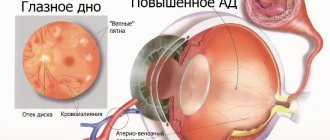
The main cause of incurable blindness is usually vascular diseases of the eyes. Obstruction of a vessel or insufficient blood flow for normal blood circulation leads to heart attacks, microinfarctions, or simply chronic oxygen deficiency in the vessels of the organ of vision.
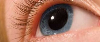
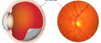
Oxygen starvation is especially dangerous for the most important layer of the eye - the photosensitive layer of the retina. The complex structure of the retina dictates especially high requirements for blood supply in its structures. If the central retinal artery is completely obstructed, vision can disappear in a few seconds!
A characteristic feature of all vascular diseases of the retina is a local circulatory disorder, leading to arterial obstruction and vein thrombosis.
Among the vascular diseases of the retina, the following are the most common:
- retinal edema;
- diabetic retinopathy;
- retinal angiopathy;
- retinal vein thrombosis;
- central retinal artery occlusion ;
- occlusion of the central retinal vein and its branches.
1 Structure of the eye and eyeball
2 Diagnosis and treatment of vascular diseases of the retina
3 Treatment of vascular diseases of the retina
Retinal swelling
Retinal edema is associated with vascular damage and accumulation of moisture and protein structures in the retina. This is not an independent disease, but a symptom of some pathology. For example, macular edema of the retina can occur as a result of diabetes mellitus, vascular and inflammatory diseases.
Diabetic retinopathy
Diabetic retinopathy is a rather insidious disease that develops against the background of elevated blood sugar levels and has no symptoms in the initial stages. If traumatic retinopathy occurs as a result of eye injury and is manifested by swelling and hemorrhage, then diabetic retinopathy develops quietly and unnoticeably.
However, if treatment is not started early, retinal detachment can occur, leading to blindness. This type of disease is characterized by neovascularization - pathological growth of blood vessels.
More detailed information about this vascular disease of the retina can be found here.
Retinal angiopathy
Angiopathy is a lesion of retinal vessels of different diameters. With angiopathy, the vessels become narrow and tortuous. Among the reasons for the development of this pathology are the following diseases: diabetes mellitus, eye injury, vasculitis, cervical osteochondrosis, high blood pressure. You can read about the symptoms of this disease and what methods are most effective for treating it here.
Retinal vein thrombosis
Symptoms of thrombosis of the central branch of the retina are simple: the appearance of blind spots in the visual field or a noticeable but painless decrease in vision in one eye. Most often, this pathology affects elderly people.
The provoking factors of this retinal vascular disease are:
- diabetes;
- high blood pressure;
- atherosclerosis;
- glaucoma;
- taking contraceptive medications, diuretics;
- leukemia, polycythemia and other conditions leading to increased blood viscosity.
When unfavorable conditions arise, the walls of the arteries thicken and become dense, which leads to compression of the adjacent vein. In the vein, blood flow slows down, forming a blood clot (thrombus).
Blood stagnation increases vascular permeability, the liquid part of the blood fills the vascular space, which leads to retinal edema and hemorrhages.
Retinal vein thrombosis can appear on the central vein and in its branches, developing in ischemic and non-ischemic types .
With non-ischemic damage, slight changes appear in the fundus of the eye, and a slight drop in visual acuity is observed.
The ischemic type is characterized by the occurrence of multiple hemorrhages and cotton wool-shaped (blind) lesions in the retina. In this case, a significant deterioration in vision occurs.
Due to the risk of various complications with such disorders, it is necessary to come for an examination by an ophthalmologist every 2 weeks.
1 Diagnosis and treatment of vascular diseases of the retina
2 Diagnosis and treatment of vascular diseases of the retina
3 Diagnosis and treatment of vascular diseases of the retina
Occlusion of the central retinal vein and its branches
Occlusion of the veins and central retinal artery, due to the speed of changes occurring, is sometimes called a “vascular catastrophe”, which results in a rapid decline in vision.
Occlusion of the central retinal vein is a deterioration in the blood circulation of the central retinal vein and its branches.
With this vascular disease of the retina, the patency of the central vein is completely disrupted, and pinpoint hemorrhages occur in all squares of the retina.
Tortuosity and dilatation of veins, cotton wool lesions, papilledema, and the appearance of neovascularization of the retina, iris, and disc are observed. Within ten days, vision (in one eye) will continue to decline, but this will not lead to complete blindness.
Central retinal artery occlusion
When the central retinal artery is occluded, a symptom such as sudden loss of vision is observed.
A person can only distinguish between light and darkness and can barely see his fingers in front of his eyes. Before the onset of the disease, the patient develops short-term episodes of complete blindness. This disease occurs 2 times more often in men than in women. Occlusion of the central retinal artery can occur in both 25-year-olds and people over 85 years of age.
When the peripheral branches of the retinal arteries become inflamed, the patient experiences partial loss of vision.
Angiospasm of the retina - what is it?
Retinal angiospasm is a dysfunction of the human visual apparatus, which is preceded by an atypical sharp narrowing of the central artery of the retina, as well as the vessels branching from it. In this case, there are no changes at the organic level in the vascular wall.
There are a number of clinical symptoms characteristic of this pathology, namely:
- The appearance of dots before the eyes, which patients often call “spots”;
- Blurred vision, difficulty focusing;
- Discomfort appearing in the periorbital region;
- Distorted perception of color, shape and size of nearby or distant objects.
In more than 90% of cases, such disorders are caused by insufficiently good blood flow in the central artery. They occur due to improper functioning of the human cardiovascular system. Most often, the disease begins to progress against the background of arterial hypertension or atherosclerosis. In approximately 25% of cases, doctors cannot determine the etiology of vascular spasms in the retina.
Ophthalmologists diagnose vasospasm in patients at any age, but in most cases in people over 40 years of age. It has been scientifically proven that men are 2 times more likely to go to medical institutions with a similar problem than women.
Spastic contraction in the middle layers of the retinal arteries in children is usually caused by pathologies in the structure of the peripheral nervous system, including autonomic dysfunction. In the matter of the etiology of this disease, modern medicine is practically powerless - there is no reliably proven assumption regarding the nature and causes of this pathology.
Angiospasm is a disease of the retina, which in adults in the vast majority of cases occurs against the background of other diseases, such as:
- Diabetes mellitus is a disruption of the endocrine system, which is caused by a relative or absolute deficiency of the pancreatic hormone insulin, as a result of which an increase in the level of glucose in the patient’s blood is observed.
- Hypertension is a malfunction of the cardiovascular system. The disease occurs against the background of dysfunction of vascular regulation centers.
- Atherosclerosis is a chronic artery disease caused by disturbances in a wide group of different compounds, i.e. lipids (fatty acids are among them), and is accompanied by an increased content of cholesterol in the inner lining of blood vessels.
Angiospasm of the retina can occur as a result of intoxication, in which an unregulated increase in the tone of the vascular wall occurs. This is possible in case of poisoning with heavy elements such as lead or carbon disulfide. Corresponding reactions are not uncommon among people employed in hazardous work environments who are diagnosed with chronic intoxication syndrome. The pathology is characterized by bilateral damage to the paired organs of vision (angiospasm of the retina of both eyes also often indicates a disorder of the nervous regulation of tone).
Adherence to bad habits is another point that is highlighted by doctors as one of the fundamental factors in the development of functional disorders of the central nervous system. Narrowing of the retinal artery can occur due to exposure to nicotine and tar in smokers, as well as alcohol in those who abuse alcoholic beverages. These harmful substances lead to temporary deformation of the walls of blood vessels, but gradually such effects provoke dilatation.
Causes of angiopathy
Changes in retinal vessels can be caused by various reasons. This pathology develops differently and is the object of study not only by an ophthalmologist, but also by other doctors, such as a neurologist, endocrinologist, etc. This is due to the fact that in the eye the veins and arteries of the retina are accessible for observation, unlike the blood supply to other human organs .
There is a huge list of reasons leading to the development of angiopathy in both eyes. Here are just a few of them:
- Diabetes.
- Smoking.
- Blood diseases.
- Osteochondrosis of the cervical spine.
- Arterial or intracranial hypertension.
- Diseases of the nervous system.
- Hypotension or hypertension.
- Autoimmune diseases.
- Injuries.
- Heredity.
- Elderly age.
All changes impair the nutrition of the retina, resulting in oxygen starvation.
Diagnosis of retinal vasospasm
Anamnestic data, the results of a visual examination in combination with additional diagnostic methods allow the ophthalmologist to establish vasospasm of the retinal vessels as a diagnosis. The list of studies within the framework of pathology detection includes:
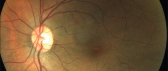
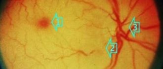
- Ophthalmoscopy. After examining the fundus of the eye, the doctor visually determines a sharp narrowing of the veins in the retina. The vessels are full of blood, but a somewhat swollen, pale pink optic disc is visible. The foveal reflex, like the macular reflex, is not diagnosed during vasospasm.
- OCT. During scanning of the central part of the retina, thickening is visible. In this case, the foveal recess is greatly smoothed, and retinal reactivity is reduced, while the shape of the curve is atypically straight.
- Non-contact tonometry method. During an attack, the specialist observes a slight increase in IOP (intraocular pressure), and after its relief, a normalization of ophthalmotonus.
- Angiography. The ophthalmologist visualizes the changes taking place in the retinal vessels. Diagnosis occurs as part of studying the characteristics of fluorescein circulation. During vasospasm, the internal blood-retinal barrier for contrast remains impenetrable.
Publications in the media
Retinal vein occlusion is a circulatory disorder in the central retinal vein or its branches. The predominant age is over 40–50 years. Etiology • Narrowing of the lumen of the vessel (hypertension, atherosclerosis) • Changes in the rheological properties of blood (DM, retinal angiitis) • Disturbance of the homeostasis system (glaucoma, eye injuries).
Classification • Thrombosis of the central retinal vein • Thrombosis of the branches of the central retinal vein • Postthrombotic retinopathy. Pathogenesis - microcirculation disorders • Compression of blood vessels • Arterial spasm of the accompanying artery. Pathomorphology • Damage to the venous endothelium • Impaired vascular wall permeability • Retinal edema.
Clinical picture • Phenomena of incomplete retinal vein thrombosis •• Venous stagnation •• Vein dilation •• Macular edema •• Hemorrhages in the posterior pole of the retina and on the periphery •• Visual functions are slightly reduced. • Retinal vein thrombosis begins with a unilateral sudden and severe decrease in vision. • With thrombosis of the central vein in the fundus of the eye, the retinal veins are sharply dilated and tortuous; multiple massive hemorrhages in the layer of nerve fibers in the form of rays directed from the optic nerve to the periphery; swelling of the retina and macula. • With thrombosis of the branches of the central retinal vein, a similar picture occurs in the area of the affected branch. • In postthrombotic retinopathy, recanalization of thrombosed vessels, development of collaterals and shunts, retinal neovascularization, microaneurysms, cystic macular degeneration, and hard exudate deposits are observed.
Diagnostics • Ophthalmoscopy • Fluorescein angiography. Differential diagnosis • Diabetic retinopathy • Hypertensive retinopathy. Treatment • In the acute stage •• Intravenous infusions of fibrinolysin 20,000 units and heparin 10,000 units (4-6 injections in total), alternate with intravenous infusion of rheopolyglucin •• Retrobulbar injections of RDG (reopolyglucin - 0.2, dexon - 0.2, heparin - 1000 units) and fibrinolysin 700–1000 units (alternate) •• Pentoxifylline 0.2 orally 3 times a day or 0.1 intravenously daily •• Etamsylate 250 mg orally 3 times a day or intramuscularly • • Acetylsalicylic acid 500 mg 3 times a day •• Antispasmodics, for example papaverine hydrochloride •• Diuretics, for example furosemide, acetazolamide • Laser coagulation of the retina •• For macular edema, barrier coagulation is indicated •• For postthrombotic retinopathy - sectoral (for thrombosis of the branches) or panretinal (for central vein thrombosis) coagulation of the retina to close ischemic areas and destroy neovascular complexes. Complications • Optic nerve atrophy • Secondary neovascular glaucoma • Cystic macular degeneration
The prognosis depends on the size of the affected area of the retinal veins, the severity of macular edema and the timeliness of treatment procedures. However, in all cases the outcome is some degree of reduction in visual acuity.
ICD-10 • H34 Retinal vascular occlusions
Angiospasm of the retina: treatment
For vasospasm, etiotropic therapy is not carried out, since it has not yet been developed in modern medicine. Methods of pathogenetic treatment of the disease are used, which are aimed at reducing pressure and dilating blood vessels undergoing spasms, as well as restoring impaired blood flow in the ischemic zone.
If the unfavorable manifestations of vasospasm are not eliminated in a timely manner, this can lead to a severe decrease in vision and, as a consequence, to a partial or complete loss of the ability to see.
Therefore, the doctor, having diagnosed the corresponding pathology in the patient, immediately after the examination prescribes antispasmodics to the patient. As part of treatment, electrophoresis and vasodilators are recommended.
Carbonic anhydrase inhibitors, as well as saluretics, help reduce intraocular pressure. In cases where the desired effect does not occur, a special irrigation system can be installed in the retrobulbar space. The pathology is also stopped by instillation of solutions of special drugs: beta-adrenergic receptor blockers. Sometimes medications are administered intramuscularly (this mainly happens when the disease is accompanied by high blood pressure).
Short-term narrowing of small branches of the central nervous system is not considered dangerous, but if the attack lasts more than 10-15 minutes, irreversible serious changes occur in the retina due to lack of trophism. Therefore, the prognosis for the treatment of the disease varies - it all depends on the form of the pathology. As for the prevention of vasospasm, it usually comes down to enhanced monitoring of blood pressure, as well as blood glucose levels.
On the Ochkov.Net website you can view a large selection of contact lenses from popular manufacturers and order them in a few clicks.
Treatment of angiopathy
Various methods are used in the treatment of retinal angiopathy. Therefore, it is important to understand exactly what form of the disease arose. After identifying the main cause, treatment is prescribed, aimed primarily at its correction, and the following is prescribed as an auxiliary treatment:
- Medications, eye drops.
- A low-calorie diet with limited simple carbohydrates and salt.
- Physiotherapeutic procedures.
- Massage of the cervical-collar area.
To prevent the appearance of angiopathy or delay its progression in case of damage to the ocular vessels, it is recommended to consult a specialist in a timely manner. Early initiation of treatment guarantees a favorable prognosis. If you have any chronic diseases or symptoms of visual impairment, do not delay visiting an ophthalmologist. Proper treatment and improved lifestyle can delay the development of the disease for a long time.
Manifestations of the disease
Retinal vascular angiopathy is painless and practically asymptomatic and is detected during an ophthalmological examination. Patients may complain of decreased visual acuity and blurred vision; progression of myopia; the appearance of lightning and flashes in the eyes.
In some cases, increased nosebleeds and temporary loss of vision are noted. An ophthalmological examination reveals retinal dystrophy.