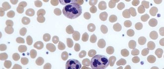
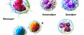
Leukocytes are white blood cells that are very important components of the blood. They differ in their structure and functions; the main feature in the structure is the presence or absence of specific granules capable of perceiving color.
Several methods are used to count white blood cells. In order to identify inflammatory diseases in the human body, patients donate blood for analysis in order to determine the number of leukocytes in the blood in the laboratory. The leukocyte formula allows you to learn about the state of the blood, its five types of leukocytes, each performing its own function in the body. There are reasons that cause a shift of leukocytes to the left or to the right .
Leukogram
Therefore, when assessing CBC (complete blood count) indicators, not only the total number of leukocytes is studied, but also the proportion of each cell type.
The percentage of all leukocyte cells is called the leukocyte formula or leukogram. Counting leukocytes in a blood smear is carried out using two methods (according to Schilling or Filipchenko). The essence of the methods is approximately identical. Using a microscope, from 100 to 200 leukocyte cells are counted and their number is arranged in a special table according to their type.
The percentage is then calculated for each type. This is the leukocyte formula (leukogram). Based on its changes (shift to the right or left), one can draw conclusions about the course of the disease, possible complications, and also make a prognosis for recovery.
Attention. It is impossible to draw final conclusions based on leukogram data alone.
Additional clinical studies are needed.
Types of leukocyte cells and their functions
Based on the presence of specific granularity, all types of leukocytes are divided into:
- granulocytic (neutrophilic (N), eosinophilic (E), basophilic (B));
- agranulocytic (lymphocytic (L), monocyte (M)).
The main function of all leukocyte cells is to provide immune responses.
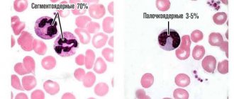
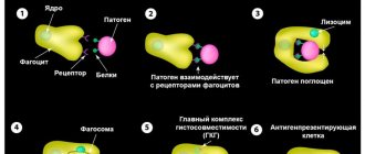
The most numerous group of leukocytes are neutrophils. Depending on the degree of maturity, young (rod) forms and mature (segmented) forms are distinguished among them. Together with monocytes, neutrophils are responsible for the processes of active phagocytosis (capture and destruction of pathogenic agents).
Thanks to monocytes, phagocytosis of destroyed and dead cells, denatured proteins, bacteria, antigen-antibody complexes, etc. occurs.
For reference. Neutrophils and monocytes can rightfully be considered the body’s main orderlies.
Lymphocytes are the most important component of immunity. Among them there are three types of cells:
- T (provide cellular immune responses);
- B (responsible for humoral immune response reactions);
- NK (destruction of viruses, tumor and mutated cells).
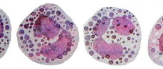
The main role of eosinophils is to phagocytose the antigen-antibody complex formed by immunoglobulin E. Together with basophils, they participate in the development of type 1 hypersensitivity reactions.
Basophils belong to the smallest group. However, they play a significant role in ensuring the inflammatory response and the development of allergic reactions.
Reasons for leukogram deviation
Deviation from normal indicators can occur in one or several parameters at once, and the indicators can either decrease or be increased.
Lymphocytes
An increase in their level is called lymphocytosis. This phenomenon is observed in most cases in the presence of syphilis, lymphoma, chickenpox, rubella, tuberculosis, some types of anemia, measles and other viral diseases.
A decrease in the rate is called lymphocytopenia. This condition may indicate the presence of infection, impaired renal function, systemic lupus erythematosus, or acute deficiency of certain vitamins.
Also, such a result may appear during treatment with corticosteroid drugs or radiation therapy methods.
You can learn more about lymphocytes here.
Neutrophils
An increase in this element is often observed when:
- Concentrations in the blood of too many different toxic elements.
- The appearance of severe bleeding, often becoming acute.
- Taking corticosteroid drugs.
- The emergence of diseases of bacterial etiology.
A decrease in the indicator as a result of the study may indicate:
- Having allergies to certain substances or drugs.
- The development of diseases of infectious etiology, their acute or chronic stages.
- The presence of autoimmune diseases.
- Exposure to ionizing radiation.
Monocytes
Their increase may indicate the presence of hematological malignancies, many types of parasites, bacterial infections in acute form, lupus erythematosus and rheumatoid arthritis in progressive form.
A decrease in monocytes with certain indicators of the total leukocyte formula indicates the presence of tuberculosis in the patient’s body.
Basophils
Their increase indicates the presence of myeloid leukemia in a person, mainly in a chronic form, and also makes it possible to determine erythremia.
Eosinophils
An increase in the indicator is typical for:
- Scarlet fever.
- Many types of allergic manifestations.
- Skin pathologies.
- Parasitic infections.
- Leukemia of the eosinophilic type.
The rate decreases with the onset of typhoid fever and its rapid progression, as well as with an increase in the activity of adrenocorticosteroids.
When deciphering the analysis, the shift of neutrophils and the number of each category according to the degree of maturation are always taken into account
In this case, special attention is paid to the ratio of mature (segmented) and immature (rod) cells. Neutrophils of other stages of maturation are absent in healthy people with normal test results, appearing only during periods of severe illness to quickly replace dying adult cells
Leukocyte formula: what is it and its norms
The leukocyte formula is the differentiation of leukocytes into 5 subclasses. Each subclass is responsible for a particular function in the body
Conducting this study is important in the differential diagnosis of inflammatory and infectious diseases, hematopathies, as well as for monitoring the treatment (chemotherapy)
Table of leukocyte formula standards for adults
| Indicators | Band neutrophils | Segmented neutrophils | Eosino-philes | Baso-phyla | Lymphocytes | Monocytes |
| Standard values | 1–6% | 47–72% | 1–5% | 0–1% | 18–37% | 3–11% |
Neutrophils (NEUT) are the most numerous class of leukocytes. It determines the body's primary defense against infections (through phagocytosis).
Reasons for increased neutrophils:
- general leukocytosis;
- chronic myeloid leukemia;
- leukemoid reaction against the background of sepsis, tuberculosis, malignant tumors with metastasis to the bone marrow.
Conditions and/or diseases in which the neutrophil count is reduced:
- viral infection;
- cytostatic therapy;
- exposure to radiation;
- aplastic and B12-deficiency anemia;
- agranulocytosis.
The percentage of eosinophils in healthy people in the peripheral blood is insignificant. But their number increases in tissues (including blood) in allergic, parasitic and tumor diseases. The minimum level of these cells is observed in the morning. The maximum is closer to midnight. Under stress, the number of eosinophils decreases sharply.
Eosinophil levels are elevated in the following conditions:
- allergic reactions with hay fever, bronchial asthma, atopic dermatitis;
- helminthiasis;
- scarlet fever;
- Wiskott–Aldrich syndrome;
- systemic collagenoses (rheumatoid arthritis, periarteritis nodosa);
- chronic myeloid leukemia;
- lymphogranulomatosis;
- the use of sulfonamides, para-aminosalicylic acid;
- eosinophilic esophagitis, gastritis, colitis in newborns fed with cow's milk.
Basophils contain histamine granules. Their main function is to participate in immediate hypersensitivity reactions.
An increase in the number of basophils may be a consequence of the following conditions:
- allergy;
- megakaryoblastic leukemia in Down syndrome;
- hematoblastosis;
- systemic mastocytosis;
- basophilic leukemia;
- condition after splenectomy;
- chronic hemolytic anemia;
- hypofunction of the thyroid gland.
An increase in the level of basophils in combination with an increase in the level of eosinophils or with their normal level indicates a myeloproliferative process.
There are 3 populations of lymphocytes: T, B and NK (natural killer cells). Each population performs its own specific function.
The level of lymphocytes increases in the following conditions:
- tuberculosis;
- whooping cough;
- chronic lymphocytic leukemia;
- chronic radiation sickness;
- thyrotoxicosis;
- bronchial asthma;
- lymphoma.
The amount of LYMP decreases in the following pathologies:
- aplastic anemia;
- some types of leukemia;
- infiltrative and miliary tuberculosis;
- glucocorticosteroid therapy;
- systemic lupus erythematosus;
- AIDS;
- sarcoidosis
Monocytes are the largest formed elements. They migrate from the blood to the tissues in response to chemotaxis. Monocytes produce biological active substances: enzymes, complement proteins, coagulation factors.
The level of monocytes increases in the following diseases:
- tuberculosis;
- syphilis;
- brucellosis;
- rubella;
- scarlet fever;
- infectious mumps;
- mononucleosis;
- lymphogranulomatosis;
- infective endocarditis.
With mononucleosis, atypical mononuclear cells are present in the peripheral blood in an amount of more than 5%.
A clinical blood test is of paramount importance in the diagnosis process. The entire further diagnostic complex begins with it (biochemical research, instrumental and specific diagnostics).
Shift of the leukocyte formula to the left: what is it, causes, explanation, how to treat
The ratio of the number of different types of leukocytes to the total number of these cells in the blood is called the leukocyte formula. The proportion of these cells will change as a result of exposure to disease in the body.
Based on these changes, the doctor can make a presumptive diagnosis. Leukocytes are white blood cells that act as protectors against pathological microorganisms that cause inflammation.
Types of white blood cells:
- Eosinophils - appear as a response to the occurrence of an irritant in the body.
- Neutrophils have a bactericidal effect.
- Lymphocytes - form protective antibodies, eliminate foreign ones.
- Monocytes - participate in the process of destroying bodies of foreign origin, forming an immune response and restoring the normal state of tissues.
- Basophils - move other white blood cells to the source of infection.
What are absolute and relative changes in a formula?
The number of some white blood cells that make up the leukocyte formula is indicated in relative values, that is, as a percentage.
For example, 20% lymphocytes means that they make up that percentage of all white blood cells. There are also absolute values that indicate the specific number of cells contained in a given volume.
In most cases, the absolute value is the more significant diagnostic factor.
When fluctuations in the concentration of leukocytes occur with a shift in the formula, both their relative and absolute values should be taken into account. In practice, most often the trend of change in indicators both to the left and to the right coincides
In practice, most often the trend of changes in indicators both to the left and to the right coincides.
In the case of a left shift, we are talking about an increase in both the absolute and relative values of the concentration of immature neutrophils. This condition is also called neutrophilia.
The norm of neutrophils in an adult is presented in the table.
| Index | Meaning | Unit measurements |
| Relative | 45-75 | % |
| Absolute in 1 µl of blood | 1900-8000 | Cells/µl |
| Absolute in 1 liter of blood | 1.9 – 8.0 x 10 9 | cells/l |
How to determine shift
An increase or decrease in the number of cells compared to the norm in the leukocyte formula indicates an infectious process in the body, so a number of additional studies are required to clarify the diagnosis
When deciphering the analysis, pay attention not only to the number of bodies, but also to their relationship with each other, especially between new and old particles
A decreased or increased content of old cells relative to new ones is called a shift in the leukocyte formula. It is determined in the laboratory by counting the leukogram and using an automatic hematology analyzer.
What causes the deviation
The reasons for deviations in the leukocyte formula are as follows.
Lymphocytosis is an increase in the content of lymphocytes in the blood. It can be caused by the following diseases:
- chicken pox;
- rubella;
- low iron levels in the blood;
- lymphoma;
- measles;
- syphilis.
A drop in the number of leukocytes is observed in the following cases:
- lupus erythematosus;
- renal failure;
- avitaminosis;
- carrying out radiation therapy;
- use of corticosteroids.
An increase in neutrophil content can be observed:
- with acute sudden loss of large amounts of blood;
- when a large amount of toxic substances enters the body;
- for bacterial infections;
- when taking corticosteroid drugs.
A decrease in the content of neutrophils in the blood indicates:
- for autoimmune disorders in the body;
- on the effects of radiation;
- for infections in the acute stage.
An increase in the number of monocytes is caused by:
- infections;
- rheumatoid arthritis;
- lupus erythematosus;
- presence of helminths.
Increased basophils - myeloid leukemia. A decrease in monocytes is tuberculosis of the lungs.
What does shift left mean?
A shift to the left means that a pathological process or other functional disorders have arisen in the body, as a result of which mature blood cells first die, and young ones are formed in their place. Therefore, the number of young leukocytes increases compared to mature ones.
Leukocytosis with a shift to the left can occur with an increased content of not only neutrophils, but also myelocytes, and this reliably confirms the presence of pathologies in the body, because in a healthy body such deviations are not observed.
This condition is caused by the following reasons:
Diagnostics
Detection of neutrophilia requires differential diagnosis. To do this, you need to consult a general practitioner. In order to obtain primary information, anamnesis is collected - how long ago the symptoms appeared, whether there was recent contact with infectious patients, whether there was an increase in body temperature, pain, skin rashes.
If there is a suspicion of acute surgical abdominal pathology, the abdomen must be palpated for tension in the muscles of the anterior abdominal wall and the presence of a positive Shchetkin-Blumberg sign. However, it must be taken into account that these signs are difficult to identify in a child under 9 years of age. To confirm the diagnosis, additional examination is prescribed, including:
- Blood tests
. The total number and percentage of all types of leukocytes are calculated. The concentration of red blood cells, platelets, and inflammatory markers (ESR, CRP) is measured. The morphology of granulocytes (toxic granularity, karyopyknosis) is studied. In a septic condition, the level of presepsin and procalcitonin is determined. The presence of autoantibodies (anti-DNA, topoisomerase, antineutrophil antibodies) is checked. - Identification of the pathogen
. To identify the pathogenic microorganism, bacteriological culture, microscopy of sputum, urine, smear from the throat and tonsils are performed. To diagnose helminthiases, a stool test is performed for worm eggs, a blood test for specific immunoglobulins, and a scraping is taken from the child’s perianal folds. - Ultrasound
. A sign of pyelonephritis on an ultrasound of the abdominal cavity is expansion, thickening of the renal collecting system, pancreatitis - enlargement, diffuse changes in the pancreatic parenchyma, cholecystitis - thickening of the walls of the gallbladder, often the presence of stones. - X-ray
. On chest x-rays with pneumonia, foci of infiltration and darkening are visible. When the ulcer is perforated, the images reveal the presence of free gas in the abdominal cavity (“sickle symptom”). In case of inflammatory diseases of the joints, x-rays show a narrowing of the joint space and marginal osteoporosis. - ECG.
Electrocardiography during myocardial infarction reveals ST segment elevation, left bundle branch block, ventricular tachycardia, and other heart rhythm disturbances. With pulmonary embolism leading to pulmonary infarction, signs of overload of the right parts of the heart are detected - a deep Q wave in III, an S wave in lead I, a high pointed P wave (P-pulmonale) in leads II, III, aVF. - Histological studies
. A definitive diagnosis of cancer can only be made on the basis of a biopsy. The main feature of solid tumors is a large number of atypical cells. In case of leukemia, in the bone marrow biopsy, hyperplasia of the granulocytic lineage and the predominance of blast cells are noted; in the tissues of the lymph node in case of lymphomas, diffuse proliferation of cells with blast morphology is noted.
How is the analysis carried out?
The number of cells is counted under a microscope or using automatic equipment - a blood analyzer. If the analysis is carried out in the form of a smear (leukocyte differentiation), then laboratory assistants initially count 100-200 blood cells, which are visible on a glass slide, and then determine the percentage of elements of each type.
Norms
For ease of counting, a smear is prepared on the glass, dried and painted. On the surface of the smear, white blood cells spread differently because the heavier particles remain at the edge, while the lighter particles spread closer to the center. Counting is carried out using two methods:
- Filipchenko's method. The resulting smear is divided into three parts. Draw a straight transverse line across the entire area of the smear.
- Schilling method. The smear is divided into four sections.
If a special analyzer is used for counting, then its diagnosis is considered more accurate. The automatic device does not decipher 100-200 cells, because up to 2000 cells can fall into its field of view.
The general clinical blood test is deciphered by the attending physician. The results of the analysis are presented in the form of a table, which indicates the quantity and name of each blood component.
Technique
Preparing for a blood test is not difficult: the patient should simply refuse to eat four hours before the procedure, and the day before he should avoid emotional and physical stress.
The material for determining the formula is venous blood. Before this, the laboratory assistant must clamp the patient’s forearm with a special belt, and then insert a thin needle into the vein located in the bend of the elbow, through which the blood will flow into the test tube. This process cannot be called painless, but most often the pain is not too strong. As a result, a drop of blood is obtained and placed on a glass plate in order to determine the proportion of leukocytes and their number using a microscope. If the clinic has modern equipment, then the particles are counted by an analyzer - a special device, and human intervention is needed only in case of serious deviations from the norm or the presence of particles of an anomalous nature.
The time it takes to obtain results mainly depends on the institution where the study is carried out. Usually you have to wait no more than a few days. The attending physician should evaluate the obtained indicators. When deciphering, you can detect a shift to the left in the leukocyte formula.
Forecast
It is impossible to predict prognosis based on neutrophilia alone. It all depends on the disease that served as the background for the occurrence of neutrophilia. For example, a transient increase in the number of neutrophils after stress, food intake, or in a child on the first day of life is absolutely benign and transient. Conversely, severe purulent-septic pathologies and oncological diseases have a fairly high incidence of deaths. Therefore, any excess of the reference values of neutrophils (especially high and persistent ones) requires contacting a doctor.
Types of leukocytes
White blood cells develop from precursor cells produced in the bone marrow. There are five different types of white blood cells, which include:
- Granulocytes - These white blood cells have granules in their cytoplasm. The granules contain chemicals and other components that are released during the immune response. The three types of granulocytes are:
- Neutrophils (neu) usually make up the largest number of circulating white blood cells. They move to an area of damaged or infected tissue where they engulf and destroy bacteria or sometimes fungi.
- Eosinophils (eos) respond to infections caused by parasites, play a role in allergic reactions (hypersensitivity), and control the extent of immune reactions and inflammation.
- Basophils (baso) usually constitute the smallest number of circulating white blood cells and are thought to be involved in allergic reactions.
- Lymphocytes exist in both the blood and the lymphatic system. They are divided into three types, but the leukoformula does not distinguish between them. All lymphocytes differentiate from common lymphoid progenitor cells in the bone marrow. The leukoformula counts all lymphocytes together. Separate specialized testing (eg, immunophenotyping) should be performed to differentiate the three types:
- B lymphocytes (B cells) are antibody-producing cells that are essential for acquired antigen-specific immune responses. Plasma cells are fully differentiated B cells that produce antibodies, immune proteins that target and destroy bacteria, viruses and other foreign antigens.
- T lymphocytes (T cells) complete their maturation in the thymus and consist of several different types. Some T cells help the body distinguish between self and non-self antigens. Others initiate and control the extent of the immune response, increasing it as needed and then slowing it down as the condition resolves. Other types of T cells directly attack and neutralize virus-infected or cancer cells.
- Natural killer cells (NK cells) directly attack and kill abnormal cells, such as cancer cells or those infected with a virus.
- Monocytes (mono), like neutrophils, move into the area of infection and engulf and destroy bacteria. They are more often associated with chronic rather than acute infections. They are also involved in tissue repair and other functions related to the immune system.
When an infection or inflammation develops somewhere in the body, the bone marrow produces more white blood cells, releasing them into the blood. Depending on the cause of the infection or inflammation, one specific type of white cell may be increased amid normal levels of other types. When the infection resolves, production of this type of cell subsides and the number returns to normal levels.
Normal indicators
Leukoformula analysis represents the proportion of all types of leukocytes. Most often, the examination is attributed in parallel to the general analysis.
Now let's look at the main indicators and components that are closely monitored during the test:
- Neutrophils are primarily used to provide adequate levels of protection. They can determine which bacteria are harmful, then influence them until they are destroyed.
- Basophils are components that appear during all kinds of allergic reactions. These components have the effect of neutralizing poisons and toxins, preventing the spread of harmful substances through the blood supply system.
- Eosinophils in the blood help destroy various parasitic bacteria. It is thanks to them that antiparasitic resistance is observed in the body.
- Monocytes are very similar in functionality to neutrophils. The main difference is the higher phagocytic effect. They also allow you to kill parasitic bacteria, while the leukocytes that die during exposure are absorbed, which leads to blood purification;
- Lymphocytes are substances that have a kind of memory; they recognize antigens and remember them. The component provides immunity to viruses and tumors.
Norms of various types of leukocytes in the leukocyte formula
For a healthy person, depending on age, there are special standards that indicate the state of the body based on the leukocyte formula.
| Age | Leukocyte ratio (cells/µl) |
| Up to 1 year | 6 – 17,5 |
| 1-2 years | 6 – 17 |
| From 2 to 4 years | 5,5 – 15,5 |
| From 4 to 6 years | 5 – 14,5 |
| From 6 to 10 years | 4,5 – 13,5 |
| From 10 to 16 years | 4,5 — 13 |
| Over 16 years | 4,5 — 11 |
Leukocyte formula of a healthy person
The leukoformula represents the total proportion of all leukocytes. There is more accurate information - leukocyte indices. This examination allows you to determine the number of different types of components of the group of leukocytes. The intoxication index is considered an extremely useful indicator; based on the test readings, the degree and severity of inflammation can be determined. It is also possible to determine the level of an allergic reaction, based on allergization, and the effectiveness of the system, due to immunoreactivity, etc.
Deviation of values from the norm
The leukocyte formula can have the values of its components depending on a particular disease.
Neutrophils
Values above normal are typical for:
- infectious diseases;
- inflammatory process in any organ;
- rheumatic diseases;
- acute inflammation of the pancreas (pancreatitis);
- burns of a large area of the body;
- some forms of bone marrow cancer.
Values below normal may occur when:
- viral infections (hepatitis B, AIDS, influenza, measles);
- massive purulent process (extensive purulent wounds, sepsis);
- aplastic anemia;
- some types of bone marrow cancer.
Shift of the leukocyte formula to the left
This term refers to an increase in the content of immature neutrophils (band neutrophils) in the peripheral blood, and in some cases the appearance of young forms of leukocytes (metamyelocytes), which are normally present only in the red bone marrow. This condition is characterized by a powerful inflammatory process, when mature neutrophils die in large numbers, and the bone marrow is forced to release immature blood cells to fight the infection.
Shift of the leukocyte formula to the right
In this case, on the contrary, the content of mature forms of neutrophils in the peripheral blood decreases. This is typical for chronic diseases of the liver, kidneys, as well as megablastic anemia.
Eosinophils
Values above normal indicate the presence of:
- allergic process (hay fever, bronchial asthma, allergies to medications);
- infection with internal parasites (worms, amoeba).
Values below normal may occur under the following conditions:
- when treated with glucocorticosteroids;
- for some endocrine diseases (Itsenko-Cushing syndrome).
Basophils
Basophils can increase when:
- malignant neoplasms of lymph nodes and bone marrow;
- polycythemia;
- anaphylactic shock, Quincke's edema and other immediate allergic reactions.
A decrease in the number of basophils is observed in patients:
- when treated with glucocorticosteroids;
- with increased thyroid function;
- in the acute phase of the infectious process.
Monocytes
Reasons for increased monocyte count:
- tuberculosis;
- sexually transmitted diseases (syphilis);
- acute infectious diseases of a bacterial nature;
- connective tissue diseases, including rheumatoid;
- cancer of various localizations (stomach, mammary glands, bone marrow, lymph nodes).
A decrease in the number of monocytes is observed with:
- treatment with corticosteroids;
- aplastic anemia.
Lymphocytes
Lymphocytes increase in the following cases:
- viral infections;
- tuberculosis;
- chronic lymphocytic leukemia;
- non-Hodgkin's lymphoma.
The reasons for a decrease in the level of lymphocytes can be:
- AIDS;
- chemotherapy and radiotherapy of malignant neoplasms;
- congenital immunodeficiencies;
- aplastic anemia;
- treatment with corticosteroids;
- after organ transplantation while taking immunosuppressive drugs;
- systemic lupus erythematosus.
Correction
Conservative therapy
There are no direct ways to normalize the number of neutrophil granulocytes. To combat neutrophilia, it is necessary to treat the underlying disease against which it developed. Short-term neutrophilia after eating, stress or physical work does not require any intervention, as it is not a sign of disease or pathological condition. Neutrophilia resulting from surgery also does not need to be treated. In the case of persistent neutrophilia, you should consult a doctor to find out the cause and prescribe differentiated treatment:
- Antimicrobial (antiparasitic) therapy
. For bacterial infections, antibiotics (amoxicillin, cefixime) are used. For generalized infections (sepsis, bacterial endocarditis), it is necessary to use a combination of 2 antibacterial drugs. In case of helminthic infestation in a child, anthelmintic drugs (mebendazole) are prescribed. - Hemorheological therapy
. For heart attacks of any location caused by thrombosis or thromboembolism, antiplatelet drugs (acetylsalicylic acid), anticoagulant drugs (low molecular weight, unfractionated heparin), and sometimes thrombolytic drugs (alteplase) are used. - Antisecretory and antienzyme therapy
. In order to reduce the secretion of hydrochloric acid in peptic ulcers, proton pump inhibitors (omeprazole, pantoprazole), H2-blockers (famotidine, ranitidine) are used. To suppress the destructive action of proteolytic enzymes of the pancreas in acute pancreatitis and pancreatic necrosis, enzyme inhibitors (aprotinin) are effective. - Anti-inflammatory treatment
. To achieve remission of rheumatological diseases, medications are prescribed that stop the inflammatory process. These include glucocorticosteroids (prednisolone), 5-aminosalicylic acid derivatives (sulfasalazine), immunosuppressants (cyclophosphamide, methotrexate). - Chemotherapy.
For the treatment of malignant tumors, chemotherapeutic drugs (cytostatics, antimetabolites, hormone antagonists) are used in combination with radiotherapy. For oncohematological diseases, a combination of several antitumor agents is necessary.
Surgery
Many diseases accompanied by neutrophilia (mainly acute abdominal pathologies) require emergency surgical intervention - laparoscopic appendectomy, laparotomy and suturing of the ulcerative defect, cholecystectomy, opening and drainage of the abscess, etc. For myeloproliferative pathologies, in case of ineffectiveness of conservative therapy, they resort to stem cell transplantation cells.
When is it necessary to evaluate the shift in leukocyte formula?
Formula calculation and shift determination may be part of the CBC when someone has general signs and symptoms of infection and/or inflammation, such as:
- Fever, chills.
- Pain and aches in the body.
- Headache.
- Various other signs and symptoms, depending on where the infection or inflammation is suspected.
Testing may be done when there are signs and symptoms that the doctor thinks may be related to a blood and/or bone marrow disorder, an autoimmune disease, or another immune disorder.
Caution should be exercised when interpreting test results. The doctor must consider the person's signs and symptoms, as well as the patient's medical history and the extent of cell enlargement or shrinkage.
Important
A number of factors can cause a short-term increase or decrease in the number of cells of one type. A persistent increase or decrease usually prompts further testing to determine the cause.
Long-term use of steroids or prolonged exposure to toxic chemicals (such as lye or insecticides) can increase the risk of abnormal differences and cause the formula to shift to the right.
Alena Paretskaya, pediatrician, medical columnist
just today
Features of the analysis
To carry out the analysis, take venous blood or blood from a finger, drop a few drops onto a glass slide, and then prepare a smear. Next, this smear is examined under a microscope, counting leukocytes relative to certain areas of the slide.
The principle of the analysis is that in a smear, as in blood, leukocyte cells are not randomly located, but adhere to certain spatial positions, the determination of which helps in calculating quantitative data. Monocytes and lymphocytes occupy a central location, and the remaining cells are located along the periphery.
Two counting methods are used:
- According to Schilling, the resulting smear is conditionally divided into 4 parts, counting the cells in each of them separately. The calculation is carried out until 100-200 cells are collected. After this, fill out a table where the quantitative and qualitative composition of leukocytes is entered, and then convert the indicators into percentages.
- According to Filippchenko, the smear is divided into three equal zones, and the counting is carried out along a conventional horizontal line. In each separate area, an equal number of cells are isolated, which are subsequently entered into the differential table.
After the numerical values have been obtained, laboratory assistants calculate their percentage. For this purpose, a special counter is used, which is capable of producing a ready-made result when entering certain data and formulas.
In modern laboratories, counting leukocytes and their types is entirely the responsibility of high-precision technology.
Within 15-20 minutes, you can obtain reliable data on the quantitative and qualitative composition of the leukocyte mass, identifying possible deviations.
In cases where there is a need, an additional morphological analysis of cells is performed, calculating their life expectancy, capacity and level of biological functioning.
What influences the result?
A healthy person's blood contains approximately the same (stable) set of leukocytes.
The leukocyte formula can change under the influence of factors such as:
- excessive physical activity, which causes injury to muscle fibers, which is accompanied by increased production of a certain type of leukocytes;
- psycho-emotional disorders, nervous outbursts, depression, apathy - any changes in mental state are reflected in the blood and its composition;
- pregnancy and menstrual cycle in women;
- the presence of bad habits - with smoking and alcohol abuse, the level of leukocytes is pathologically increased, and the largest number is not lymphocyte cells, but nephtrophils, which indicates chronic intoxication;
- frequency of blood transfusions and significant blood loss;
- removal of the spleen, part of the liver or kidney.
Leukocytes are also affected by certain medications that artificially inhibit them or, conversely, provoke their active synthesis.
Therefore, before donating blood, you definitely need to prepare.
Preparing for analysis
A blood test with a leukocyte formula is performed in the morning. The most optimal time for blood collection is from 7 to 9 am. During this time, you should not eat food or drink drinks such as coffee, alcohol and milk. For women, the analysis is not performed during menstruation, or 2-3 days before it begins.
If it is necessary to systematically use medications, stop taking them 12 hours before blood sampling. During pregnancy, the leukocyte formula may have some deviations caused by physiological processes. Therefore, slides with smears are marked with the appropriate abbreviation in order to compare the output values with the norms characteristic of the pregnancy period.
Explanation and description
The composition of the blood can change throughout life, so the leukocyte formula is assessed taking into account age-related changes.
At birth in children, most of the leukocytes are neutrophils (about 65-70% of the total mass). Lymphocytes account for only 25-30%. This indicates that all organs and systems are activated in the newborn’s body, which will henceforth function as a separate organism that does not receive nutrition from the mother’s body. In the first days of life, hormonal changes occur, as a result of which the first leukocyte crossover is observed, in which the percentage of lymphocytes and neutrophils equalizes. By the first month of life, children's bodies produce more lymphocytes than neutrophils, forming reliable protection for the body. In the total mass, lymphocytes occupy 65%, leaving only 15-20% for neutrophils. This provides the child of the first year of life with reliable immunity, which he needs in understanding the world.
After a year, when children’s immunity is definitely formed, there is a gradual decrease in the lymphocyte mass in favor of neutrophils, which is also controlled by biological processes and is necessary for the development of all types of immunity. By the age of four, a second crossover is observed, in which lymphocytes again align with neutrophils, forming a powerful barrier to microbes and pathogens. After this, the number of lymphocytes gradually increases, and the remaining leukocyte cells are produced as needed.
By the age of 6, the composition of a child’s blood is close to the blood of an adult, where the majority of the total mass is occupied by neutrophils and lymphocytes.
During the period of hormonal changes, the leukocyte formula may have slight deviations from the norm by 10-15%, which is not pathological and directly depends on the biological processes in the body.
Leukogram at birth and in the first month of life
When born, the child’s body begins to adapt to environmental conditions, which is reflected in physiological processes. From here, the following normal indicators are identified for the first week of life, the decoding of which is included in the table.
| Name of leukocytes | Normal value in % of total leukocyte mass |
| neutrophils | 65 |
| lymphocytes | 20-35 |
| monocytes | 3-5 |
| basophils | 0-1 |
| eosinophils | 1-2 |
By the first month of life, the picture changes somewhat, allowing the body to receive reliable protection from lymphocytes.
| Name of leukocytes | Normal value in % of total leukocyte mass |
| lymphocytes | 65-70 |
| neutrophils | 20-25 |
| monocytes | 3-6 |
| basophils | 1-2 |
| eosinophils | 0,5-1 |
Leukocyte formula from 1 to 3 years
During this period, the number of lymphocytes and neutrophils is unstable and can change not only during the day, but also under certain conditions: prolonged exposure to the sun, hypothermia, chronic or genetic diseases.
| Name of leukocytes | Normal value in % of total leukocyte mass |
| neutrophils | 32-52 |
| lymphocytes | 35-55 |
| monocytes | 10-12 |
| basophils | 0-1 |
| eosinophils | 1-4 |
Indicators from 4 to 6 years
Neutrophils again take precedence over lymphocytes, so the decoding with normal indicators will look like this.
| Name of leukocytes | Normal value in % of total leukocyte mass |
| neutrophils | 36-52 |
| lymphocytes | 33-50 |
| monocytes | 10-12 |
| basophils | 0-1 |
| eosinophils | 1-4 |
Leukocyte formula norms after 6-7 years
After 6 years, the leukocyte composition norm is identical to that of an adult.
| Name of leukocytes | Normal value in % of total leukocyte mass |
| lymphocytes | 19-35 |
| neutrophils | 50-72 |
| monocytes | 3-11 |
| eosinophils | 1-5 |
| basophils | 0-1 |
During the period of hormonal changes, a shift of the leukogram by 10-15% is allowed.

