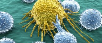
Leukocytes are a group of blood cells that are characterized by the absence of color, the presence of a nucleus and the ability to move. The name is translated from Greek as “white cells”. The group of leukocytes is heterogeneous. It includes several varieties that differ in origin, development, appearance, structure, size, core shape, and functions. White blood cells are produced in the lymph nodes and bone marrow. Their main task is to protect the body from external and internal “enemies”. Leukocytes are found in the blood and in various organs and tissues: in the tonsils, intestines, spleen, liver, lungs, under the skin and mucous membranes. They can migrate to all parts of the body.
Types of leukocytes
White cells are divided into two groups:
- Granular leukocytes - granulocytes. They contain large, irregularly shaped nuclei, consisting of segments, the more of which the older the granulocyte is. This group includes neutrophils, basophils and eosinophils, which are distinguished by their perception of dyes. Granulocytes are polymorphonuclear leukocytes. You can learn more about granulocytes from this article.
- Non-granular - agranulocytes. These include lymphocytes and monocytes, which contain one simple oval-shaped nucleus and do not have a characteristic granularity.
Leukocytes are blood cells whose main function is to fight infectious agents.
Determining the number of red blood cells is an integral part of the leukocyte formula and is not performed separately.
Synonyms Russian
White blood cells, white blood cells.
English synonyms
White Blood Cell Count, WBC count, Leukocyte count, White count.
Units
*10^9/l (10 in st. 9/l).
What is this analysis used for?
To detect infection, inflammation or cancer, their presence is indicated by an increase in the number of white blood cells. A significant decrease in their number may indicate a decrease in immunity.
When is the study scheduled?
In case of suspected infection, inflammation or conditions in which the number of white blood cells changes, as well as to monitor the effectiveness of treatment for such diseases.
What biomaterial can be used for research?
Venous or capillary blood.
General information about the study
Leukocytes are blood cells that are formed in the bone marrow. Their main function is to fight infection and tissue damage. There are five types of leukocytes, which differ in appearance and functions: eosinophils, basophils, neutrophils, lymphocytes and monocytes. They are present in the body in relatively stable proportions and, although their numbers can vary significantly throughout the day, they normally remain within reference values.
White blood cells are formed from bone marrow stem cells and, during the process of maturation, go through a series of intermediate stages, during which the cell and the nucleus it contains become smaller. Only mature leukocytes should enter the bloodstream. They do not live long, so they are constantly renewed. The production of white blood cells in the bone marrow increases in response to any tissue damage, as part of the normal inflammatory response. The goal of the inflammatory response is to limit the damage, remove the causative factor and restore the tissue.
Different types of leukocytes have slightly different functions, but they are capable of coordinated interactions using certain substances - cytokines.
A significant increase in the number of white blood cells (over 100 x 1012/L) can make the blood more viscous, which can lead to headaches, increased blood pressure and visual disturbances. If the number of neutrophil leukocytes decreases and becomes less than 1 x 10¹²/l, then the risk of infections increases and their course becomes more severe. In this case, the infection can be caused by microbes that are normally “friendly” to the body.
What is it used for and when is the study prescribed?
This test is usually included in a routine general blood test.
An increased white blood cell count (leukocytosis) helps identify infection and inflammation.
A significant increase in the number of leukocytes (more than 50-100 thousand x 1012/l), as a rule, indicates a malignant bone marrow tumor and requires urgent medical attention.
A decrease in the number of white blood cells (leukopenia) is much less common than leukocytosis. It most often indicates a viral infection, but can be a sign of more dangerous diseases such as AIDS or aplastic anemia.
The use of radiation therapy or certain drugs (in particular, cytostatics) can lead to a decrease in the number of leukocytes, so their number is monitored for timely correction of therapy.
Among other things, this study is carried out in the treatment of leukemia to evaluate the effectiveness of therapy.
What do the results mean?
Reference values
| Floor | Age | Reference values |
| Female | Less than 1 year | 6 - 17.5 *10^9/l |
| 1-2 years | 6 - 17 *10^9/l | |
| 2-4 years | 5.5 - 15.5 *10^9/l | |
| 4-6 years | 5 - 14.5 *10^9/l | |
| 6-10 years | 4.5 - 13.5 *10^9/l | |
| 10-16 years | 4.5 - 13 *10^9/l | |
| 16-18 years old | 4.5 - 11 *10^9/l | |
| Over 18 years old | 3.98 - 10.4 *10^9/l | |
| Male | Less than 1 year | 6 - 17.5 *10^9/l |
| 1-2 years |
Where are they formed and how long do they live?
The bulk of white cells, namely granulocytes, are produced by red bone marrow from stem cells. From the maternal (stem) cell, a precursor cell is formed, then passes into a leukopoetin-sensitive cell, which, under the influence of a specific hormone, develops along the leukocyte (white) series: myeloblasts - promyelocytes - myelocytes - metamyelocytes (young forms) - rods - segmented. Immature forms are found in the bone marrow, mature forms enter the bloodstream. Granulocytes live for about 10 days.
The lymph nodes produce lymphocytes and a significant proportion of monocytes. Some agranulocytes from the lymphatic system enter the blood, which transports them to the organs. Lymphocytes live a long time - from several days to several months and years. The lifespan of monocytes ranges from several hours to 2-4 days.
General characteristics
Leukocytes in the blood are a group of cells that are responsible for the human body’s resistance to various pathogenic bacteria, viruses, helminths, parasites and other pathological microorganisms.
They also fight not only infectious agents, but also any foreign object:
- malignant or benign neoplasms of any location;
- transplanted donor organ;
- a foreign object that may accidentally enter the body.
The place of formation of leukocytes is blood stem cells, which are localized in the red bone marrow. In order to fully carry out their work, they undergo a large number of transformations, during which their structure and functions change.
In addition to blood, they are also found in fluids such as:
- urine;
- cerebrospinal fluid;
- pleural effusion;
- feces;
- gastric juice.
However, their concentration in such cases will be significantly lower, for example, for urine analysis, from 4 to 6 leukocytes are acceptable, and no more than 8 white blood cells should be present in the cerebrospinal fluid.
An increase or decrease in such blood components in any of the above structures most often indicates the occurrence of a disease.
In addition to the main task, the functions of leukocytes include:
- release of specific substances to combat various tumors;
- absorption and digestion of the pathogenic agent;
- relief of hemorrhages;
- acceleration of wound healing.
As stated above, white blood cells have several subtypes.
- What foods can increase white blood cells in the blood - List of the 10 most effective
Thus, there are the following types of leukocytes:
- neutrophils - aimed at destroying bacterial infection;
- lymphocytes – responsible for the immune system and immune memory;
- monocytes - absorb and digest particles of foreign cells;
- eosinophils - fight allergen carriers;
- basophils - assist other particles in detecting foreign agents, however, they perform all their “duties” outside the bloodstream - in the internal organs.
It follows from this that subtypes of leukocytes perform their own mission.
Types of leukocytes
All types of such substances, in addition to their functions, differ in the following indicators:
- dimensions;
- core shape;
- way of development.
It is also worth noting about the structural features of each type of white blood cell. For example, neutrophils, eosinophils, basophils and monocytes are born from myeloblasts, the precursor of which is myelopoiesis. This happens under the influence of a stimulator cell in the bone marrow.
The lifespan of leukocytes is on average 2-4 days, and they are often destroyed in the liver, spleen and foci of inflammatory processes. The only exceptions are lymphocytes, some of which live in the human body from birth to death.
In neutrophils, eosinophils and basophils, the entire life cycle takes place in the bone marrow, which is why their immature cells are normally completely absent from the blood. Monocytes continue to exist in the spleen, liver and skeletal system, where they degenerate into macrophages and dendrocytes. Lymphocytes have a longer “life” in the spleen, lymph nodes and thymus.
Leukocytes received their common name - white blood cells - because, unlike red blood cells, they are colorless.
From all of the above, it follows that if there are no leukocytes in the blood, the human body simply will not be able to function.
- How long do leukocytes live and where are they formed? Types and functions of leukocytes
Structure
The structure of leukocytes of different types is different, and they look different. What they all have in common is the presence of a core and the absence of their own color. The cytoplasm can be granular or homogeneous.
Neutrophils
Neutrophils are polymorphonuclear leukocytes. They are round in shape and have a diameter of about 12 microns. There are two types of granules in the cytoplasm: primary (azurophilic) and secondary (specific). Specific small, lighter and make up about 85% of all granules, contain bactericidal substances, the protein lactofferin. Ausorophilic ones are larger, they contain about 15%, they contain enzymes, myeloperoxidase. In a special dye, the granules are colored lilac, and the cytoplasm is colored pink. The granularity is fine, consists of glycogen, lipids, amino acids, RNA, enzymes, due to which the breakdown and synthesis of substances occurs. In young forms, the nucleus is bean-shaped, in rod-nuclear forms it is in the form of a stick or a horseshoe. In mature cells - segmented - it has constrictions and looks divided into segments, which can be from 3 to 5. The nucleus, which can have processes (appendages), contains a lot of chromatin.
Eosinophils
These granulocytes reach a diameter of 12 microns and have a monomorphic coarse granularity. The cytoplasm contains oval and spherical granules. The granularity is stained pink with acidic dyes, the cytoplasm becomes blue. There are two types of granules: primary (azurophilic) and secondary, or specific, filling almost the entire cytoplasm. The center of the granules contains a crystalloid, which contains the main protein, enzymes, peroxidase, histaminase, eosinophil cationic protein, phospholipase, zinc, collagenase, cathepsin. The eosinophil nucleus consists of two segments.
Basophils
This type of leukocyte with polymorphic granularity has dimensions from 8 to 10 microns. Granules of different sizes are stained with a basic dye in a dark blue-violet color, the cytoplasm is stained pink. The grain contains glycogen, RNA, histamine, heparin, and enzymes. The cytoplasm contains organelles: ribosomes, endoplasmic reticulum, glycogen, mitochondria, Golgi apparatus. The core most often consists of two segments.
Lymphocytes
By size they can be divided into three types: large (from 15 to 18 microns), medium (about 13 microns), small (6-9 microns). The latter are in the blood most of all. Lymphocytes are oval or round in shape. The nucleus is large, occupies almost the entire cell and is painted blue. A small amount of cytoplasm contains RNA, glycogen, enzymes, nucleic acids, and adenosine triphosphate.
Monocytes
These are the largest white cells, which can reach a diameter of 20 microns or more. The cytoplasm contains vacuoles, lysosomes, polyribosomes, ribosomes, mitochondria, and the Golgi apparatus. The nucleus of monocytes is large, irregular, bean-shaped or oval, may have bulges and indentations, and is colored reddish-violet. The cytoplasm acquires a gray-blue or gray-blue color under the influence of the dye. It contains enzymes, saccharides, and RNA.
The main types of mature leukocytes:
First of all, it is logical to mention that there are five main types of mature leukocytes in the blood. They are determined in tests in the form of a leukocyte formula, so the level of leukocytes in the blood is assessed not only as a whole. The content of these cells is also always calculated. These include (in descending order of quantity):
• Neutrophils
• Lymphocytes
• Monocytes
• Eosinophils
• Basophils.
They have different functions, but they work in collaboration, influence each other, transmit information among themselves, etc. High or low white blood cells in the blood, belonging to one type or another, indicate various diseases, so determining their number is very important in medical practice.
Content
Leukocytes in the blood of healthy men and women are contained in the following ratio:
- segmented neutrophils – from 47 to 72%;
- band neutrophils – from 1 to 6%;
- eosinophils – from 1 to 4%;
- basophils – about 0.5%;
- lymphocytes – from 19 to 37%;
- monocytes – from 3 to 11%.
You can learn about the white blood cell count in pregnant women from this article.
The absolute level of leukocytes in the blood in men and women normally has the following values:
- band neutrophils – 0.04-0.3X10⁹ per liter;
- segmented neutrophils – 2-5.5X10⁹ per liter;
- young neutrophils – absent;
- basophils – 0.065X10⁹ per liter;
- eosinophils – 0.02-0.3X10⁹ per liter;
- lymphocytes – 1.2-3X10⁹ per liter;
- monocytes – 0.09-0.6X10⁹ per liter.
You can read about the number of blood leukocytes in children here.
Pathological leukocytosis: main causes
In some cases, exceeding the norm of leukocytes indicates the development of pathology:
- almost all infections (mainly associated with bacteria and fungi, less often with viruses);
- inflammatory processes of various nature;
- allergy;
- leukemia, lymphoma and other malignancies;
- myocardial infarction;
- penetration of parasites;
- receiving various types of injury (physical, electrical, thermal).
Having completed the physiological cycle, leukocytes enter the spleen, where they are destroyed and excreted from the body. Therefore, when the spleen is removed and its functions are disrupted, leukocytosis is also observed. Moreover, it has a persistent and pronounced character (in this case, the level of leukocytes of all types increases).
As for leukopenia (reduced number of white blood cells in the blood), it can be one of the signs of such diseases and disorders:
- exhaustion due to severe pathologies, prolonged fasting;
- viral infections;
- autoimmune processes;
- immunodeficiency, including those associated with HIV;
- severe purulent-septic conditions;
- bone marrow damage, especially after taking medications or other illnesses;
- radiation sickness;
- radiation therapy, chemotherapy;
- deficiency of vital substances - proteins, vitamins, minerals;
- insufficient activity of the thyroid gland;
- certain forms of anemia;
- certain forms of leukemia.
Functions
The general functions of leukocytes are as follows:
- Protective – consists in the formation of specific and nonspecific immunity. The main mechanism is phagocytosis (capture of a pathogenic microorganism by a cell and deprivation of its life).
- Transport - lies in the ability of white cells to adsorb amino acids, enzymes and other substances found in plasma and transport them to the right places.
- Hemostatic - involved in blood clotting.
- Sanitary – the ability, with the help of enzymes contained in leukocytes, to dissolve tissues that have died due to injury.
- Synthetic – the ability of some proteins to synthesize bioactive substances (heparin, histamine and others).
Each type of leukocyte has its own functions, including specific ones.
Neutrophils
The main role is to protect the body from infectious agents. These cells capture bacteria into their cytoplasm and digest them. In addition, they can produce antimicrobial substances. When an infection enters the body, they rush to the site of penetration, accumulate there in large quantities, absorb microorganisms and die themselves, turning into pus.
Eosinophils
When infected with worms, these cells penetrate the intestines, are destroyed and release toxic substances that kill the helminths. In allergies, eosinophils remove excess histamine.
Basophils
These leukocytes take part in the formation of all allergic reactions. They are called first aid for bites of poisonous insects and snakes.
Lymphocytes
They constantly patrol the body in order to detect foreign microorganisms and out-of-control cells of their own body, which can mutate, then quickly divide and form tumors. Among them there are informants - macrophages, which constantly move throughout the body, collect suspicious objects and deliver them to lymphocytes. Lymphocytes are divided into three types:
- T-lymphocytes are responsible for cellular immunity, come into contact with harmful agents and destroy them;
- B lymphocytes identify foreign microorganisms and produce antibodies against them;
- NK cells. These are real killers that maintain normal cellular composition. Their function is to recognize defective and cancer cells and destroy them.
Types and norm of leukocyte content:
White blood cells in the blood are only a few micrometers in size and, of course, cannot be seen with the naked eye. However, when a blood smear is examined under a microscope, it becomes obvious that white blood cells do exist and are very different from each other.
In general, if you trace the path from a stem cell to mature leukocytes, you can count several dozen forms of cells of different “ages”. However, we will consider only those varieties that are of the greatest interest to doctors - mature cells. There are five types.
Basophils are white blood cells with an S-shaped nucleus that are stained blue with standard dyes. They take part in the development of allergic reactions. Among all leukocytes, of which the blood contains 3-9 × 10⁹/l, basophils occupy a share of 0.5-1.5%.
Eosinophils are slightly more numerous. They perceive acidic dyes, so they appear pink-orange in a normally stained blood smear. Inside them there is granularity visible under high magnification. There are 1-4% of eosinophils in the blood, and they also participate in the allergy mechanism.
Monocytes are cells with a large bean-shaped nucleus of a purple color, and there is granularity inside them. They are capable of forming outgrowths of the cytoplasm - “pseudopods”, with the help of which these leukocytes can move in the blood. The number of monocytes makes up 4-8% of all types of white blood cells.
Lymphocytes are small cells with a round nucleus and no granularity in the cytoplasm. Despite their not very impressive size, lymphocytes perform many functions and are the main ones in the implementation of cellular and humoral immunity. Their number is quite large - 20-40%.
Neutrophils are the most numerous (up to 80%) type of white blood cells. They have blue cytoplasm with pink granules and a large purple nucleus, sometimes divided into segments.
How to count
To count leukocytes, an optical device is used - the Goryaev camera
White cell (WBC) levels are determined during a clinical blood test. Leukocyte counting is carried out using automatic counters or in a Goryaev chamber, an optical device named after its developer, a professor at Kazan University. This device is highly accurate. It consists of thick glass with a rectangular recess (the chamber itself), where a microscopic mesh is applied, and a thin cover glass.
The calculation is as follows:
- Acetic acid (3-5%) is tinted with methylene blue and poured into a test tube. Blood is drawn into a capillary pipette and carefully added to the prepared reagent, after which it is mixed thoroughly.
- The coverslip and chamber are wiped dry with gauze. The coverslip is rubbed against the chamber to create colored rings, fill the chamber with blood and wait for a minute until cell movement stops. Count the number of leukocytes in one hundred large squares. Calculated using the formula X = (a x 250 x 20): 100, where “a” is the number of leukocytes in 100 squares of the chamber, “x” is the number of leukocytes in one μl of blood. The result obtained from the formula is multiplied by 50.
Origin of white blood cells:
Leukocytes in children and adults are found and studied mainly in the blood, but they are formed outside of it - in a special organ. It is quite impressive in size and weight (weighs about 2.5 kg), and its peculiarity is that it is distributed throughout the body.
We are talking about red bone marrow, which is located inside the tubular and spongy bones of the human body. This is where the production of blood elements occurs. All white blood cells come from a single stem cell. It divides, giving rise to several cell sprouts; its descendants develop, mature, pass into the blood, and eventually begin to realize their functions in a mature state.