Pathologies of the cardiovascular system require regular monitoring and special care for the patient. They are dangerous due to the rapid development of an acute condition and sudden death of the patient. To maintain a stable condition, the patient must live according to the regimen prescribed by the doctor and control his emotional state, but this is not always possible without outside support. People with cardiovascular pathologies often suffer from increased anxiety and fear of death, they are afraid of being alone for a long time or of being without vital medications at hand. Such patients require the help of a professional caregiver at home or in a boarding house. Peace, proper routine, care and attention to their needs have a calming effect on the wards and improves their well-being.
Principles of caring for patients with cardiovascular diseases
To prevent the development of acute conditions in the patient, the nurse carries out a whole range of preventive measures every day. This is painstaking work that requires discipline and patience from both the caregiver and the ward, but the result is worth it. Strengthening the heart muscle leads to an overall improvement in physical and psychological condition.
Proper care for patients reduces the risk of pathologies of the cardiovascular system. It assumes:
- daily monitoring of blood pressure and pulse,
- compliance with diet and medication,
- regular cleansing of the body,
- therapeutic massage to improve blood circulation,
- moderate physical activity,
- prevention of thrombosis and thromboembolism in bedridden patients,
- creating a calm environment and psychological support.
The caregiver must be able to recognize signs of dangerous conditions and have emergency medical skills. The life of the ward may depend on his qualifications.
Symptoms of Cardiovascular Diseases
Arrhythmia
An accelerated or slowed heartbeat is not dangerous in itself, but it may be the first manifestation of a serious pathology. Treated with special medications
Dyspnea
Stagnation of venous blood leads to excess carbon dioxide and lack of oxygen in cells. An acute attack is called cardiac asthma and in severe cases is accompanied by pulmonary edema. A patient with shortness of breath needs fresh air first. In addition, tourniquets on the legs and inhalation of humidified oxygen may be required.
Edema
Fluid retention in the body occurs due to overflowing of the veins and blood particles leaving their walls. Swelling reduces the protective properties of the skin, causing a feeling of nausea and bloating in the abdomen. To normalize fluid outflow, diet and diuretics are required.
Pain in the heart area
This is a sign of poor blood supply to the organ. With prolonged oxygen starvation, biochemical changes occur in the heart muscle, manifested by pressing sensations, weakness, and profuse sweating. In mild cases, the pain is relieved with a validol or nitroglycerin tablet. However, an attack can be caused by a developing myocardial infarction. If the syndrome does not go away even after taking medications, the patient requires emergency hospitalization
Increased blood pressure
Regular increases in blood pressure cause the development of hypertension and concomitant dysfunction of the brain and kidneys. To avoid this, the patient needs to get enough sleep, measure blood pressure twice a day, give up bad habits and take medications regularly. If there is a sharp increase in blood pressure (hypertensive crisis), you need to call an ambulance
Collapse
This is a sharp decrease in blood pressure, provoked by acute vascular insufficiency, myocardial infarction, severe bleeding, including internal bleeding, poisoning or infection. In this state, the patient’s breathing and pulse become more frequent, and the extremities become cold. Possible loss of consciousness. The patient needs to be warmed and positioned so that blood flows to the brain. Caffeine-containing medications are recommended to raise blood pressure.
Lecture “Nursing process for congenital heart defects in children”
Lecture 1. Nursing process for diseases of the circulatory system (CVD)
Lecture outline:
1) Definition, causes, risk factors for congenital heart disease,
2) Disturbed needs, problems with congenital heart defects.
3) Early clinical signs of congenital heart disease,
4) Features of diagnosis and treatment of congenital heart defects.
5) Nursing care for children with congenital heart defects.
6)
1. Consolidation of new material (conduct a frontal survey, explaining questions that are difficult for them to understand)
2. Self-preparation task (include new terms with explanations in the glossary, in the diary: groups of medications and emergency care for a hypoxemic crisis)
A heart defect is a persistent pathological change in the structure of the heart that impairs its function. Congenital heart defects (CHD) and large vessels are formed as a result of impaired embryogenesis in the 2-8th week of pregnancy or endocarditis suffered during intrauterine development. Among malformations of internal organs, congenital heart defects occupy the second place (after central nervous system anomalies) and occur in 0.3-0.8% of newborns. Of the total number of patients with congenital defects in the population, about 60% are children under 14 years of age. In the absence of surgical treatment, from 55 to 70% of patients die in the first year of life, therefore, with age, cardiac development anomalies are less common. The frequency of this pathology in early childhood, its severe course, and often unfavorable outcome make it particularly important to early detect congenital heart defects and great vessels, their accurate topical diagnosis and timely referral of patients for surgical treatment to specialized institutions
Viral diseases play an important role in the development of congenital heart disease.
mothers (rubella, measles, mumps, chicken pox, influenza), as well as toxoplasmosis in pregnant women.
Heart defects occur in close relatives and are often accompanied by chromosomal diseases and developmental abnormalities
.
Radiation exposure, the age of the parents, exposure of pregnant women to toxic and chemical substances, and the use of certain medications play a certain role in their occurrence.
(methotrexate, phenobarbital, etc.).
Depending on the state of hemodynamics
In the pulmonary circulation (LCC) and systemic circulation (BCC),
four groups of congenital heart disease are distinguished:
Group I
- vices with enrichment of a small circle;
II -
with depletion of the small circle;
Group III
- with depletion of the great circle;
Group IV
- without hemodynamic disturbances.
Congenital heart disease can appear immediately after birth or after some time and is recognized by characteristic clinical signs:
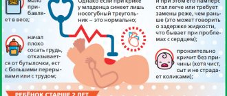
In a newborn, cyanosis appears when sucking.
Patients develop cyanosis (permanent, transient or temporary), shortness of breath, noise over the heart and blood vessels.
The boundaries of the heart increase. There is a tendency to respiratory infections and prolonged repeated pneumonia. Children are lagging behind in physical development.
During UPS, three phases are distinguished.
• First phase
(primary adaptation
) is characterized by the body’s adaptation to hemodynamic disturbances.
• After 2-3 years, the second phase begins -
the phase of relative compensation
. During this period, the child’s condition, physical development and motor activity improve significantly.
• Third phase
- terminal
.
It occurs when compensatory possibilities are exhausted and dystrophic and degenerative changes in the heart muscle develop. The third phase inevitably ends with the death of the patient. Risk factors
birth of a child with congenital heart disease are:
-mother’s age over 35 years, occupational hazards, alcoholism, radiation, hypovitaminosis
- endocrine disorders in spouses,
- toxicosis in the first trimester and threats of termination of pregnancy
- history of stillbirth
-presence of other children with congenital malformations,
- a woman taking endocrine drugs to maintain pregnancy, etc., diseases of the genital area.
| Characteristic | Vice | |
| Without cyanosis – white defects | With cyanosis – blue defects | |
| With blood flow enrichment MCC (hypovolemia, hypertension | VSD, ASD, PDA | |
| Depletion of blood flow ICC | Isolated pulmonary stenosis | Tetralogy of Fallot, Ebstein's disease |
| With BKK depletion | Coartation of the aorta, aortic stenosis | |
General clinic
:
• In case of severe hemodynamic disturbances – retardation in physical development
• Often shortness of breath, pallor or cyanosis
• Deformation of the nail phalanges (watch glasses, drumsticks)
• Deformation of the chest (heart hump)
• Expansion of the borders of the heart, systolic murmur is heard
Diagnostics
– instrumental research methods
1. X-ray examination (with enrichment of the ICC - signs of blood stagnation, changes in the shape of the heart due to hypertrophy of its various parts)
2. ECG, Doppler-Echo-CG (will show hypertrophy of various calvings
hearts)
3. Ultrasound of the heart, probing
4. Angiography, phonocardiography (will record murmurs, character, localization)
Defects with enrichment of the pulmonary circulation.
They are characterized by the discharge of blood into
the right parts of the heart
and
the pulmonary artery as a result of the presence of pathological communication between the pulmonary and systemic circulation.
1.Open ductus arteriosus (PDA).
During fetal development, the ductus arteriosus connects the aorta
to
the pulmonary artery and balances the pressure in the pulmonary and systemic circulation. In the first days after the birth of the child, it closes. Preservation of its function after 3 months of life is regarded as congenital heart disease. A patent ductus arteriosus of small diameter is not accompanied by hemodynamic disorders. With a wide ductus arteriosus, in the first days of life, cyanosis may be observed due to the discharge of blood from the pulmonary artery into the aorta, since in a newborn the right ventricle of the heart is more powerful than the left.
Subsequently, due to the difference in pressure in
In the systemic and pulmonary circulation, blood is discharged from the aorta
into
the pulmonary artery, which leads to overflow of the pulmonary circulation and overload of the left chambers of the heart.
With the development of pulmonary hypertension, overload of the right ventricle is also observed.
Clinical manifestations of the defect are shortness of breath, increased
fatigue, pain in
areas of the heart.
The maximum blood pressure is normal, the minimum pressure is low, especially in a standing position.
Pulse jumping. Palpation reveals a trembling of the chest, most pronounced in the second intercostal space on the left, the borders of the heart are expanded mainly to the left and upward, and with the development of pulmonary hypertension, the right parts of the heart also enlarge. In the second intercostal space to the left of the sternum, systolic and then systolic and diastolic (“machine”
) noise, which is carried out on the aorta, cervical vessels and in the interscapular area.
The principle of treatment is ligation or dissection of the duct after 6 months of life.
2.Atrial septal defect (ASD).
One of the most common congenital heart disease. The degree of hemodynamic disorders depends on the size of the defect. An open oval window and a small midline defect are usually not
have clinical manifestations. Large defects lead to the development of typical symptoms of the disease.
Patients complain of shortness of breath and fatigue. The skin is pale. The borders of the heart are expanded transversely, more to the right. In the II-III intercostal space to the left of the sternum, a systolic murmur is heard, the II sound on the pulmonary artery is strengthened and split.
Treatment principle
– suturing the defect
or plastic surgery in the 1st year of life.
3.Ventricular septal defect (VSD).
By race
extent takes first place, accounting for about 20-30% of all CHDs.
Hemodynamic disorders are determined by the discharge of blood from the left ventricle to the right. The size of the shunt depends on the location and size of the defect.
There are two forms of VSD:
1) high defects
, localized in the membrane part of the septum;
2) small defects
located in the muscle part.
With high VSD there are
shortness of breath, cough, weakness, fatigue, frequent respiratory infections, retardation in physical development. The skin is pale, and mild cyanosis occurs with anxiety and crying. Chest deformity often develops. The transverse size of the heart increases, its upper border moves upward. During auscultation in the 3rd-4th intercostal space, a prolonged systolic murmur is heard from the sternum, radiating to the entire cardiac region and back, the 2nd sound on the pulmonary artery is intensified and split.
Small defect in the muscle part
septum practically does not cause hemodynamic disturbances, since during systole the defect decreases in size (
Tolochinov-Roger disease
).
A diagnosis of congenital heart disease can be made by a rough, grinding systolic murmur in the IV-V intercostal space
to the left of the sternum or on the sternum.
The principle of treatment
is suturing or plastic surgery of the defect at the age of 3-5 years.
Defects with depletion of the pulmonary circulation.
They arise as a result of narrowing of the pulmonary artery, often combined with pathological discharge of blood from the right ventricle into the systemic circulation (shunt from right to left).
1.Isolated pulmonary artery stenosis
.
There are various anatomical variants of this defect, the most common being pulmonary valvular stenosis.
The main manifestations of the defect include shortness of breath, expansion of the borders of the heart mainly to the right, a rough systolic murmur in the second intercostal space to the left of the sternum, which extends to the left subclavian region and the carotid arteries. The first tone at the apex is enhanced, the second sound at the pulmonary artery is weakened or absent. Treatment principle
– dissection
valves only in severe cases in infancy.
2. Fallot's disease (triad, tetrad, pentad)
.
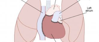
The most common form of “blue” heart defects that occurs with cyanosis is tetralogy of Fallot.
The defect includes a combination of four anomalies:
• pulmonary stenosis (A),
• interventricular defect
transposition of the aorta to the right (B)
and right ventricular hypertrophy(D)
.
Due to pulmonary artery stenosis, part of the blood when the right ventricle contracts through the ventricular septal defect enters the left ventricle and then into the aorta. This leads to insufficient saturation of arterial blood with oxygen and the development of cyanosis.
Clinically, the defect manifests itself immediately after birth
or in the first month of life. Its important signs are shortness of breath and cyanosis, most noticeable on the mucous membranes of the lips and oral cavity, in the area of the nail bed of the fingers. The extreme severity of cyanosis is blue-blue skin color, gray sclera with injected vessels.
Characteristic of tetralogy of Fallot is the patient's posture
: the child squats or lies on his side with his legs tucked to his stomach, since in this position he is less bothered by shortness of breath.
Due to blood stasis in tetralogy of Fallot
in the capillaries of the skin,
the terminal phalanges of the fingers of the limbs thicken in the form of drumsticks
,
the nails become convex, taking the shape of a watch glass.
Children are lagging behind in physical development. The borders of the heart remain normal or slightly expanded to the left. A rough systolic murmur is heard along the left sternal border
, II tone on the pulmonary artery is weakened.
In the peripheral blood, the level of hemoglobin and the number of red blood cells increase.
In case of decompensation of the defect
Hypoxemic (blue breath) attacks
appear They arise as a result of spasm of the outflow tract of the right ventricle and the stenotic pulmonary artery, which leads to complete shunting of blood into the aorta.
In this case, against the background of ordinary acrocyanosis, patients experience an attack of shortness of breath, tachycardia, and cyanosis increases. The child is excited. Fainting often occurs. Subsequently, a hypoxic coma may develop, accompanied by loss of consciousness and convulsions.
Treatment principle
– 2 stages. At an early age, the imposition of an anastomosis between the vessels of the ICC and BCC. At 6-7 years of age, plastic surgery of the IVS defect and dissection of the fused pulmonary artery leaflets.
Defects with depletion of the systemic circulation
. 1.
Coarctation of the aorta
.
With this defect, there is a narrowing of the thoracic aorta below the mouth of the left subclavian artery. The degree and extent of narrowing varies. The vessels of the lower half of the body receive little blood. Above the site of narrowing, hypertension is observed, spreading to the vessels of the head, shoulder girdle, and upper extremities.
Patients experience headaches, dizziness, a feeling of pulsation in the neck and head, and tinnitus. The upper half of the body is better developed than the lower.
Ischemic pain in the abdomen and calf muscles and increased fatigue when walking are noted.
Kidney function is impaired.
One of the main clinical symptoms of aortic coarctation is the difference in blood pressure in the upper and lower extremities. The pulsation of the vessels of the lower extremities is weakened and transmitted with a delay, which contrasts sharply with the jumping pulse in the arteries of the arms and carotid arteries.
The borders of the heart are expanded to the left. The auscultatory picture is uncharacteristic: the localization of the noise depends on the level of narrowing, the second sound in the aorta is enhanced.
Treatment principle
(below)
Treatment of congenital heart disease
.
The only treatment for congenital heart disease is surgery
.
Surgical treatment is divided into radical and palliative.
After radical interventions for defects of the interatrial and interventricular septa and patent ductus arteriosus, hemodynamic disturbances normalize.
Palliative operations alleviate the condition of patients and prevent early death. The most favorable period for the operation is phase II of the defect (age from 3 to 12 years).
It is possible to perform surgical treatment at an earlier age.
Conservative therapy for congenital heart disease
provides:
1) emergency care for heart failure and hypoxemic crises;
2) maintenance treatment; 3) prevention of thrombosis; 4) treatment of anemia.
Maintenance therapy is carried out
by prescribing
cardiac glycosides
in small doses,
riboxin, ATP, cocarboxylase, and potassium preparations.
With congenital heart disease there is a high risk of thrombosis, therefore the use of acetylsalicylic acid
and disaggregants.
Children with congenital heart disease should have higher levels of hemoglobin and red blood cells
. If the indicators correspond to the age norm, this condition is regarded as anemia and requires the prescription of iron and copper supplements.
Emergency care for hypoxemic crisis
(hypoxia - lack of oxygen in organs and tissues).
The basis of treatment of hypoxemic crises are:
oxygen therapy, correction of acidosis and drug effects on
spasm of the initial sections of the pulmonary artery.
Help begins with the administration of promedol.
If there is no effect, obzidan is used.
Oxygen therapy is advisable
with constant positive expiratory pressure.
Correction of metabolic acidosis is carried out with a 4% sodium bicarbonate solution.
For convulsions, sodium hydroxybutyrate is indicated,
simultaneously having an antihypoxic effect.
The use of cardiac glycosides and diuretics is contraindicated! Prolonged and severe attacks with loss of consciousness require emergency hospitalization in the cardiac surgery department.
Care.
For children with congenital heart disease, a proper daily routine with maximum exposure to fresh air is important.
It is necessary to provide the child with a balanced diet, control the amount of food, and limit salt and liquid as indicated.
Children should be protected from infections and gentle hardening should be carried out. Patients with congenital heart disease without severe heart failure can engage in physical therapy or, under medical supervision, attend physical education classes at school, in a preparatory group.
Exercises that require great physical exertion, participation in sports clubs and participation in competitions (even chess!) are contraindicated.
Children with congenital heart disease should recover in rheumatological sanatoriums (Khvalynsk).
Prevention and medical examination:
• RSS among pregnant women (exclude harmful working conditions)
• Healthy lifestyle
• Prohibition of contact with infectious patients
• Monitoring the use of medications The tasks of the outpatient service include
: early detection of children with congenital heart disease;
• systematic monitoring of them;
• determination, together with a cardiac surgeon, of the optimal timing of surgical intervention;
• timely prescription of conservative therapy;
• resolving security issues, carrying out rehabilitation.
Children who are diagnosed with congenital heart disease in the maternity hospital or clinic should be observed by a local pediatrician together with a cardiac surgeon.
During dispensary observation
The topical diagnosis of the defect is clarified by electro- and phonocardiography, x-ray examination of the heart and blood vessels, angiocardiography, ultrasound examination and cardiac probing.
Patient problems due to congenital heart disease:
| № | Real | Potential | Priority |
| 1 | Dyspnea | • Tachycardia • Anxiety • Difficulties in self-care • Pale skin • cyanosis | Dyspnea |
| 2 | Heartache | • Fear of death • Fright • Sleep disturbance • Feelings of despair and hopelessness | |
| 3 | Increased fatigue | • Decreased physical activity • Emotional lability • Negative attitude • to medical manipulations |
Questions for self-control
1. What causes and conditions/diseases of a pregnant woman can lead to the formation of congenital heart disease in the fetus?
2. At what stage of pregnancy does organogenesis occur and the formation of heart defects in the fetus is possible?
3. Which congenital heart defects are classified as defects with enrichment of the pulmonary circulation?
4. Which congenital heart defects are related to defects with depletion of the pulmonary circulation?
5. What is coarctation of the aorta?
6. By what signs can one suspect a congenital heart defect in a child?
7. How is emergency care provided during a hypoxemic crisis?
8. What are the methods of examination/diagnosis (laboratory and instrumental) in children with congenital heart disease?
9. What indicators of the CBC are specific in case of congenital heart disease Tetralogy of Fallot?
10.List the methods of treatment for congenital heart disease.
11.The most favorable period of the course of congenital heart disease for radical surgery.
12.Name the names of the drugs and the group of drugs used in the treatment of congenital heart disease, indicating the pharmacological action (for example: Analgin, an analgesic drug).
Homework assignment
learn lecture notes No. 1, read the textbook “Nursing care in pediatrics” by Katolikova O.S. pp. 157-163, repeat the AFO of the circulatory system.
1. Textbook “Features of providing nursing care to children”:
textbook/K.I.Grigoriev, R.R.Kildiyarova – M.: GEOTAR-Media, 2016.272p.
2. Textbook “Nursing care in pediatrics”: MDK.02.01. Nursing care for various diseases and conditions / Katolikova O.S. – Rostov n/D-Phoenix. 2015.-539p.
Diet for cardiac diseases
The patient must strictly follow the diet prescribed by the doctor. The diet should include sufficient quantities of foods rich in potassium and proteins:
- lean meat,
- vegetables,
- cereals,
- eggs,
- fish,
- greenery,
- high protein bread,
- potato,
- butter,
- cottage cheese.
It is recommended to eat 5-6 times a day in small portions. You need to have dinner no later than 2-3 hours before bedtime. Fluid intake should be limited by half, and salt should be replaced with spices, as it retains fluid in the body.
Physiotherapy
Caring for patients with cardiovascular diseases involves mandatory physical activity. The doctor selects a set of exercises individually depending on the patient’s health condition. The doctor will recommend special poses to relieve spasms, increase chest volume and relax muscles. Exercises should be performed at a slow pace, controlling breathing and smoothly changing positions. The sudden transition may cause shortness of breath or irregular heart rhythm.
It is recommended to start doing simple light exercises immediately after sleep, without getting out of bed. This is necessary to prepare the cardiovascular system for the climb. You need to train 3-4 times a day. The duration of classes is 10-25 minutes.
In addition to warming up for joints, the complex includes:
- balance exercises,
- training with a stick and ball,
- breathing practices.
In addition, daily walking for 30 minutes to 2 hours is necessary. Moderate aerobic exercise strengthens the heart muscle and blood vessels, normalizes blood pressure and metabolism.
Living conditions in boarding houses "Doverie"
Our specialists will provide patients suffering from various pathologies of the cardiovascular system with comprehensive care. Careful observation and timely assistance will prevent the development of acute conditions in our patients.
We accept elderly people with heart disease for rehabilitation, for example, we provide rehabilitation for patients with myocardial infarction, offer permanent residence, and also organize patronage. The Trust network of boarding houses for the elderly with rehabilitation has created comfortable conditions for cardiac patients. Our guests can count on:
- for medical care at any time of the day;
- regular observation by a doctor;
- dietary tasty food;
- comfortable rooms with functional furniture;
- favorable psychological environment;
- safe area for walking;
- a variety of pleasant leisure activities.
Friendly staff not only provide physical care for patients, but also help people with cardiovascular diseases quickly adapt to new living conditions and remain socially active even when lying down.
Relatives of our wards can visit them at a convenient time and always stay in touch via phone or instant messengers. We invite you to take a tour of any of our boarding houses so that you can personally see the conditions in which our guests live and communicate with our specialists. Leave a request on the website. Our manager will answer all your questions.
Nursing care for patients with acquired heart defects
One of the main objectives is to reduce the risk of complications for the patient. To achieve this goal, they decide in accordance with the criteria: activity of the rheumatic process; presence of signs of circulatory disorders (even degree I); form of vice.
Nursing care
1. Therapeutic and protective regime (ENT) - a regime depending on the severity of the disease, quartzing, wet cleaning, ventilation, changing underwear and bed linen, feeding the bedpan and washing, changing position in bed;
2. Diet: meals 5-6 times a day, lean meats (beef, rabbit, veal), cereals, soups, pasta are allowed, but smoked, salted, pickled, fatty, spicy are prohibited, 1-1.2 l liquids per day.
3. Monitoring vital functions (VF) - blood pressure, respiratory rate, pulse, body temperature, skin color;
4. Preparation for studies (ECG, EchoCG, phonography, OAC, BHA, OAM, angiography)
5. Fulfilling doctor's prescriptions (beta blockers, anticoagulants, cardiac glycosides, calcium channel blockers).
6. The nurse conducts conversations with patients:
• about the causes, clinical manifestations and complications of PPS,
• about organizing the correct daily routine with limited physical activity,
• the need to follow doctor’s orders,
• the need to strictly follow a diet,
• about bad habits
• about the rules of administration, side effects of medications and their effects
• about the need for exercise therapy
• about supporting the psycho-emotional state
• about the rules of preparation for research
• about the need to attend health school
7. The nurse teaches patients and/or their relatives:
• blood pressure measurement;
• determination of pulse;
• determination of daily diuresis, water balance;
• keeping a diary of general condition;
• self-care (changing body position in bed to prevent bedsores, changing underwear and bed linen).
The nurse's role is to conduct a health school for patients. The goal of the school is to improve the quality of life, stabilize the course of the disease, and prevent the development of complications.
In the process of educational activities, nurses can use reminders, booklets, audio and video materials.
Recently, the role of nursing staff has increased in creating and maintaining positive changes in their lifestyle. Therefore, nursing care is very important to improve the quality of life of patients.
CHAPTER II. IDENTIFYING THE FEATURES OF NURSING CARE FOR PATIENTS WITH PPS ON THE BASE OF GBUZ SOKB. V. D. Seredavina
To conduct the study, we used data from statistical reports ..... department of the State Budgetary Healthcare Institution SOKB named after. V. D. Seredavina
A comparative analysis was carried out based on the data obtained, which were systematized in the form of tables and diagrams.


