Sokolov-Lyon Index (SLI)
Sokolov-Lyon index (SLI) = SV1 (mm) + RV5 (mm) or RV6 (mm).
Normal: up to 48 mm in persons under 40 years of age and up to 38 mm in persons over 40 years of age
To determine this index, it is necessary to add the amplitude (depth) of the SV1 tooth to the amplitude (height) of RV5 or RV6. The resulting figure, expressed in millimeters, will be the index.
Importance and clinical significance
The gold standard for detecting ventricular hypertrophy is an echocardiogram. However, this research is expensive and requires trained personnel to conduct and analyze it.
The importance of the Sokolow index is that it is based on the measurement of the electrocardiogram, which is the basic test of clinical examination, and is also inexpensive, easy to perform, and can be analyzed by any physician.
Based on the electrocardiogram, the doctor diagnoses ventricular hypertrophy, and the patient is referred to a cardiologist for in-depth study.
Cornell voltage product (CVP)
Cornell voltage product (CVP) = [RaVL(mm) + SV3 (mm)] × QRS (ms)
Normal: less than 2440 mm/ms.
The Cornell voltage product is calculated a little more complicated: it is necessary to multiply the duration of the QRS complex, expressed in milliseconds, by the sum of R -aVL (mm) + S -V3 (mm). The difficulty is that you have to multiply large numbers and you can’t do it without a calculator. If the obtained value exceeds 2440, then we are talking about LVMH.
ECG
1: fourth intercostal space to the right of the sternum.
2: fourth intercostal space to the left of the sternum.
3: in the middle of the line connecting V2 to V4.
4: at the junction of the fifth intercostal space with the midclavicular line.
5: parallel to V4, but along the anterior axillary line.
6: parallel to the previous ones, but along the midaxillary line.
In this study, time, expressed in seconds, is measured in the horizontal plane. In this case, the voltage, expressed in volts, is in the vertical plane.
Therefore, on the graph paper on which the electrocardiogram is printed, a square measuring 1 mm in the horizontal plane corresponds to 0.04 seconds and 0.1 millivolts in the vertical plane.
OTHER CRITERIA
RV4 < RV5 > RV6 or RV4 < RV5 < RV6 - shift of the transition zone to the left
Now we will discuss the criterion of LVMH determined by the dynamics of the R wave in the precordial leads. The fact is that in non-hypertrophied myocardium the total excitation vector is directed to the apex, therefore the highest R wave will be recorded in lead V4. In the case of hypertrophy, (but not always) the vector shifts to the left, partly due to thickening of the lateral wall of the left ventricle, and partly due to changes in the boundaries of the heart. Because of this, the maximum R begins to be determined not in V4, as is normal, but in lead V5 or V6.
Many “old school” cardiologists consider this sign to be the most important, however, this is not so. The reason lies in the fact that cardiologists are accustomed to seeing mostly sick people who have hypertrophy in 80%-90%, but if they examine the entire population, then an erroneous conclusion about LVMH will be given to every second patient with such changes on the ECG. So, be careful with this criterion, although it has greater sensitivity than the above indices (ISL and KVP), but its specificity is no more than 60%, while for the above indices it approaches 100%.
Diagnostic ECG criteria for LV hypertrophy
There are a lot of diagnostic criteria for LV hypertrophy, but the Sokolov-Lyon index and the Cornish voltage index are most often used.
For a reliable diagnosis of LV hypertrophy, voltage criteria must be supplemented by non-voltage signs.
Voltage criteria in standard leads:
Voltage criteria in chest leads:
Non-voltage signs of LVH:
What else may indicate LVH:
Example 1: LV hypertrophy in a patient with hypertension
This ECG can detect several criteria for LV hypertrophy:
Example 2: LV hypertrophy in a patient with atrial fibrillation
Source
SYSTOLIC OVERLOAD OF THE LEFT VENTRICLE
A separate concept in the LVMH section is “systolic overload of the left ventricle.” It must be said that in fact, the ventricle does not experience any special overload either at the time of ECG recording or during any other period of time, except for that which is associated with high blood pressure. There are situations when a person with blood pressure is 150/90 mm Hg. we see “systolic overload”, but in others, even with a pressure of 220/120 mm Hg, there is no hint of such changes. In addition, normalization of blood pressure never led to the disappearance of “overload”.
Systolic overload as an ECG phenomenon occurs in the case of severe hypertrophy, which leads to disruption of the repolarization wave, but we will not go into details, the main thing is that you understand the meaning of this term and do not confuse hemodynamics with electrical activity.
So, how does this “phenomenon” manifest itself? And it manifests itself in the form of disturbances in the repolarization processes of a very specific type (see further on the ECG), occurring in the area of the lateral wall of the left ventricle, always in combination with high R waves or, more often, in combination with positive criteria for LVMH (ISL or CVP).
In conclusion, it is worth noting that in my (and not only my) opinion, in the presence of positive criteria for ISL and CVP, we need to talk about quantitative signs of hypertrophy, while a shift of the transition zone to the left should be called qualitative signs of hypertrophy.
ECG diagnosis of LV hypertrophy
Left ventricular hypertrophy
LV hypertrophy occurs in response to an increase in pressure in it, for example, with hypertension or aortic valve stenosis.
On the ECG, this is manifested by an increase in the amplitude of the R wave in the “left” leads (I, aVL and V4-V6) and an increase in the depth of S in the “right” leads (III, aVR, V1-V3).
Thickening of the walls of the LV leads to a prolongation of its depolarization (manifested by an increase in the rise time of the R wave) and to a change in repolarization (changes in the T wave and ST segment in the lateral leads).
Limitations of criteria, IMPORTANT! IMPORTANT! IMPORTANT!
1. In the presence of complete or incomplete blockade of the left leg, the criteria do NOT work!
2. In the presence of complete blockade of the right leg, the criteria do NOT work!
3. If there are scar changes, the criteria do NOT work!
4. If the criteria are negative, this does NOT mean that there is no hypertrophy! Low sensitivity.
An attempt to identify left ventricular myocardial hypertrophy by ignoring points 1-3 is a mistake, which, however, is also made by cardiologists!
Well, let's see how it all looks.
Sokolov and Lyon
North American cardiologists Maurice Sokolov and Thomas Lyon based their professional careers on the study of hypertension and its complications.
One of the most common conditions associated with this disease is left ventricular hypertrophy, which is nothing more than a thickening of the muscular wall of the left ventricle of the heart.
These specialists described a way to calculate the state of the heart muscle by measuring the voltage of certain waves on the electrocardiogram. The technique was published in 1949 in a scientific journal. American Heart Journal and it was called "Ventricular complex in left ventricular hypertrophy obtained with unipolar precordial and limb electrodes."
The method is called the Sokolow-Lyon Index, and is one of the criteria currently used to determine left ventricular hypertrophy in patients with heart disease, especially hypertension.
EXAMPLE 1
On this ECG you can see almost all the criteria for left ventricular myocardial hypertrophy (LVMH).
1. Sokolov-Lyon index (ISL) = SV1 (19 mm) + RV5 (22 mm) = 41 mm (norm up to 38 mm)
2. Cornell voltage product (CVP) = [RaVL(13 mm) + SV3 (10 mm)] × QRS (100 ms) = 2300 ms*mm (the norm is up to 2440, this negative criterion was slightly missed)
3. Transition zone offset - RV4 < RV5 < RV6.
4. Systolic overload - we see ST depression in V5 and V6 and biphasic, predominantly negative, T, that is, a violation of repolarization processes.
Content:
The Sokolow Index is a criterion used to diagnose left ventricular chamber enlargement of the heart based on the measurement of the waves that represent the left ventricle on the electrocardiogram.
The electrocardiogram is printed on a millimeter sheet. From the sum of the small squares that the wave's amplitude occupies, its voltage is obtained. The Sokolov index is the result of the sum of the amplitude of the S wave in precordial vectors V1 plus the R wave in V5 or V6. Thus: S V1 + RV 5 or 6 = Sokolov index.
A result greater than 35 mm indicates that the patient has left ventricular hypertrophy, meaning that their left ventricular heart chamber is thicker than normal.
This type of abnormality is a consequence of diseases such as hypertension, which causes the heart muscle to become overworked due to the high pressure it must overcome to pump blood.
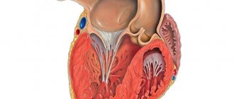
Portrait of hypertrophy
Left ventricular hypertrophy
Before we begin to describe the electrocardiographic signs of hypertrophy (enlargement) of the right and left ventricles, let us dwell on some aspects of pathophysiology and anatomy. We will try to imagine and describe together what a hypertrophied myocardium looks like. Perhaps this will help someone to remember the signs of hypertrophy (enlargement) faster and easier. So, normally the left ventricle (LV) is almost 3 times larger in mass than the right. This fact is explained by the fact that the LV operates under conditions of high resistance. He needs to overcome the vascular resistance that is created in the aorta, the efferent vessel.
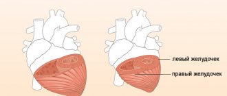
The right ventricle (RV) is much easier; it does not encounter such vascular resistance. Therefore, he does not need the same muscle mass as his neighbor. A prerequisite for the formation of remodeling of the right and left ventricle (LV) is an additional increased resistance that one or the other has to overcome. When the pressure in the pulmonary circulation (pulmonary blood flow) increases, the pancreas experiences stress. When the pressure in the aorta increases or when there is an obstacle to the normal functioning of the LV for other reasons, this chamber of the heart experiences increased load.
Right ventricular hypertrophy
In order to cope with the situation and perform its pumping function, as before, the muscle fibers of the right or left ventricle thicken and lengthen. It seemed like these were good compensatory mechanisms. This chamber of the heart will become stronger and will continue to work. But everything has its own margin of safety. And the ventricles are no exception in this situation. For some time, these chambers of the heart operate at their previous level, but then, sooner or later, the compensatory mechanisms become exhausted. The heart muscle begins to lose its pumping function. What new characteristics does the right or left ventricle acquire in the case of established hypertrophy? Muscle fibers lengthen and thicken.
The camera has increased in size and weight. Sclerotic (overgrowth of connective tissue) and dystrophic processes in the myocardium due to depletion of energy reserves in the cell. An increase in the electromotive force of the heart and an increase in the excitation vector of the ventricle. The thicker the myocardium, the more energy the heart needs to expend in order for the electrical impulse to excite the entire thickness of the myocardium. Accordingly, the time of excitation of the ventricle increases. All these points lead to changes in the electrocardiogram, indicating the presence of hypertrophy.
Treatment methods
Despite the fact that thickening of the left ventricular wall can only be completely eliminated through surgery, conservative therapy is most often carried out, since this pathology is not so dangerous that surgery is prescribed for all patients.
Treatment tactics depend on the disease that caused the problem.
Conservative therapy: medications
For hypertension
Use one of the following medications, not all at the same time.
| Group of drugs | Effect | Examples of drugs |
| ACE inhibitors | Lower blood pressure | Lisinopril, Enalapril, Diroton |
| Beta blockers | Reduce blood pressure and slow pulse | Metoprolol, Anaprilin, Atenolol |
| Calcium channel blockers | Dilate arteries, including coronary ones, reduce pulse | Diltiazem, Verapamil, |
For atherosclerosis of the aorta
| Group of drugs | Effect | Examples of drugs |
| Statins | Regulate blood cholesterol levels | Lovastatin, Simvastatin, Rosuvastatin |
| Endotheliotropic drugs | Nourishes the inner walls of blood vessels and strengthens them, stops the deposition of cholesterol on the walls of blood vessels | Pyricarbate, Policosanol |
| Antiplatelet agents | Prevents the formation of blood clots | Aspirin, Clopidogrel |
For complications
| Group of drugs | Effect | Examples of drugs |
| Antiarrhythmic | Relieves attacks of arrhythmia | Disapyramide, Quinidine |
| Nitrates | Relieves attacks of heart pain | Nitroglycerine |
Operations
If left ventricular hypertrophy is caused by heart defects, it will have to be treated with surgery.
Surgical treatment of LVH can be of two types:
- Aortic valve replacement. It is used for stenosis (narrowing). With this disease, surgery is simply necessary, because without treatment, the life expectancy of 95% of patients is no more than 5 years.
- Aortic stenting is indicated for aortic atherosclerosis. If an atherosclerotic plaque narrows the vessel by more than 50%, surgical intervention is necessary. At an earlier stage of the disease, you can get by with taking medications.
Aneurzyme is an expansion - give up all bad habits;
- get rid of excess weight if you have it;
- do physical therapy if you lead a sedentary lifestyle;
- avoid stress;
- If your job involves heavy physical labor, change it.
Treatment of the disease that caused the left ventricular wall to thicken is usually sufficient. But if left ventricular hypertrophy is severe, surgery may be prescribed to remove excess tissue from the enlarged heart.
Lifestyle and diet
If you have been diagnosed with this heart abnormality, first of all:
If the enlargement of the left ventricle is caused by arterial hypertension or atherosclerosis of the aorta, follow the diet prescribed by your doctor.
Athletes with left ventricular hypertrophy will need to consult a sports physician. If the pathology is severe, you may be excluded from sports.
Folk remedies
They will help fight LVH caused by hypertension.
Do not under any circumstances replace traditional treatment with folk remedies. Consult your physician before using alternative medicine recipes.
| Lily of the valley drops | Take 1 tablespoon of lily of the valley flowers, pour a glass of natural vodka or an aqueous solution of alcohol, and seal tightly. Leave for 2 weeks in a cool, dark place. Dilute 15 drops of the product in 0.5 glasses of water and take three times a day. |
| St. John's wort | Take 50 g of St. John's wort, add 1 liter of water, boil for 30 minutes. Take a third of a glass three times a day. |
| Blueberry | Take 1 tbsp. l. shoots of the plant, pour 200 ml of water, boil for 10 minutes. Take 1 tbsp. l. three times a day. |
| Herbal collection | Take 1.5 tbsp. l. motherwort, 1 tbsp. l. wild rosemary, 1 tbsp. l. cucumbers. Pour 1 liter of water, boil for 5 minutes. Cover and place in a warm, dark place for 4 hours. Drink 0.5 glasses three times a day a quarter of an hour before meals. |