The T wave on the ECG is normal in children and adults
The beginning of the T wave coincides with the repolarization phase, that is, with the reverse transition of sodium and potassium ions through the membrane of heart cells, after which the muscle fiber becomes ready for the next contraction. Normally, T has the following characteristics:
- begins on the isoline after the S wave;
- has the same direction as the QRS (positive where R is dominant, negative when S is dominant);
- smooth in shape, the first part is flatter;
- amplitude T up to 8 cells, increases from 1 to 3 chest leads;
- may be negative in V1 and aVL, always negative in aVR.
In newborns, T waves are low in height or even flat, their direction is opposite to the adult ECG. This is due to the fact that the heart turns in direction and takes a physiological position by 2 - 4 weeks. At the same time, the configuration of the teeth on the cardiogram gradually changes. Typical features of a pediatric ECG:
- negative T in V4 persists up to 10 years, V2 and 3 – up to 15 years;
- adolescents and young adults may have negative T waves in the 1st and 2nd chest leads; this type of ECG is called juvenile;
- height T increases from 1 to 5 mm; in schoolchildren it is 3–7 mm (as in adults).
We recommend reading about how an ECG is performed. You will learn about the principles of operation of an electrocardiograph, preparation for an ECG, methods of conducting, and interpretation of indicators. And here is more information about what myocardial ischemia looks like on an ECG.
Causes and clinical significance of the ECG phenomenon of ST segment elevation
W. Brady et al. analyzed the results of emergency physicians' assessment of 448 ST-segment elevation ECGs. Erroneous assessment of the ECG in the form of overdiagnosis of acute myocardial infarction (MI) with the subsequent administration of thrombolytic therapy to patients was detected in 28% of cases with cardiac aneurysm (AC), in 23% with early ventricular repolarization syndrome (EVRS), in 21% with pericarditis and in 5% – with left bundle branch block (LBBB) without signs of MI. Assessment of the ECG phenomenon, which consists of ST segment elevation, is complex and includes an analysis of not only the characteristics of ST changes and other ECG components, but also the clinical picture of the disease. In most cases, a detailed analysis of the ECG is sufficient to differentiate the main syndromes leading to ST segment elevation. ST changes can be a variant of a normal ECG, reflect non-coronarogenic changes in the myocardium and cause acute coronary pathology requiring emergency thrombolytic therapy. Thus, therapeutic tactics for patients with ST segment elevation are different. 1. Normal Concave ST segment elevation is acceptable in the limb leads up to 1 mm, in the chest leads V1–V2, sometimes V3 up to 2–3 mm, in leads V5–V6 up to 1 mm (Fig. 1). 2. Myocardial infarction with ST segment elevation (MI) MI is necrosis of a portion of the heart muscle that occurs as a result of absolute or relative insufficiency of coronary circulation. Electrocardiographic manifestations of ischemia, damage and necrosis of the myocardium depend on the location, depth of these processes, their duration, and the size of the lesion. It is believed that acute myocardial ischemia manifests itself mainly by changes in the T wave, and damage - by displacement of the ST segment, necrosis - by the formation of a pathological Q wave and a decrease in the R wave (Fig. 2, 4). The ECG of a patient with MI undergoes changes depending on the stage of the disease. At the stage of ischemia, which usually lasts from several minutes to 1–2 hours, a high T wave is recorded above the lesion. Then, as ischemia and damage spread to the subepicardial regions, ST segment elevation and T wave inversion are detected (from several hours to 1–3 days .). The processes occurring at this time can be reversible, and the ECG changes described above may disappear, but more often they move to the next stage, with the formation of necrosis in the myocardium. Electrocardiographically, this is manifested by the appearance of a pathological Q wave and a decrease in the amplitude of the R wave. 3. Prinzmetal's angina (AP) With the development of spasm of the epicardial artery and subsequent transmural damage to the myocardium, a rise in the ST segment is noted in the leads reflecting the affected area. In SP, the spasm is usually short-lived, and the ST segment returns to baseline without subsequent myocardial necrosis. With SP, the characteristic features are cyclical attacks of pain, a monophasic appearance of the ECG curve and cardiac arrhythmias. If the spasm continues long enough, an MI develops. The cause of vasospasm of the coronary arteries is endothelial dysfunction. ST segment elevation in SP and developing MI does not differ significantly, since it is a reflection of one pathophysiological process: transmural ischemia due to occlusion of the epicardial artery caused by transient spasm in the first condition and persistent thrombosis in the second (Fig. 3, 4). Patients with SP are predominantly young women who do not have classical risk factors for coronary heart disease (CHD), excluding smoking. SP is associated with such manifestations of angiospastic conditions as Raynaud's syndrome and migratory headaches. What these syndromes have in common is the possibility of developing arrhythmia. For the diagnosis of SP, tests with physical activity are not very informative. The most sensitive and specific provocative test is the intravenous administration of 50 mcg of ergonovine at 5-minute intervals until a positive result is obtained, while the total dosage of the drug should not exceed 400 mcg. A test with ergonovine is considered positive when an attack of angina and ST segment elevation on the ECG occur. To quickly relieve the symptoms of vasospasm caused by ergonovine, nitroglycerin is used. The dynamics of ST segment changes in SP can be monitored by long-term ECG recording using the Holter method. In the treatment of SP, vasodilators are used - nitrates and calcium antagonists; b-blockers and high doses of acetylsalicylic acid are contraindicated. 4. Cardiac aneurysm (AC) AC usually forms after transmural MI. Bulging of the ventricular wall causes stretching of adjacent areas of the myocardium, which leads to the appearance of a zone of transmural damage in the surrounding areas of the myocardium. On the ECG, AS is characterized by a picture of transmural MI, and therefore QS, occasionally Qr, is observed in most ECG leads. For AS, a “frozen” ECG is specific, which does not undergo dynamic changes in stages, but remains stable for many years. This frozen ECG has signs observed in stages II and III of ST-segment elevation MI (Fig. 5). 5. Early ventricular repolarization syndrome (ERRS) ERR is an ECG phenomenon consisting of registration of ST segment elevation up to 2–3 mm with a convexity downward, usually in many leads, most significantly in the chest leads. The transition point of the descending part of the R wave into the T wave is located above the isoline; often a notch or wave is determined at the place of this transition (“camel hump”, “Osborne wave”, “hat hook”, “hypothermic hump”, “J wave”) , the T wave is positive. Sometimes, as part of this syndrome, there is a sharp increase in the amplitude of the R wave in the chest leads, combined with a decrease and subsequent disappearance of the S wave in the left chest leads. ECG changes may decrease during exercise testing and regress with age (Fig. 6). 6. Acute pericarditis (AP) A characteristic ECG sign of pericarditis is a concordant (unidirectional with the maximum wave of the QRS complex) ST segment displacement in most leads. These changes are a reflection of damage to the subepicardial myocardium adjacent to the pericardium. In the ECG picture of AP, a number of stages are distinguished: 1. Concordant ST shift (ST elevation in those leads where the maximum wave of the ventricular complex is directed upward - I, II, aVL, aVF, V3-V6, and ST depression in the leads where the maximum wave is in QRS it is directed downward - aVR, V1, V2, sometimes aVL), turning into a positive T wave (Fig. 7). 2. The ST segment approaches the isoline, the T wave is smoothed out. 3. The T wave becomes negative in most leads (except aVR, where it becomes positive). 4. Normalization of the ECG (smoothed or slightly negative T waves can persist for a long time). Sometimes with pericarditis, involvement of the atrium myocardium in the inflammatory process is observed, which is reflected on the ECG in the form of a displacement of the PQ segment (in most leads - PQ depression), the appearance of supraventricular arrhythmias. With exudative pericarditis with a large amount of effusion on the ECG, as a rule, there is a decrease in the voltage of all teeth in most leads. 7. Acute cor pulmonale (ACP) During ACP, the ECG shows signs of overload of the right side of the heart for a short time (occurs with status asthmaticus, pulmonary edema, pneumothorax, the most common cause is thromboembolism in the pulmonary artery basin). The most characteristic ECG signs are: 1. SI-QIII - the formation of a deep S wave in lead I and a deep (pathological in amplitude, but usually not widened) Q wave in lead III. 2. Elevation of the ST segment, turning into a positive T wave (monophasic curve), in the “right” leads – III, aVF, V1, V2, combined with depression of the ST segment in leads I, aVL, V5, V6. In the future, the formation of negative T waves in leads III, aVF, V1, V2 is possible. The first two ECG signs are sometimes combined into one - the so-called McGean-White sign - QIII-TIII-SI. 3. Deviation of the electrical axis of the heart (EOS) to the right, sometimes the formation of EOS type SI–SII–SIII. 4. Formation of a high pointed P wave (“P-pulmonale”) in leads II, III, aVF. 5. Right bundle branch block. 6. Block of the posterior branch of the left bundle branch. 7. Increase in the amplitude of the R wave in leads II, III, aVF. 8. Acute signs of right ventricular hypertrophy: RV1>SV1, R in lead V1 more than 7 mm, RV6/SV6 ratio ≤ 2, S wave from V1 to V6, shift of the transition zone to the left. 9. Sudden appearance of supraventricular cardiac arrhythmias (Fig. 8). 8. Brugada syndrome (BS) BS is characterized by syncope and episodes of sudden death in patients without organic heart disease, accompanied by ECG changes in the form of permanent or transient right bundle branch block with ST segment elevation in the right precordial leads (V1–V3) . Currently, the following conditions and diseases that cause SB are described: fever, hyperkalemia, hypercalcemia, thiamine deficiency, cocaine poisoning, hyperparathyroidism, hypertestosteronemia, mediastinal tumors, arrhythmogenic right ventricular dysplasia (ARVD), pericarditis, MI, SP, mechanical obstruction of the right outflow tract ventricle tumors or hemopericardium, pulmonary embolism, dissecting aortic aneurysm, various anomalies of the central and autonomic nervous system, Duchenne muscular dystrophy, Frederick's ataxia. Drug-induced SB has been described during treatment with sodium channel blockers, mesalazine, vagotonic drugs, α-adrenergic agonists, β-adrenergic blockers, 1st generation antihistamines, antimalarials, sedatives, anticonvulsants, neuroleptics, tri- and tetracyclic antidepressants, lithium preparations. The ECG of patients with SB is characterized by a number of specific changes that can be observed in complete or incomplete combination: 1. Complete (in the classic version) or incomplete blockade of the right bundle branch. 2. Specific form of ST segment elevation in the right precordial leads (V1–V3). Two types of ST segment elevation have been described: “saddle–back type” and “coved type” (Fig. 9). The rise of the “coved type” significantly prevails in symptomatic forms of SB, while the “saddle-back type” is more common in asymptomatic forms. 3. Inverted T wave in leads V1–V3. 4. Increasing the duration of the PQ interval (PR). 5. The occurrence of paroxysms of polymorphic ventricular tachycardia with spontaneous cessation or transition to ventricular fibrillation. The last ECG sign mainly determines the clinical symptoms of this syndrome. The development of ventricular tachyarrhythmias in patients with SB often occurs at night or early in the morning, which makes it possible to associate their occurrence with activation of the parasympathetic component of the autonomic nervous system. ECG signs such as ST segment elevation and prolongation of the PQ interval may be transient. H. Atarashi proposed taking into account the so-called “S-terminal delay” in lead V1 - the interval from the top of the R wave to the top of the R wave. An extension of this interval to 0.08 s or more in combination with an ST rise in V2 of more than 0.18 mV is a sign of an increased risk of ventricular fibrillation (Fig. 10). 9. Stress cardiomyopathy (tako-tsubo syndrome, SCM) SCM is a type of non-ischemic cardiomyopathy that occurs under the influence of severe emotional stress, more often in elderly women without significant atherosclerotic lesions of the coronary arteries. Damage to the myocardium is manifested in a decrease in its contractility, most pronounced in the apical sections, where it becomes “stunned.” EchoCG reveals hypokinesis of the apical segments and hyperkinesis of the basal segments of the left ventricle (Fig. 11). In the ECG picture of SCM, a number of stages are distinguished: 1. Elevation of the ST segment in most ECG leads, absence of reciprocal depression of the ST segment. 2. The ST segment approaches the isoline, the T wave is smoothed out. 3. The T wave becomes negative in most leads (except aVR, where it becomes positive). 4. Normalization of the ECG (smoothed or slightly negative T waves can persist for a long time). 10. Arrhythmogenic dysplasia/cardiomyopathy of the right ventricle (ARVD) ARVD is a pathology that is an isolated lesion of the right ventricle (RV); often familial, characterized by fatty or fibrofatty infiltration of the ventricular myocardium, accompanied by ventricular arrhythmias of varying severity, including ventricular fibrillation. Currently, two morphological variants of ARVD are known: fatty and fibrofatty. The fatty form is characterized by almost complete replacement of cardiomyocytes without thinning of the ventricular wall; these changes are observed exclusively in the pancreas. The fibrofatty variant is associated with significant thinning of the pancreatic wall, and the process may involve the left ventricular myocardium. Also, with ARVD, moderate or severe dilatation of the pancreas, aneurysms, or segmental hypokinesia may be observed. ECG signs: 1. Negative T waves in the chest leads. 2. Epsilon (ε) wave behind the QRS complex in leads V1 or V2, which sometimes resembles incomplete RBBB. 3. Paroxysmal right ventricular tachycardia. 4. The duration of the QRS interval in lead V1 exceeds 110 ms, and the duration of the QRS complexes in the right precordial leads may exceed the duration of the ventricular complexes in the left precordial leads. The ratio of the sum of QRS durations in leads V1 and V3 to the sum of QRS durations in V4 and V6 has great diagnostic value (Fig. 12). 11. Hyperkalemia (HK) ECG signs of increased potassium content in the blood are: 1. Sinus bradycardia. 2. Shortening of the QT interval. 3. Formation of tall, pointed positive T waves, which, combined with a shortening of the QT interval, creates the impression of ST elevation. 4. Widening of the QRS complex. 5. Shortening, with increasing hyperkalemia - lengthening of the PQ interval, progressive impairment of atrioventricular conduction up to complete transverse block. 6. Reduced amplitude, smoothing of the P wave. With an increase in potassium levels, the complete disappearance of the P wave. 7. Possible depression of the ST segment in many leads. 8. Ventricular arrhythmias (Fig. 13). 12. Left ventricular hypertrophy (LVH) LVH occurs with arterial hypertension, aortic heart defects, mitral valve insufficiency, cardiosclerosis, and congenital heart defects (Fig. 14). ECG signs: 1. RV5, V6>RV4. 2. SV1+RV5 (or RV6) >28 mm in persons over 30 years of age or SV1+RV5 (or RV6) >30 mm in persons under 30 years of age. 13. Overload of the right and left ventricles The ECG with overload of the LV and RV looks identical to the ECG with hypertrophy, however, hypertrophy is a consequence of prolonged overstrain of the myocardium with excess blood volume or pressure, and changes in the ECG are permanent. One should think about overload when an acute situation occurs; changes on the ECG gradually disappear with the subsequent normalization of the patient’s condition (Fig. 8, 14). 14. Left bundle branch block (LBBB) LBBB is a conduction disorder in the main trunk of the left bundle branch before its division into two branches or simultaneous damage to two branches of the left bundle branch. Excitation spreads in the usual way to the RV and in a roundabout way, with a delay - to the LV (Fig. 15). The ECG shows a widened, deformed QRS complex (more than 0.1 s), which in leads V5–V6, I, aVL looks like rsR', RSR', RsR', rR' (the R wave predominates in the QRS complex). Depending on the width of the QRS complex, left bundle branch block can be complete or incomplete (incomplete LBBB: 0.1 s 15. Transthoracic cardioversion (EIT) Cardioversion may be accompanied by transient ST segment elevation. J. van Gelder et al. reported that in In 23 of 146 patients with atrial fibrillation or flutter after transthoracic cardioversion, ST segment elevation of more than 5 mm was determined, and there were no clinical or laboratory signs of myocardial necrosis. Normalization of the ST segment was observed on average within 1.5 minutes (from 10 s to 3 min.).However, patients with ST-segment elevation after cardioversion have a lower ejection fraction than patients without ST-segment elevation (27 and 35%, respectively).The mechanism of ST-segment elevation is not completely clear (Fig. 16).16. Wolff-Parkinson-White syndrome (WWS) WSWS - conduction of an impulse from the atria to the ventricles along the accessory bundle of Kent-Palladino, bypassing the normal conduction system of the heart. ECG criteria for WSWS: 1. Shortened PQ interval to 0.08–0.11 s. 2. D-wave – an additional wave at the beginning of the QRS complex, caused by the excitation of the “non-specialized” ventricular myocardium. The delta wave is directed upward if the R wave predominates in the QRS complex, and downward if the initial part of the QRS complex is negative (Q or S wave predominates), except for WPW syndrome, type C. 3. Bundle branch block (widening of the QRS complex more than 0.1 s). With WPW syndrome, type A is carried out by a pulse of atrial to the ventricles, it is carried out on the left beam of Kent - Palatino, for this reason the excitation of the left ventricle begins earlier than the right, and the blockade of the right leg of the GIS bundle is fixed on the ECG. With WPW syndrome, type B impulse from the atrial to the ventricles is drawn on the right bunch of Kent -Paladino. For this reason, the excitation of the right ventricle begins earlier than the left, and on the ECG the blockade of the left leg of the bundle of Gis is fixed. With WPW syndrome, type C, an impulse from the atrial to the side wall of the left ventricle goes along the left beam of Kent - Palatino, which leads to the excitation of the left ventricle earlier than the right, and on the ECG the blockade of the right leg of the Gis beam and a negative d - wave in V5– V6. 4. The tooth r is normal and duration. 5. The tendency to attacks of supraventricular tachyarrhythmia (Fig. 17). 17. The atrial flutter (TP) of TP is accelerated, superficial, but the correct rhythm of the reduction of atrial with a frequency of 220–350 per min. As a result of the presence of a pathological focus of excitement in atrial muscles. Due to the appearance of functional atrioventricular blockade, most often 2: 1 or 4: 1, the frequency of ventricular contractions is much less than the frequency of atrial contraction. ECG -criteria of the atrial flutter: 1. F -waves located in equal intervals, with a frequency of 220–350 per min., The same height, width and shape. The waves of F are well expressed in the leads of II, III, AVF, are often superimposed on the ST segment and imitate its elevation. 2. There are no isoelectric intervals - the waves of flutter form a continuous wave -like curve. 3. The typical shape of the waves f is “sawn -shaped”. The ascending knee is steep, and the descending one descends gradually hollow down and passes without an isoelectric interval into a steep ascending knee of the next wave F. 4. A partial AV blockade of varying degrees is almost always observed (more than 2: 1). 5. Complex QRS of the usual shape. Due to layering of waves F, ST interval and tooth T is deformed. 6. The R -R interval is the same with a constant degree of atrioventricular blockade (the correct form of the atrial flutter) and different - with the changing degree of AV blockade (the irregular form of flutter atrial) (Fig. 18). 18. Hypothermia (Osborne, GT syndrome) characteristic ECG -criteria GT are the occurrence of teeth in the area of the doughs of Osborne, elevation of the ST segment in the leads of II, III, AVF and left breast V3 - V6. Osborne’s teeth are directed in the same direction as the QRS complexes, while their height is directly proportional to the degree of GT. As the body temperature decreases, along with the described changes in ST - T, a slowdown in heart rate, lengthening of PR and QT intervals (the latter - mainly due to the ST segment) are revealed. As the body temperature decreases, the amplitude of the Osbourne tooth increases. At body temperature below 32 ° C, atrial fibrillation is possible, ventricular arrhythmias often occur. At body temperature 28-30 ° C, the risk of ventricular fibrillation increases (maximum risk at a temperature of 22 ° C). At body temperature 18 ° C and below, asystole occurs. GT is defined as a decrease in body temperature to 35 ° C (95 ° F) and below. It is customary to classify GT as light (at body temperature 34–35 ° C), moderate (30–34 ° C) and heavy (below 30 ° C) (Fig. 19). Thus, the Osborne tooth (hypothermic tooth) can be considered as a diagnostic criterion of pronounced central disorders. The amplitude of the Osbourne tooth was correlated back with a decrease in body temperature. According to our data, the severity of the Osbourne tooth and the value of the QT interval determine the prognosis. The lengthening of the QT C> 500 ms interval and the pronounced deformation of the QRST complex with the formation of the Osbourne tender significantly worsen life forecast. 19. Positional changes positional changes in the ventricular complex sometimes imitate the signs of them on the ECG. Positional changes differ from them by the absence of the dynamics of the ST and TT segment characteristic of the infarction, as well as a decrease in the depth of the Q to Zuber q during the registration of ECG at the height of inhalation or exhalation. The conclusion on the basis of the analysis of domestic and foreign literature, as well as my own data, I would like to emphasize that the elevation of the ST segment does not always reflect coronary pathology, and the practitioner often has to conduct differential diagnosis of many diseases, including rarely encountered.
Literature 1. Alpert D., Francis G. Treatment of myocardial infarction // Practical guide: Transl. from English – M.: Praktika, 1994. – 255 p. 2. Heart disease: A guide for doctors / Ed. R.G. Oganova, I.G. Fomina. – M.: Litterra, 2006. – 1328 p. 3. Dzhanashiya P.Kh., Kruglov V.A., Nazarenko V.A., Nikolenko S.A. Cardiomyopathies and myocarditis. – M., 2000. – P. 66–69. 4. Zhdanov G.G., Sokolov I.M., Shvarts Yu.G. Intensive therapy of acute myocardial infarction. Part 1 // Bulletin of intensive care. – 1996. – No. 4. – P.15–17. 5. Isakov I.I., Kushakovsky M.S., Zhuravleva N.B. Clinical electrocardiography. – L.: Medicine, 1984. 6. Clinical arrhythmology / Ed. prof. A.V. Ardasheva - M.: Publishing House "Medpraktika-M", 2009. - 1220 p. 7. Kushakovsky M.S. Cardiac arrhythmias. - St. Petersburg: Hippocrates, 1992. 8. Kushakovsky M.S., Zhuravleva N.B. Arrhythmias and heart block (atlas of electrocardiograms). – L.: Medicine, 1981. 9. Limankina I.N. On the issue of cerebrocardiac syndrome in mentally ill patients. Current issues in clinical and social psychiatry. – Ed. SZPD, 1999. – pp. 352–359. 10. Mravyan S.R., Fedorova S.I. ECG phenomenon of ST segment elevation, its causes and clinical significance // Clinical Medicine. – 2006. – T. 84, No. 5. – P. 12–18. 11. Orlov V.N. Guide to electrocardiography. – M.: Medical Information Agency, 1999. – 528 p. 12. Guide to electrocardiography / Ed. honorable activities sciences of the Russian Federation, prof. Zadionchenko V.S. – Saarbrucken, Germany. Publisher: LAP LAMBERT Academic Publishing GmbH&Co. KG, 2011. – P. 323. 13. Sedov V.M., Yashin S.M., Shubik Yu.V. Arrhythmogenic dysplasia/cardiopathy of the right ventricle // Bulletin of Arrhythmology. – 2000. – No. 20. – P. 23–30. 14. Topolyansky A.V., Talibov O.B. Emergency cardiology: Directory / Ed. ed. prof. A.L. Vertkina. – M.: MEDpress-inform, 2010. – 352 p. 15. Antzelevitch C., Brugada P., Brugada J. et al. Brugada syndrome: 1992–2002: a historical perspective // J Am Coll Cardiol 2003; 41:1665–1671. 16. Atarashi H., Ogawa S., Harumi K. et al. Characteristics of patients with right bundle branch block and ST–segment elevation in right precordial leads // Am J Cardiol 1996; 78:581–583. 17. Brugada R., Brugada J., Antzelevitch C. et al. Sodium channel blockers identify risk for sudden death in patients with ST–segment elevation and right bundle branch block but structurally normal hearts // Circulation 2000; 101:510–515. 18. Duclos F., Armenta J. Permanent Osborn wave in the absence of hypothermia // Rev Esp Cardiol 1972 Jul–Aug; Vol. 25 (4), pp. 379–82. 19. Durakovic Z.; Misigoj–Durakovic M.; Corovic N. Q–T and JT dispersion in the elderly with urban hypothermia // Int J Cardiol 2001 Sep–Oct; Vol. 80 (2–3), pp. 221–6. 20. Eagle K. Osborn waves of hypothermia // N Engl J Med 1994; 10: 680. 21. Fazekas T., Liszkai G., Rudas LV Electrocardiographic Osborn wave in hypothermia. // Orv Hetil 2000 Oct 22; Vol. 141(43), pp. 2347–51. 22. Gussak I., Bjerregaard P., Egan TM, Chaitman BR ECG phenomenon called the J wave: history, pathophysiology, and clinical significance // J Electrocardiol 1995; 28:49–58. 23. Heckmann JG, Lang CJ, Neundorfer B. et al. Should stroke caregivers recognize the J wave (Osborn wave)? // Stroke 2001 Jul; Vol. 32 (7), pp. 1692–4. 24. Igual M., Eichhorn P. Osborn wave in hypothermia // Schweiz Med Wochenschr 1999 Feb 13; Vol. 129(6), pp. 241. 25. Kalla H., Yan GX, Marinchak R. Ventricular fibrillation in a patient with prominent J (Osborn) waves and ST segment elevation in the inferior electrocardiographic leads: a Brugada syndrome variant? // J Cardiovasc Electrophysiol 2000; 11:95–98. 26. Osborn JJ Experimental hypothermia: Respiratory and blood pH changes in relation to cardiac function // Am J Physiol 1953; 175:389–398. 27. Otero J., Lenihayn DJ The normothermic Osborn wave induced by severe hypercalcemia // Tex Heart Inst J 2000; Vol. 27 (3), pp. 316–7. 28. Sridharan MR, Horan LG Electrocardiographic J wave of hypercalcemia // Am J Cardiol. 29. Strohmer B., Pichler M. Atrial fibrillation and prominent J (Osborn) waves in critical hypothermia // Int J Cardiol 2004 Aug; Vol. 96(2), pp. 291–3. 30. Yan GX, Lankipalli RS, Burke JF et al. Ventricular repolarization components on the electrocardiogram: Cellular basis and clinical significance // J Am Coll Cardiol 2003; 42:401–409.
ECG changes and their meanings
Most often, changes are suspected of coronary heart disease, but such a disorder may be a sign of other diseases:
- thromboembolism,
- myocarditis, pericarditis,
- tumors, infections and injuries,
- ventricular hypertrophy,
- intoxication, including cardiac glycosides, antiarrhythmic drugs, aminazine, nicotine,
- stress, neurocirculatory dystonia,
- diseases of the endocrine system,
- potassium deficiency,
- decreased blood circulation in the brain,
- osteochondrosis.
Therefore, to make a diagnosis, all clinical signs and changes in the cardiogram are taken into account as a whole.
Two-phase
On the cardiogram, T first decreases below the isoline, and then crosses it and becomes positive. This symptom is called the “roller coaster” syndrome. May occur in the following pathologies:
- left ventricular hypertrophy;
- Hiss bundle branch block;
- increased calcium levels in the blood;
- intoxication with cardiac glycosides.
Biphasic T wave with left ventricular hypertrophy
Smoothed
Flattening of the T wave can be caused by:
- taking alcohol, Cordarone or antidepressants;
- diabetes mellitus or eating a lot of sweets;
- fear, excitement;
- cardiopsychoneurosis;
- hypokalemia;
- myocardial infarction in the scarring stage.
Decrease in indicator
A reduced T is indicated by its amplitude, which is less than 10% of the QRS complex. This symptom on the ECG causes:
- coronary insufficiency,
- cardiosclerosis,
- obesity,
- elderly age,
- hypothyroidism,
- dishormonal cardiomyopathy,
- myocardial dystrophy,
- taking corticosteroids,
- anemia,
- tonsillitis.
The T wave on the ECG is smoothed
The T wave can be smoothed in the same conditions as an absent wave, since both definitions characterize low-amplitude oscillations. It should be taken into account that violation of the rules for ECG registration can also cause smoothing of T. It also occurs in metabolic diseases - low function of the thyroid gland (myxedema, hypothyroidism). It can be found in completely healthy people throughout the day in several cardiac cycles (according to Holter monitoring).
Inversion
Inversion (turning over) of the T wave means a change in its position relative to the isoline, that is, in leads with a positive T, it changes its polarity to negative and vice versa. Such deviations can also be normal - in the right chest leads with a juvenile ECG configuration or a sign of early repolarization in athletes.
T wave inversion in leads II, III, aVF, V1-V6 in a 27-year-old athlete
Diseases that are accompanied by T inversion:
- myocardial or cerebral ischemia,
- influence of stress hormones,
- bleeding in the brain,
- attack of tachycardia,
- violation of impulse conduction along the branches of the Hiss bundle.
Negative T wave
For coronary heart disease, a characteristic sign is the appearance of negative T waves on the ECG, and if they are accompanied by changes in the QRS complex, then the diagnosis of a heart attack is considered confirmed. In this case, changes in the cardiogram depend on the stage of myocardial necrosis:
- acute – abnormal Q or QS, ST segment above the line, T positive;
- subacute – ST on the isoline, negative T;
- in the scar stage, weakly negative or positive T.
A negative T wave in leads V5-V6 (highlighted in red) indicates ischemia.
A variant of the norm may be the appearance of negative T wave with frequent breathing, excitement, after a heavy meal, which contains a lot of carbohydrates, as well as with individual characteristics in some healthy people. Therefore, the detection of negative values cannot be considered a serious illness.
Pathological conditions that are accompanied by negative T waves:
- heart disease - angina pectoris, heart attack, cardiomyopathy, inflammation of the myocardium, pericardium, endocarditis, mitral valve prolapse;
- violation of hormonal and nervous regulation of cardiac activity (thyrotoxicosis, diabetes mellitus, diseases of the adrenal glands, pituitary gland);
- pulmonary heart;
- after paroxysmal tachycardia or frequent extrasystoles;
- subarachnoid hemorrhage.
Subarachnoid hemorrhage is accompanied by negative T waves
Absence of T wave on ECG
The absence of T on the ECG means that its amplitude is so low that it merges with the isoelectric line of the heart. This happens when:
- drinking alcohol;
- against the backdrop of excitement, anxiety;
- cardiomyopathy in patients with diabetes mellitus;
- neurocirculatory dystonia (with a sudden change in body position or after rapid breathing);
- insufficient intake of potassium or its loss through sweat, urine, intestinal contents (diarrhea);
- scarring of myocardial infarction;
- use of antidepressants.
High rate
Normally, in those leads where the highest R is recorded, the maximum amplitude is noted; in V3 - V5 it reaches 15 - 17 mm. Very high T can occur when the influence on the heart of the parasympathetic nervous system, hyperkalemia, subendocardial ischemia (first minutes), alcoholic or menopausal cardiomyopathy, left ventricular hypertrophy, and anemia predominate.
Changes in the T wave on the ECG during ischemia: a - normal, b - negative symmetrical "coronary" T wave, c - high positive symmetrical "coronary" T wave, d, e - biphasic T wave, f - reduced T wave, g - smoothed T wave, z - weakly negative T wave.
Flat
A slightly inverted or flattened T may be either a normal variant or a manifestation of ischemic and dystrophic processes in the heart muscle. It occurs with complete blockade of the conduction pathways in the ventricles, myocardial hypertrophy, acute or chronic pancreatitis, taking antiarrhythmic medications, and hormonal and electrolyte imbalance.
Coronary
When the heart muscle is hypoxic, the fibers located under the inner membrane, the endocardium, are most affected. The T wave reflects the ability of the endocardium to maintain a negative electrical potential, therefore, in case of coronary insufficiency, it changes its direction and becomes this shape:
- isosceles;
- negative (negative);
- pointed.
These signs characterize the ischemic wave, or it is also called coronary. Manifestations on the ECG are maximum in those leads where the greatest damage is localized, and in mirror (reciprocal) leads it is sharp and isosceles, but positive. The more pronounced the T wave, the deeper the degree of myocardial necrosis.
Rise of the T wave on the ECG
Moderate physical stress, hyperkalemia, infectious processes in the body, thyrotoxicosis, and anemia lead to an increase in the amplitude of T waves. Elevated T without changes in well-being can occur in healthy people, and can also be a symptom of vegetative-vascular disorders with a predominance of vagal tone.
Depression
A reduced T wave can be a manifestation of cardiomyodystrophy; it occurs with pneumonia, rheumatism, scarlet fever, acute inflammatory process in the kidneys, cor pulmonale and hypertrophic increase in the muscular layer of the myocardium.
The T wave is positive
Normally, T waves in leads should be positive: first, second standard, aVL, aVF, V3-V6. If it appears where in healthy people it is negative or close to the isoelectric line, then this indicates a lack of blood flow through the arteries of the heart (myocardial ischemia), blockade of the branches of the His bundle. Temporary changes are caused by stress, an attack of rapid heartbeat, and intense exercise in athletes.
T wave on an ECG - norm and pathology
Positive, negative and biphasic T waves on the ECG The T wave reflects on the ECG the process of repolarization of the ventricular myocardium. Depending on the summing vector, it can be positive, negative or biphasic. But besides the vector, its amplitude and morphological shape are also important, we’ll talk about this a little further.
In most leads where there is a high R wave, the T wave is also positive. In this case, the largest R wave corresponds to the largest T wave in amplitude, and vice versa.
- the T wave is always positive in leads I, II, aVF, V3-V6.
- The T wave can be positive, biphasic or negative. in leads III, aVL and V1-V2
- The T wave is always negative in lead aVR
Example ECG No. 1 - Normal variant
Variant of a normal ECG with a negative T wave in leads V1-V2
Example ECG No. 2 - Normal variant
Example of a normal ECG with a negative T wave in lead III. Perhaps it would be more accurate to say that the tooth is two-phase. Note that in the enhanced lead AVF the T wave is positive! as it should be normally
Example ECG No. 3 - Pathological ECG
There are negative T waves in leads V5-V6, which may indicate a previous infarction, although this finding is not specific.
Example ECG No. 4 - Pathological ECG
In leads V3-V4, the T waves are clearly negative, which is not normal. In leads V5-V6, the first phase of the wave is positive, and the second is negative - this is a two-phase or bi-phase T wave, which in these leads is also pathological
Example ECG No. 5 - Pathological ECG
There is a pathological, negative T wave in leads III and AVF.
In addition to the polarity, the T wave has several other important characteristics, such as shape and amplitude, but this article does not set itself the task of covering all the nuances, but only to learn how to find the pathological T wave.
Nevertheless, we will look at several examples of ECGs that every doctor should remember. We are talking about hyperkalemia. There is no high-quality ECG in the archive for this coin, so this recording was borrowed from another resource.
Example ECG No. 6 - Hyperkalemia
Pay attention to the special shape of the T waves. For example, in leads V2, V3, I. The T waves are tall with a sharp apex, if you see an ECG like this, then it is worth checking the level of potassium in the blood - this may be an emergency condition.
If you find any error, please select a piece of text and press “ Ctrl+Enter”
Nonspecific T wave changes
Nonspecific changes in the T wave include all its deviations from the norm that cannot be associated with any disease. There are such ECG descriptions:
- variant of the norm;
- with strong compression of the limbs with electrode cuffs;
- after taking cardiac glycosides, diuretics, and some drugs to lower blood pressure;
- with frequent and intense breathing;
- due to abdominal pain;
- associated with an imbalance of the main blood electrolytes (sodium, potassium, calcium, magnesium) with vomiting, diarrhea, dehydration, and drinking alcohol on the eve of diagnosis.
In the absence of symptoms (heart pain, shortness of breath, rapid resting pulse, rhythm disturbances, swelling, enlarged liver), such changes are considered minor and do not require treatment. If there are signs of cardiac disease, then daily Holter ECG monitoring is necessary to clarify the diagnosis. It will show whether the restoration of cardiac polarity will be impaired during normal physical activity.
In some cases, nonspecific disturbances in the shape and size of the T wave occur when:
- insufficient nutrition of the myocardium (ischemic disease);
- increased blood pressure, especially with concomitant hypertrophy (thickening of the heart muscle) of the left ventricle;
- violation of intraventricular conduction (branch block).
A synonym for nonspecific changes in the T wave is a doctor’s conclusion: a violation of ventricular repolarization.
Disturbance of repolarization processes
General information
The process of repolarization is the phase during which the original resting potential of the nerve cell membrane is restored after the passage of a nerve impulse . During the passage of a nerve impulse, a temporary change in the molecular structure of the membrane occurs, as a result of which ions can freely pass through it. During repolarization, ions diffuse in the opposite direction to restore the previous electrical charge of the membrane, after which the nerve is ready for further transmission of impulses through it. Among the most common causes of disruption of repolarization processes are:
- coronary heart disease;
- hypertrophy and overstrain of the ventricular myocardium;
- violation of the depolarization sequence;
- electrolyte imbalance;
- pharmacological effects;
Disorders of repolarization processes in adolescents have become much more common. There are no data regarding long-term follow-up of such adolescents. At the same time, there may be cases where quite pronounced disturbances in cardiac repolarization in a teenager occurred from the age of 7-8, sometimes with positive dynamics on the ECG without special treatment. Positive dynamics on the ECG indicate the functional (metabolic) genesis of disturbances in the repolarization process. Adolescents with such deviations must be examined in a hospital using complex treatment (corticotropic hormone, panangin, anaprilin, cocarboxylase, vitamins) with mandatory dispensary observation.
Numerous studies have shown that repolarization disorders can be caused by hundreds of reasons. All of them are divided into several main groups, depending on their characteristics:
- diseases associated with disruption of the neuroendocrine system. It is she who partly regulates the functioning of all organs of the cardiovascular system;
- heart disease (ischemia, hypertrophy, electrolyte imbalance);
- pharmacological effects, taking medications that have a negative effect on the heart.
There are also known nonspecific causes that can provoke changes in repolarization processes. In this case, they mean diagnosing it in adolescents. Doctors do not yet know the exact list of factors that provoke these disorders. But practice shows that in adolescence, impaired repolarization of the ventricular myocardium occurs quite often, in most cases it goes away on its own, without requiring drug treatment.
Causes of disruption of repolarization processes
Numerous studies have shown that repolarization disorders are divided into several main groups, depending on their characteristics. Diseases associated with disruption of the neuroendocrine system. It is she who partly regulates the functioning of all organs of the cardiovascular system. Heart disease - these include ischemia, hypertrophy, electrolyte imbalance;
There are also known nonspecific causes that can provoke changes in myocardial repolarization processes. In this case, they mean diagnosing it in adolescents. Doctors do not yet know the exact list of factors that provoke these disorders. But practice shows that in adolescence, impaired repolarization of the ventricular myocardium occurs quite often, in most cases it goes away on its own, without requiring drug treatment.
Symptoms
What is dangerous in this situation is the almost complete absence of symptoms of the disease. Often, repolarization of the left ventricle is detected only during an ECG, which the person was referred for for a completely different reason. The ECG will show repolarization disturbances. According to the electrocardiogram, the doctor is able to make the following diagnosis: a violation of the repolarization processes occurring in the myocardium:
- changes in the P wave indicate the presence of atrial depolarization;
- the QRS complex will show depolarization of the ventricular myocardium. On the cardiogram you can see that Q and S are negative, R is positive. In this case, there may be several last teeth;
- The T wave provides information about the features of ventricular repolarization; based on its deviations from the norm, disorders are diagnosed.
From the general picture of the disease, its form is very often isolated - early myocardial repolarization syndrome. This means that all processes of restoring the electrical charge start earlier than they should be. On the cardiogram such a change will be displayed as follows:
- the ST segment begins to rise from the J point;
- in the descending portion of the R wave, peculiar notches may be found;
- against the background of ST elevation, concavity is observed. It is directed upward;
- The T wave is characterized by narrowness and asymmetry.
It is clear that there are many more nuances, and only a qualified specialist can read them from the ECG results, as well as prescribe effective treatment.
Treatment of disorders of repolarization processes
Treatment will depend primarily on the main cause of the cause that provoked these disorders. If it is identified, then the main goal will be its elimination with re-diagnosis after the patient has completed the course of treatment. If there is no root cause as such, therapy is carried out in the following directions:
- vitamin preparations (they will support the full functioning of the heart and will be able to ensure the supply of all the vitamins and microelements it needs);
- corticotropic hormones (the main active ingredient is cortisone, which has a beneficial effect on the processes occurring in the body);
- cocarboxylase hydrochloride (helps restore carbohydrate metabolism, improve trophism of the central and peripheral nervous system, has a beneficial effect on the cardiovascular system);
- anaprilin or panangin, drugs from the group of beta blockers.
Before selecting a drug and its dosage, the doctor must carefully study the test , obtaining a full assessment of the patient’s health status. Only if the disorder really poses a threat to health, for example, if early myocardial ventricular repolarization syndrome is diagnosed, will the doctor select the most effective therapy.
In most cases, the basis for treating disorders of repolarization processes are vitamin preparations and means to maintain heart function. If we talk about beta blockers, they are used only in extreme cases.
Double-humped T wave
Double-humped T waves are their shape, in which instead of one dome-shaped tip, 2 waves appear on the ECG. Such changes most often occur with a lack of potassium. This is manifested by the appearance of a distinct U wave, which is normally indistinguishable. With a pronounced lack of a microelement, this rise is so pronounced that the wave reaches the T level and can even overtake it in amplitude.
Possible causes of double-humped T include:
- the use of diuretics that remove potassium;
- laxative abuse;
- diarrhea, vomiting due to infection;
- long-term use of antibiotics, hormones;
- profuse sweating;
- diseases of the kidneys, adrenal glands, intestines;
- overdose of vitamin B12 and folic acid.
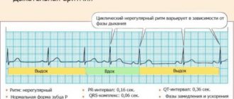
Discordant T wave
A T wave is called discordant if its direction is opposite to the ventricular QRS complex. It happens with bundle branch block, as well as during the period of restoration of blood circulation in the heart muscle after a heart attack.
Discordant T-waves may also appear in cases of severe hypertrophy of the left ventricular myocardium, as well as Wellens syndrome - blockage of the left anterior coronary artery. The latter condition is characterized by attacks of angina-type pain, a high risk of heart attack and the absence of other significant ECG changes, except for the direction of T, and normal blood tests.
Tall T waves in precordial leads
Tall T waves in the chest leads are accompanied by angina pectoris. It can be both stable and progressive, that is, threatening the development of myocardial infarction. In this case, it is important to take into account the clinical picture and other ECG changes. A typical sign of ischemic waves is their symmetry.
High T can also manifest itself as:
- hyperkalemia (excessive intake of potassium, taking drugs that inhibit its excretion);
- anemia;
- circulatory disorders in the brain;
- left ventricular hypertrophy.
T wave alternation
T wave alternans is understood as any change during exercise: on a treadmill, exercise bike, or administration of medications compared to the ECG at rest. One of the options is to analyze the daily recording (monitoring) of the cardiogram.
The doctor may discover that the shape, direction, duration of T, and its amplitude (height) have changed. But there are also micro-changes that are found when analyzed with special equipment - signal-averaged ECG.
By identifying T wave alternans, the electrical instability of the heart muscle is determined. This means that under the influence of stress or stress, a life-threatening arrhythmia with cardiac arrest can occur. Studying the characteristics of T is necessary if you have:
- changes in the duration of the QT interval;
- cardiomyopathy due to arrhythmia;
- ventricular tachycardia;
- ventricular fibrillation.
For information on changes in the T wave on an ECG, watch this video:
Normal QT interval
Normally, the QT interval does not have a constant value. The distance from the beginning of Q to the end of T depends on:
- gender and age of the subject;
- time of day;
- states of the nervous system;
- use of medications, especially analogues of stress hormones (Adrenaline, Dopamine, Hydrocortisone);
- calcium, magnesium and potassium levels in the blood.
The most significant dependence can be traced to heart rate. Therefore, the calculation formulas that take this indicator into account have been continued. The faster the heart rate, the shorter the QT. When mathematically analyzing ECG data from healthy people, an approximate pattern was derived and is reflected in the table.
| QT characteristic | Men, ms. | Women, ms. |
| Normal | 360-390 | 370-400 |
| Bit longer | 391-450 | 401-460 |
| Lengthened | 451-470 | 461-480 |
| Significantly lengthened | From 470 | From 480 |
| Shortened | 360-330 | 370-340 |
| Significantly shorter than normal | Up to 330 | Up to 340 |
What is the ST segment?
The ST segment is one of the sections of the electrocardiogram - a recording on which the electrical impulses of the heart are recorded. The work of this organ is regulated with the help of a pacemaker in the sinus node, which produces 60-90 impulses every minute.
The latter cover various areas of the heart muscle (myocardium) with excitation and are recorded on paper tape in the form of an ECG curve.
The segment of the graph, marked with the Latin letters ST, displays the part of the heart cycle when slow repolarization of the ventricles occurs, that is, their relaxation before subsequent contraction. Cardiac muscle cells cannot remain excited for a long time.
The leading role in the processes of extinction of myocardial activity is played by the coordinated work of the potassium-sodium pump, the return of sodium ions to their original state. Since electrical activity is low during this period, the ST segment should normally be in a straight line between two adjacent teeth.
Its duration (length on the graph) depends on the heart rate; the higher it is, the shorter the segment. The exact duration is difficult to measure and has little clinical significance.
Shortening of the QT interval on the ECG
Shortening the QT interval on the ECG is dangerous, as it provokes complex types of rhythm disturbances. This syndrome can be a congenital feature, and also appears when:
- treatment with cardiac glycosides in the usual dose, progresses with its increase;
- increased concentrations of potassium and calcium in the blood;
- fever;
- a shift in the blood reaction to the acidic side (acidosis).
Short QT syndrome can be constant and repeated from cycle to cycle or paroxysmal due to changes in heart rate. Patients with such disorders are prone to dizziness, lightheadedness, and sudden loss of consciousness. In severe cases, there is a risk of sudden cardiac arrest.
Nonspecific ST-T changes
Nonspecific ST-T changes include all minor violations of ST height, smoothing or the opposite direction of T. They “do not reach” obvious pathologies, but the doctor pays attention to them when deciphering them. This can be important, since if there are complaints of heart pain, further examination is necessary. It is also carried out with risk factors:
- high pressure,
- smoking,
- elderly age,
- high cholesterol,
- sedentary lifestyle.
The main causes of nonspecific symptoms include:
- imbalance of electrolytes (potassium, magnesium, calcium);
- use of medications;
- angina pectoris;
- infectious diseases, pulmonary pathology;
- pain attack;
- consumption of large amounts of food, alcoholic beverages;
- left ventricular hypertrophy;
- cerebrovascular accident.
Since all these factors are diverse, when making a diagnosis, the doctor takes into account the symptoms and, if necessary, prescribes blood tests, an ECG using the Holter method (24-hour monitoring), and stress tests with exercise.
Heart ST depression
Depression of the ST segment, or its decrease relative to the isoline by more than 1 mm, is a clinically significant sign. Such a change can be caused by both nonspecific and specific reasons, which are discussed below.
Non-cardiac and physiological causes include:
- errors on the part of medical staff when taking an ECG - incorrect application of electrodes or their poor contact with the patient’s skin;
- water and electrolyte disturbances that occur when drinking sea water in large quantities, dehydration, alcohol abuse, salt-free diet, anorexia, heavy physical activity, use of diuretics, as well as certain pathologies (diseases of the kidneys, gastrointestinal tract, thyroid gland, burns, significant blood loss);
- drinking very cold water;
- hyperventilation (increased air exchange in the human body).
What does ECG mean in decoding?
Nonspecific ST-T changes on the ECG in the form of its decrease relative to the isoelectric line are primarily associated with cardiac ischemia, as the most dangerous condition that can lead to death.
However, this change is nonspecific and can also occur with other disorders of the heart. To correctly interpret this deviation, differential diagnosis is necessary.
Why is the ST segment reduced in adults and children?
A decrease in the ST area on the ECG in adults and children can occur in the following cases:
- Myocardial infarction with absence of ST elevation. In this case, depression of the segment is observed in two or more leads (by lead is meant the difference in biopotentials between two points of the body, for example, right hand - left hand). This type of coronary insufficiency is less common than elevation of the segment infarction (about 28% of all cases), but in recent years there has been an increase in incidence. The cause of this disorder is most often blockage of the coronary artery by a thrombus, which leads to a significant deterioration in the blood supply to the heart muscle and the development of necrosis in the tissues. The development of this condition can be triggered by stress, anemia, and infectious and inflammatory pathologies.
- Unstable angina (58% of all cases) is an intermediate condition in coronary heart disease, when the risk of developing myocardial infarction is high. The most common cause is atherosclerotic plaques in the coronary vessels of the heart, closing their lumen by ¾ or more. Risk factors are arterial hypertension, tachycardia, physical and emotional stress, consumption of heavy food and alcohol, which increases the load on the heart and causes a lack of oxygen.
- Inflammatory lesions of the heart muscleappearing against the background of infectious diseases (including influenza and respiratory pathologies).
- Toxic effects of ethanol in alcohol abuse. In this case, there is an accumulation of fatty tissue in the heart muscle and an expansion of all the cavities of the heart. At the initial stage, this disorder can be reversible, that is, giving up alcoholic beverages contributes to a significant improvement in function.
- Changes in hormonal levels during menopause in women. The main factor in menopausal cardiomyopathy is a decrease in estrogen production, which contributes to the formation of protein deficiency in the myocardium.
- Dilated cardiomyopathy , in which stretching of the cavities of the heart develops. At the same time, heart failure progresses, the rhythm and conductivity of this organ are disrupted.
- Enlargement of the right chambers of the heart due to embolism (blockage) of the pulmonary artery, severe attack of bronchial asthma, pneumonia, accumulation of air in the pleural cavity.
- Intoxication with cardiac glycosides. Heart failure does not occur immediately, but as the drug accumulates in the blood.
- Paroxysmal tachycardia , detected in elderly people.
In pregnant women, disorders associated with the ST segment are most often caused by physiological reasons - changes in the anatomical position of the heart. Therefore, if such a condition is not accompanied by complaints or a history of cardiac pathologies, then treatment is not required. In doubtful cases, the doctor may prescribe an echocardiogram.
Types of segment reduction
There are 3 types of ST depression:
- obliquely ascending, occurring against the background of tachycardia and being a variant of the norm;
- horizontal, often serving as a sign of ischemic processes in the myocardium;
- oblique, also a symptom of cardiac muscle dysfunction.
To accept the above-described phenomena as normal, the doctor must first exclude possible pathological causes.
Symptoms of deviation
Patients may experience the following symptoms as subjective symptoms:
- Myocardial ischemia:
- severe pain, feeling of pressure, heaviness behind the sternum;
- in women, pain can often radiate to the neck and arm;
- dyspnea;
- arrhythmia;
- general weakness;
- cough;
- in more rare cases - nausea, vomiting, bloating, dizziness, darkening of the eyes, cold sweat, swelling.
- Unstable angina:
- painful attacks lasting 1-10 minutes, similar in nature to the previous case;
- increased symptoms when walking, physical and emotional stress;
- signs subside when taking nitroglycerin.
- Alcoholic cardiomyopathy:
- heartache;
- dyspnea.
- Menopausal cardiomyopathy:
- pressing pain in the upper region of the heart;
- At the same time, other symptoms occur - facial redness, sweating, feeling hot;
- increased symptoms are not associated with physical activity and are not relieved by nitroglycerin.
- Intoxication with glycosides:
- nausea, vomiting, diarrhea;
- severe arrhythmia;
- tachycardia.
Differential diagnosis
Nonspecific ST-T changes require differential diagnosis taking into account medical history, additional studies and analysis of other indicators on the ECG.
To detect acute myocardial infarction, rapid determination of cardiac markers in the blood (globular protein troponin, oxygen-binding protein myoglobin, creatine kinase-MB) is used. For angina pectoris, this test shows negative results.
With alcoholic cardiomyopathy, making a diagnosis usually does not cause difficulties even after interviewing the patient. Painful attacks usually occur the day after alcohol abuse.
Pulmonary embolism is accompanied by a sharp increase in pressure in the right parts of the heart and their expansion, coronary insufficiency occurs, and on the ECG graph the electrical axis of the heart deviates to the right.
In case of an overdose of cardiac glycosides, the ECG shows a rarer heart rhythm, short electrical systole, and a trough-shaped appearance of the ST segment.
Emergency (first) aid for depression
As medical statistics show, about 70% of patients with acute myocardial infarction die in the first 6 hours.
First aid consists of the following activities:
- Call an emergency medical team.
- If the patient is conscious, give him 1 tablet of nitroglycerin under the tongue.
- Provide the patient with peace and fresh air, unfasten his clothes.
- If the patient is alone in the house, open the door to provide free access to the ambulance team, sit down and cough forcefully to induce blood flow to the heart.
Treatment of conditions accompanied by ST depression
Treatment of the most severe and dangerous condition - myocardial infarction - is carried out using the basic means indicated in the table below.
| Drug name | Dosage per day | Average price, rub. |
| Painkillers, narcotic analgesics, intravenously (dispensed strictly according to prescription) | ||
| Morphine | 3-10 mg | 120 |
| Fentanyl | 0.05-0.1 mg | 300 |
| Moradol | 1-2 mg | 250 |
| Thrombolytic agents (dissolution of blood clots) | ||
| Streptokinase | 150 million IU | 1500 |
| Urokinase | 2 million IU | 1400 |
| Anticoagulant therapy (preventing blood clots) | ||
| Heparin | 5 ml (25 thousand units) | 260 |
| Agents for limiting foci of necrosis in the heart muscle (cardioselective beta blockers) | ||
| Metoprolol | 15-50 mg | 25 |
| Atenolol | 10-50 mg | 20 |
All patients also undergo oxygen therapy - oxygen supply through a mask or catheter, which is inserted into the nasal passages. This procedure allows you to saturate the blood with oxygen and reduce the degree of ischemic damage to the myocardium.
ST segment elevation
ST segment elevation occurs in the following diseases:
| Diseases | Description |
| Myocardial ischemia, infarction | In the leads from the site of the lesion it is increased, passes into T, a pathological Q wave may appear, and in the opposite ST it decreases. Then the T first becomes negative and then the ST returns to normal. |
| Accumulation of fluid in the pericardial sac | It differs from a heart attack in that there are changes in many leads and there is no decrease in the opposite leads, there is no Q. First, ST returns to normal, then T. |
| Aneurysm of the left ventricular wall (bulging due to muscle destruction) | There is a deep Q, all changes in ST and T are persistent. |
Increasing the segment is a variant of the norm. In this case:
- the ST dome is directed downward, turns into a unipolar (concordant) T;
- T extended;
- changes can be traced in all leads and cycles.
Rising (elevation) can be caused by an increased concentration of potassium in the blood, inflammation (myocarditis) and a tumor process in the heart.
We recommend reading about the normal ECG in children. From the article you will learn about why children undergo an ECG, indications for examination, rules of conduct, decoding of indicators in children of different ages. And here is more information about ECG for myocarditis.
ST segment elevation
Nonspecific ST-T changes on the ECG in the form of elevation, or a higher position of the segment, in some cases are also a variant of the norm. This deviation can reach 3.5 mm in persons under 40 years of age. However, this nonspecific sign also serves as a criterion for damage to the basal sections or septum of the heart.
What does the ECG mean?
Pathological upward displacement of the ST segment above the isoelectric line is accompanied by a change in its shape:
- combination of a pronounced rise with an unrounded surface;
- horizontal flow with convexity upward;
- the excess of the amplitude of the T wave over the rise of the segment is no more than 1 mm.
Nonspecific ST-T changes on ECG
Normally, the ST is a straight horizontal or gently rising line that merges with the T, or has a convexity that faces downwards (crescent shape). In healthy people, segmental displacement with a deep S wave and a high T wave in the right precordial leads may also be observed. In doubtful cases, the patient undergoes a dynamic electrocardiographic study.
Causes in adults, children
The reasons for the higher position of this segment may be the following violations:
- Acute myocardial infarction. This is the most common pathology in which ST elevation is observed. During an attack, the segment on the ECG graph has a convex shape (“cat’s back”) and smoothly passes into the left knee of the positive T wave. Subsequently, the segment acquires a curved shape, and the T wave becomes negative, and a deep pathological Q wave appears. The rise of this section of the graph indicates about ischemic myocardial damage with complete blockage of one of the coronary arteries.
- Prinzmetal's angina. This disorder is rare in patients with chest pain - in 2-3% of cases. This deviation is based on spasm of the coronary arteries, which occurs due to an imbalance between constrictor and dilating factors (constricting and relaxing blood vessels) and the increased sensitivity of the coronary vessels to them.
- Dressler syndrome , developing in 1-3% of patients after a major myocardial infarction. The cause of this disease is autoimmune aggression and damage to the connective tissue lining of the heart.
- Early ventricular repolarization syndrome , detected in 1-9% of patients. At risk are athletes and men leading a sedentary lifestyle, people with diseases of the connective tissue and cardiovascular system. This condition can be provoked by taking alpha-adrenergic agonists, hypothermia, and lipid metabolism disorders. The severity of the disease also depends on genetic predisposition.
- Post-infarction ventricular aneurysm , when a section of the wall can gradually protrude outward during the ejection of blood into the aorta. Ultimately, part of the left ventricle expands and heart failure develops.
- Brugada syndrome , which is a genetic disorder of the heart rhythm.
Clinical manifestations
Nonspecific ST-T changes on the ECG during elevation may be accompanied by the following symptoms:
- Myocardial infarction:
- a few weeks before an acute condition - angina pectoris and rest, increased frequency of attacks;
- Otherwise, the symptoms are similar to those of a heart attack with segment depression.
- Dressler's syndrome:
- pain or discomfort in the heart;
- general malaise;
- temperature increase;
- chills;
- increased sweating;
- shallow breathing;
- red or pink skin rash;
- cough with moist wheezing.
- Early ventricular repolarization syndrome is often asymptomatic, and arrhythmia occurs in some patients.
- Prinzmetal's angina is angina that occurs regardless of external factors.
- Brugada syndrome - frequent short-term fainting.
Diagnostics
Diagnosis of acute myocardial infarction in this case is made in the same way as in the previous case. A dynamic ECG study, determination of cardiac markers, and echocardiography are performed. For Prinzmetal's angina, coronary angiography is done to study blood flow in the coronary arteries.
Dressler's syndrome is detected using laboratory blood tests (increased ESR, leukocytosis, eosinophilia, C-reactive protein), echocardiography (marked dilatation of the left ventricle, fluid in the pericardial cavity), radiography (pleural effusion). Left ventricular aneurysm is also determined using echocardiography.
Features of treatment
Features of the treatment of the above diseases are the following:
- Prinzmetal's angina – use of calcium channel blockers (Nifedipine, Diltiazem, Verapamil), long-acting nitrovasodilators (Cardiket, Monomak and others);
- Dressler's syndrome - diuretic, antiarrhythmic, antibacterial drugs, anticoagulants, cardiac glycosides, low dosage NSAIDs (Aspirin cardio, Diclofenac), glucocorticosteroids (Prednisolone);
- early ventricular repolarization syndrome - antiarrhythmic drugs (specific therapy has not been developed);
- Ventricular aneurysms – surgical treatment.
ST Down Shift
A pronounced downward ST shift is a sign of insufficient myocardial nutrition - coronary heart disease. It is clinically manifested by angina pectoris, heart attack, and post-infarction cardiosclerosis. Similar changes, but without clear localization, are characteristic of:
- overdose of cardiac glycosides;
- use of diuretics;
- tachycardia;
- increased and frequent breathing;
- hypertrophy of the ventricles of the heart;
- intraventricular conduction disorders.
The T wave reflects the process of ventricular repolarization after their contraction. This is the most labile wave on the ECG; its changes may be the first sign of impaired blood supply to the myocardium in coronary heart disease. To make a diagnosis, you need to compare the clinical symptoms and other signs on the cardiogram.