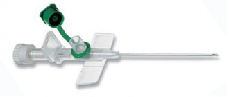
In cases where a one-time procedure of intravenous drip infusion is not sufficient, and long sessions of such therapy are necessary, a special medical device is used to ensure quick and safe access to the vein - a peripheral intravenous catheter.
Despite the fact that the catheter is designed to remain in the vein for a long time, caring for it is essential to avoid serious complications. Here we will talk about self-installation of the device, as well as measures for its maintenance at home.
- Consequences of improper catheter maintenance
How does a venous catheter work? Types of peripheral intravenous catheters
A peripheral venous catheter is a special device designed to be inserted into a vein to provide access to the bloodstream. For the manufacture of such devices, polymers are used - polytetrafluoroethylene, polyurethane, fluoroethylenepropylene copolymer.
Peripheral intravenous catheters, based on the design of providing access to the bloodstream, are divided into 2 types:
- Polymer ones include a flexible PVC tube, which is inserted into the vein using a steel guide needle and is the main element that provides access to the bloodstream for the entire time the catheter is used.
- Metal, where a medical steel needle is the main and only element that provides access to the venous bed during the entire period of use of the peripheral catheter.
To understand how a venous catheter works, let’s look at it in more detail in the figure below:
- The cap is made of transparent polymer and serves to ensure the sterility of the catheter tube and stylet. It has no other purpose.
- A catheter tube is a flexible PVC needle that has a smooth rounding at the end, which prevents injury to the walls of the bloodstream when the device is already in the vein. Present in all models of polymer catheters, hence this type of instrument takes its name.
- The adapter is a non-removable structural part that connects the catheter tube to the channel in the device body (main unit).
- The wings are paired plates designed to secure the catheter to the skin using strips of adhesive tape. Non-removable - the attachment to the main unit is a monolithic connection. They may be color coded according to the size, capacity and purpose of the device.
- The injection port is intended for bolus (syringe) administration of drugs. It is used both during the period of intravenous infusion of the main drug, i.e. simultaneously with a dropper, and separately. Has a valve that prevents the reverse flow of liquids. After catheterization of the vein, this port should always be closed, except for maintenance purposes such as injections or flushing of the device. This design detail is not necessarily present in every model. If it is present, it is always located at the top of the main catheter assembly. If not available, all maintenance is performed through the main infusion port or the most appropriate part of the IV during the period of action. At the end there is a luer-slip or luer-lock connector.
- The main port is a technological hole that serves to connect the catheter to the dropper. As a rule, it is not equipped with a check valve, which does not interfere with the free flow of blood when removing the stiletto needle from the vein when installing the device. At the end there is a connector of one of two types - luer-slip or luer lock.
- Plugs are present on all ports of this instrument. They may be color coded according to the purpose of the device itself. There are two types of fasteners compatible with the connector - luer-slip (standard) and luer-lock (screw-on). The main purpose is to block ports. When installing a catheter into a venous bed, the main port cover must be open and closed only after the device is correctly installed and flushed with solution.
- A stiletto is a conductor made of medical steel, which is an exact copy of a conventional injection needle with an angled cut at the end. Provides venipuncture and insertion of a flexible catheter tube into a vein, then removed from the main node. In metal catheters - where there is no polymer venous tube - it is a non-removable main needle and is included in the design of the main assembly.
- The support plate is a small polymer strip that provides support for the thumb both during insertion of the stylet during venipuncture and when it is removed from the main catheter assembly.
- The indicator chamber is the main assembly of the stylet. Always made of transparent material, so with proper venipuncture, blood is always visible on its walls. At the end opposite from the needle there is a luer-lock or luer-slip connector, onto which a plug cap is placed. An indicator capacity is present in all models of venous catheters - both polymer and metal. In polymer ones it is the body of the stylet, in metal ones it is part of the main assembly of the catheter itself.
The vast majority of device models include these 10 structural parts. Exceptions are metal catheters, where there is no PVC tube as the main part for access to the bloodstream, and some polymer ones, where there is no additional injection port.
Polymer intravenous catheters. Dimensions and selection principle
Catheters are available in different diameters and lengths, which must be indicated on the packaging. Each type has its own color marking. Its throughput depends on the diameter of the device.
| Color code | Size | Throughput (ml/min) | Application area |
| Orange | 14G (2.0x45mm) | 270 | Rapid transfusion of large volumes of fluid or blood products. |
| Grey | 16G (1.7x45mm) | 180 | Rapid transfusion of large volumes of fluid or blood products. |
| White | 17G (1.4x45mm) | 125 | Rapid transfusion of large volumes of fluid or blood products. |
| Green | 18G (1.2x32-45mm) | 80 | Transfusion of blood products (erythrocyte mass) as planned. |
| Pink | 20G (1.0x32mm) | 54 | Long-term intravenous therapy (from 2-3 liters per day). |
| Blue | 22G (0.8x25mm) | 31 | Long-term intravenous therapy, pediatrics, oncology. |
| Yellow | 24G (0.7x19mm) | 13 | Oncology, pediatrics, thin sclerotic veins. |
| Violet | 26G (0.6x19mm) | 12 | Oncology, pediatrics, thin sclerotic veins. |
The main principle of selection is to choose the smallest catheter of all that provides the necessary throughput into the largest vein available for catheterization.
The types and sizes of devices are determined by the doctor in accordance with the purposes for which they will be used, but there are general recommendations:
- Infusion of large volumes of drugs or blood products in a relatively short time. The most commonly used is orange (14G), which has a diameter of 2.0 and a length of 45 mm. This is the largest catheter with a throughput of 270 ml per minute. Gray (16G) with a flow rate of 180 ml/min, or white (17G) with a flow rate of 125 ml/min can also be used here.
- For a course of elimination of BCC deficiency, a green catheter (87G) is usually used, capable of passing 80 ml per minute. It is recognized as the most optimal for routine transfusion of blood products after the main danger to the body has already been eliminated.
- For daily infusions of up to 2–3 liters of medicinal solutions, it is recommended to use pink (20G), which passes liquids at a rate of 54 ml per minute.
- Long-term intravenous therapy (for example, for children or cancer patients) involves the use of a blue catheter (22G), allowing up to 31 ml of solution per minute.
- In oncology, pediatrics, and also for thin sclerotic veins, yellow (24G at a rate of 13 ml/min) or violet (26G at a rate of 12 ml/min) catheters are usually used.
What is a “butterfly needle” and how does it work? Recommendations for selection
The “butterfly needle” is also a catheter for small veins. Not suitable for long-term use, because the main part is a short (19 mm) needle made of medical steel with double sharpening, and not the PVC tube usual for such instruments.
The mounting of the device itself most often comes with the Luer-Lock type, but sometimes you come across models with a standard port.
It is a convenient device equipped with special rough wing fasteners that allow you to easily attach it to the skin using a patch. The color of the “Wings” corresponds to a special marking, which, in general, does not correspond to the color marking of other types of catheterization devices.
Below is the marking of German products according to the ISO standard:
| Color code | Size | Needle diameter (mm) | Needle length(mm)/tube(mm) |
| Pink | 18G | 1.20 | 19 / 300 |
| White | 19G | 1.10 | 19 / 300 |
| Yellow | 20G | 0.90 | 19 / 300 |
| Green | 21G | 0.80 | 19 / 300 |
| Black | 22G | 0.70 | 19 / 300 |
| Blue | 23G | 0.60 | 19 / 300 |
| Violet | 24G | 0.55 | 19 / 300 |
| Orange | 25G | 0.50 | 19 / 300 |
| Brown | 26G | 0.45 | 19 / 300 |
| Grey | 27G | 0.40 | 19 / 300 |
The selection of a butterfly needle is somewhat different from other types of catheters. Taking into account the viscosity and volume of the drug, it is recommended to use the following devices, based on their size and age of the patients:
- Infants 0-12 months: 22G (0.7mm) black.
- Children 1–3 years: 22G (0.7 mm) black or 25G (0.5 mm) orange.
- Children 4-14 years old: 22G (0.7 mm) black, 25G (0.5 mm) orange, 24G (0.55 mm) purple or 23G (0.6 mm) blue.
- Children over 14 years of age and adults: General parameters apply, including 21G (0.8 mm) with green markings.
Butterfly needles with a diameter of 20G, 19G, 18G are not recommended for use for pediatric veins due to the small diameter of the vessels and the large diameter of the metal part of the needle. The choice of the thickness of the 20G butterfly needle must be agreed with the doctor and can be used if a high rate of infusion of the drug or its large volume.
Attention!
Please note that our recommendations are general and for a strict prescription you need to consult a doctor.
The butterfly needle design is different from other catheter designs, mainly because it is a metal type of device.
Let's look at it in more detail in the figure below:
- The protective cap serves to ensure sterility. It has no other functions. If the device is immediately connected to the dropper, then the system is filled with it. In this case, the protective cap must be removed and installed back only when the air is completely displaced by the solution and the latter begins to flow freely through the needle.
- The injection needle is made of medical steel. In this device, it is the only one, i.e. it is not a stylet-conductor necessary for introducing a flexible PVC tube into the bloodstream.
- The adapter is a non-removable structural part that serves to reliably connect the needle with the through channel in the wing body.
- The wings are designed to secure the catheter to the skin using adhesive tape.
- Extension tube. It also serves as an indicator chamber: when the vein is entered correctly, it immediately fills with blood. This flexible thin tube has a standard length of 300 mm and is made of transparent polyvinyl chloride.
- Injection port (adapter). It can be either with a standard adapter or with a Luer-Lock type of fastening at the end (more often in models for blood sampling). Serves to connect the device to a syringe or infusion system.
- A blank cover in one form or another is present in any device model. It is also an injection valve. At the time of catheterization, the plug should always be open until the air under blood pressure is completely released from the tube, then it is closed, or the device is connected directly to the dropper and the entire system is filled with the medicine along with it.
The butterfly needle has only one port for administering the drug.
Like any other steel catheter, this device should not be left in the vein for more than 24 hours to avoid the risk of blood clots and infiltrates. Rigid fixation is required, associated with significant restrictions on the patient’s mobility.
The procedure for venipuncture with such a catheter is no different from the standard one generally accepted for other similar devices.
Modern devices
In medical practice, catheters with steel needles are currently practically not used, because the comfort and safety of the patient come to the fore. Unlike the metal model, a plastic peripheral intravenous catheter can follow the bends of the vein. Thanks to this, the risk of injury is significantly reduced. The likelihood of blood clots and infiltrates is also minimized. In this case, the time that such a catheter remains in the vessel increases significantly.
Patients who have such a plastic device installed can move calmly without fear of damaging their veins.
How to choose a site for catheterization?
First of all, straight sections of the distal veins, the largest and softest to the touch, and well filled, are considered for catheterization. As a rule, preference is given to the lateral and medial saphenous veins of the arm - these are the intermediate venous beds of the elbow and forearm. Less commonly, if venipuncture of the former is impossible, metacarpal and digital ones are used.
It should be noted that inserting a catheter yourself is an extremely difficult task, especially for inexperienced people, since most manipulations require both hands, so you should at least invite someone to help. Training the skill of gaining access to the bloodstream using improvised means will also not be superfluous.
Choosing a site for catheterization is no different from the procedures for placing intravenous injections or drips. You can learn more about this by studying visual photos and diagrams here.
Materials used
The first plastic models were not very different from steel catheters. Polyethylene could have been used in their manufacture. The result was thick-walled catheters, which irritated the inner walls of blood vessels and led to the formation of blood clots. In addition, they were so rigid that they could even lead to perforation of the vessel walls. Although polyethylene itself is a flexible, inert material that does not form a loop, it is very easy to process.
Polypropylene can also be used in the production of catheters. Thin-walled models are made from it, but they are too rigid. They were mainly used to access arteries or to insert other catheters.
Later, other plastic compounds were developed, which are used in the production of these medical devices. Thus, the most popular materials are: PTFE, FEP, PUR.
The first of them is polytetrafluoroethylene. Catheters made from it glide well and do not lead to thrombus formation. They have a high level of organic tolerance and are therefore well tolerated. But thin-walled models made of this material can be compressed and form loops.
FEP (fluoroethylene propylene copolymer), also known as Teflon, has the same positive characteristics as PTFE. But, in addition, this material allows for better control of the catheter and increases its stability. A radiopaque contrast medium can be injected into such an intravenous device, which will allow it to be seen in the bloodstream.
PUR material is a well-known polyurethane. Its hardness depends on temperature. The warmer it is, the softer and more elastic it becomes. It is often used to make central intravenous catheters.
Preparing to insert a catheter
Before you begin installing a catheter yourself, you need to prepare the following set of tools and materials:
- Peripheral catheter of the required size.
- Syringe.
- An ampoule with saline solution or a pre-prepared heparin solution.
- Venous tourniquet.
- Bandage.
- Patch.
- Preferably sterile gloves.
- Sterile wipes.
- Any medical antiseptic or 70% alcohol.
- Tray for used materials.
Once you have prepared everything you need, you can begin to install the catheter into the vein.
If you intend to attach a drip, then at the time of catheterization the system should be completely assembled and filled with a medicinal solution.
Possible complications
The algorithm for placing a peripheral venous catheter is quite simple. However, the risk of complications is very high, since the skin is injured.
- Phlebitis is sepsis of a blood vessel associated with mechanical stress or infection.
- Thrombophlebitis.
- Thrombosis.
- Equipment bending.
A common problem is infection, especially with penetration into the circulatory system. Sometimes it can even be deadly. To prevent the spread of pathogenic bacteria, various preventive measures are used, including surface treatment or impregnation of the catheter with antiseptic or antibacterial compounds.
Algorithm of actions for installing a catheter in a vein
To install a catheter into a vein yourself, you must follow the following algorithm:
- Wash your hands thoroughly, let them dry, and treat them with an antiseptic. Then put on gloves.
- Examine the arm and select the most appropriate site for venipuncture. In no case should there be visible mechanical or thermal damage or allergic reactions in this area. In this case, you should avoid places of physiological bends (for example, the ulnar fossa) in order to avoid injury to the vein with a needle when moving your hand.
- Apply a tourniquet approximately 10–15 centimeters above the catheterization site and “work” vigorously with your fist.
- Palpate the vein and, if it is well filled, clench your fist and stop further movements with your hand.
- Open the catheter packaging and remove the protective cap.
- Fix the vein with your free hand, lightly pressing and stretching the area of skin a few centimeters below the intended puncture point.
- Inject into the vein at an angle of 15–20 degrees until blood appears in the indicator chamber of the catheter.
- Then, reducing the angle of inclination of the device, we advance the guide needle into the venous bed almost to the base.
- Loosen the tourniquet.
- Place a sterile napkin under the remaining outside part of the catheter and, lightly pressing the area of skin above the vein with the catheter in it with your finger, with a confident movement of the other hand, pull the guide needle out of the device port.
- Immediately close the catheter port with the valve.
- Securely secure the catheter with a plaster or bandage.
The procedure is quite responsible, so beginners should not undertake it. It is better to ask a qualified medical professional to carry it out. If this is not possible, and you have to do everything yourself, then you need to at least practice using improvised means.
General recommendations
There are certain rules for working with devices for intravenous drug administration.
All health care workers who will select or install an intravenous catheter should know them. The algorithm for their use provides that the first installation is carried out from the non-dominant side at a distal distance. That is, the best option is the back of the hand. Each subsequent installation (if long-term treatment is necessary) is done on the opposite hand. The catheter is inserted higher along the vein. Compliance with this rule allows you to minimize the likelihood of developing phlebitis.
If the patient undergoes surgery, it is better to install a green catheter. It is the thinnest of those through which blood products can be transfused.
Connection of a venous catheter with a dropper. Setting up injections
The main thing is to follow the rules of asepsis. Whichever of these manipulations you perform, be sure to treat your hands and all external surfaces of the venous catheter with which you will interact with an antiseptic before and after the injection or connecting the IV.
The administration of injections was mainly clarified by considering the issues of catheter design. It remains to be recalled that for this purpose a special additional injection port is used, which is located at the top of the main unit of the device - where the wings are located. You can also find it by the colored plug above the valve.
To give an injection, you need to draw the medicinal solution into the syringe in the usual way, and then, after first removing the injection needle and the cap from the device port, press the plunger, creating pressure inside the catheter chamber. In the same way, you can wash the device as part of standard care.
If there is no injection port, then the drug for bolus infusion can be injected into the only available port of the catheter - the infusion port, through which the IV is connected. If it is necessary to add a drug during an infusion session, you should draw the required medicine into the syringe and, without removing the needle, inject the solution into the injection unit of the system itself.
Before each connection of a venous catheter with a dropper, the infusion port of the former is washed, especially if several hours have passed since the previous procedure.
We attach the pre-assembled and filled dropper to the port immediately after flushing. The port cover should be treated with an antiseptic and stored under sterile conditions for the entire duration of the procedure.
After the end of the next session, the infusion system is disconnected in the same way as it was connected, and the port is washed again and closed with a cap.
How to independently assemble and fill a dropper and how to use it correctly is described in detail here.
Advantages and disadvantages of ports
Manufacturers produce several types of devices designed for intravenous administration of medicinal solutions. According to many, it is preferable to use cannulas equipped with a special port. But it is not always the case. They are necessary if the treatment involves additional infusion of medications.
If this is not required, a regular intravenous catheter can be installed.
- Why do the veins in the arms hurt in the elbow bend?
A photo of such a device makes it possible to see that it is very compact. Devices without additional ports are cheaper. But this is not their only advantage. When using them, there is less chance of contamination. This is due to the fact that the injection element of this system is separated and changed daily.
In intensive care and anesthesiology, preference is given to ported catheters. In all other areas of medicine, it is enough to establish the usual option.
By the way, in pediatrics they can install a catheter with a port for injecting medications even in cases where children do not need to have an IV installed. This is how antibiotics can be injected, replacing injections into the muscle with intravenous administration. This not only increases the effectiveness of treatment, but also facilitates the procedure. It is easier to install a cannula once and almost imperceptibly inject the medicine through the port than to give painful injections several times a day.
Rules of care
It is important to remember that each type of catheter has its own, strictly defined period during which it can remain in the vein without risk to the patient’s health. For steel catheters, the installation time should not exceed 24 hours, for polymer catheters - no more than 3 days. At this time, systematic special treatment of the device is necessary, which allows not only to prevent bacteria from entering the bloodstream, but also to prevent occlusion (blocking) of the device ports, which inevitably occurs due to the “growth” of plasma clots and drug residues.
Consequences of improper catheter maintenance
What happens if you do not maintain the catheter promptly and correctly? At first glance, this device is completely harmless and is designed to reduce the number of invasive interventions in the patient’s body, but improper and untimely care of the catheter or the surface of the arm at the catheterization site can lead to serious consequences.
The main dangerous complications typical of catheterization are phlebitis and abscess.
Phlebitis with PVK is inflammation of the walls of the venous bed as a result of prolonged mechanical or chemical exposure.
Mechanical irritation is explained by the fact that a foreign object - a metal or plastic needle - has been in the vein for a long time. During any procedural manipulations or simply careless movements of the patient, mechanical impact on the vein walls cannot be avoided. The longer the catheter is in the bloodstream, the higher the risk of phlebitis.
Chemical irritation occurs as a result of the administration of certain drugs. This may depend on the concentration or feed rate of solutions.
An abscess with PVK is a purulent inflammation localized at the site of direct catheterization and resulting from non-compliance with asepsis rules.
An airtight dressing can also have a negative impact due to the accelerated growth of bacteria and fungi in such an environment. In this regard, it is not recommended to apply airtight dressings, except in cases where the catheterization site is close to open or burn wounds or tracheostomies.
In hospital settings, specialists constantly conduct bacteriological cultures of drug residues and patient blood from the surfaces of the catheter and skin at the site of venipuncture. This is done to conduct analysis and prevent the risk of infectious infection of the venous bed, as well as tissues near the catheterization site.
When using the device, it is necessary to regularly inspect the catheterization site and the vein above the catheter. At the slightest sign of redness or swelling, the device must be removed immediately. This can be done simply by lightly pressing the skin at a distance of 7–10 cm above the catheter, smoothly pulling it towards you, and after the needle comes out, bend your arm at the elbow, pressing a swab soaked with an antiseptic to the venipuncture site.
Flushing the catheter ports
Typically, the catheter has two ports covered with caps of different colors - white and colored (according to the target labeling). The colored valve closes the port for direct administration of medications using conventional injection syringes, and the white valve closes the port for infusions, i.e. this is where the dropper is connected.
To avoid infection of the bloodstream, which inevitably arises when using a catheter for a long time, it is necessary to flush the device. Typically, a heparinized solution or saline solution is used for this, which is drawn into a sterile disposable syringe, after which the needle is removed.
Immediately before this manipulation, the valve is removed and pressing on the syringe plunger ensures flushing of the port.
Depending on the total time, frequency of direct use and composition of the drugs, catheters can be washed regularly, from one to three times a day, or after each infusion session. The latter is most preferable.
The fact is that the cavity of the device port is a comfortable environment for the development of decomposition bacteria, and blood clots and drug residues, growing, can cause clogging of the tube, which reduces the capacity of the catheter initially declared by the manufacturer.
For rinsing, either pure saline solution (0.9% sodium chloride solution) is used, or the same solution, but in combination with heparin. The combined solution is prepared at the rate of 100 IU (0.02 ml) of heparin per 1 ml of 0.9% NaCl.
There is no need to prepare a solution for flushing in advance; we focus only on the internal volume of the venous catheter, i.e. 5–10 ml will be enough.
Varieties of plastic models
Doctors can choose which catheter to place in a patient. You can find models on sale with or without additional injection ports. They can also be equipped with special fixing wings.
To protect against accidental punctures and prevent the risk of infection, special cannulas have been developed. They are equipped with a protective self-activating clip, which is installed on the needle.
For the convenience of injecting medications, an intravenous catheter with an additional port can be used. Many manufacturers place it above the wings designed for additional fixation of the device. When administering medications into such a port, there is no risk of cannula displacement.
When purchasing catheters, you should follow the recommendations of doctors. After all, these devices, although externally similar, can differ significantly in quality. It is important that the transition from needle to cannula is atraumatic, and that there is minimal resistance when inserting the catheter through the tissue. The sharpness of the needle and its sharpening angle are also important.
Experts often recommend purchasing models from such manufacturers as the BD concern, B. Braun, HMD, Wallace Ltd.
An intravenous catheter with a Braunulen port has become the standard for developed countries. It is equipped with a special valve, which prevents the possibility of reverse movement of the solution introduced into the injection compartment.


