Rheoencephalography of cerebral vessels
REG (rheoencephalography) is a non-invasive method for diagnosing cerebral vessels, recording changes in the electrical resistance of vascular walls when a high-frequency pulse passes through them.
Provides an opportunity to check the vessels of the head, their reactivity, tone, elasticity, level of resistance of vascular tissue, possible blockage and characteristics of blood supply. REG is indicated for suspected diseases:
- vascular pathologies;
- circulatory disorders;
- atherosclerosis;
- stroke;
- vascular stenosis;
- clarification of the effect of drugs;
- traumatic brain injuries.
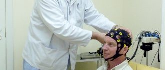
To perform rheoencephalography, rheographs with a number of channels from 2 to 6 are used, recording the corresponding number of vessels. The patient is sitting or lying down. Electrodes are installed on his head using rubber bands. A special paste can be applied underneath them to improve contact with the skin and reduce resistance. Electrodes are attached depending on the vessel being examined. So, if you need to check the internal carotid artery, the electrode is placed on the bridge of the nose and mastoid process.
Some electrodes send electrical signals, others receive them and transmit them to the rheograph. The latter processes and transmits to a monitor or print in the form of a graph - a rheoencephalogram. The procedure lasts several minutes.
During the examination, the patient may be given a small dose of nitroglycerin under the tongue, Papaverine, or asked to change body position or turn his head. These measures will allow you to see a change in tone and blood supply.
The advantage of rheoencephalography is its simplicity, safety for the patient, and the ability to monitor blood flow for a long time. Disadvantages include the inability to measure cerebral blood flow and determine the exact cause of circulatory disorders.
Before the study, you should not smoke, drink alcohol, coffee, or energy drinks. If the patient is taking medications, the doctor must be informed about this. Some may need to be stopped for a while.
Rheoencephalography is not prescribed for children under 7 years of age, as well as for wounds and injuries at the places where the electrodes are attached.
Duplex scanning
This is one of the types of ultrasound examination. The result is a color image of the blood flow in the vessel. The method is completely safe, non-invasive and does not expose humans to x-ray radiation.
Since the combination of Dopplerography and B-mode has more capabilities than Doppler ultrasound. This diagnostic method can be performed an unlimited number of times.
The examination takes place in an outpatient clinic, as with other ultrasound examinations, a gel is applied to the patient’s skin, the procedure lasts about half an hour. However, the resulting picture is highly informative. The vessels are visible in both longitudinal and transverse sections, but only in an area that is not very significant in area. Both veins and arteries are equally clearly visible.
Experts believe that the only competitor to such scanning can be X-ray contrast angiography, which is invasive and is not suitable for everyone due to health reasons.
If the goal is to track the dynamics of recovery and it is necessary to determine the condition of the vessels several times, then it is better to resort to the duplex method.
Contraindications to the use of in-depth studies
Hardware methods, despite their information content and the most gentle approaches, still have a number of contraindications. These include:
•Allergies to contrast solution;
•Kidney, heart, liver failure;
•Infectious diseases and inflammations;
•Chronic diseases in the acute stage;
•Psychical deviations.
Diagnosing the condition of blood vessels in pregnant and lactating women, elderly patients, and people with bleeding disorders requires extreme caution.
Warning: Undefined array key “module” in /customers/0/e/9/germanklinik.de/httpd.www/control/config/inc/templates_c/front^7198fc69815638cccb44869b7c86cae91ff823b0_0.file.DefaultGK.tpl.php on line 488 Warning: Attempt to read property “value” on NULL in /customers/0/e/9/germanklinik.de/httpd.www/control/config/inc/templates_c/front^7198fc69815638cccb44869b7c86cae91ff823b0_0.file.DefaultGK.tpl.php on line 488 Warning : Undefined array key “extension” in /customers/0/e/9/germanklinik.de/httpd.www/control/config/inc/templates_c/front^7198fc69815638cccb44869b7c86cae91ff823b0_0.file.DefaultGK.tpl.php on line 490 Warning: Attempt to read property “value” on NULL in /customers/0/e/9/germanklinik.de/httpd.www/control/config/inc/templates_c/front^7198fc69815638cccb44869b7c86cae91ff823b0_0.file.DefaultGK.tpl.php on line 490
Thank you! We have received your request for consultation.
If you want to fill out a Request for Treatment, continue filling out. Having all the necessary data, we will be able to find the best solution to your problem in the shortest possible time
Methods for studying cerebral vessels
A specialist can make a reliable diagnosis using ultra-sensitive medical equipment that provides reliable data. A doctor’s mistake can cause drug therapy to be ineffective, and precious time will be lost.
- How to check the blood vessels of the brain, what methods are used
Before the manipulations, the blood is examined. This is necessary to identify pathologies that affect its coagulation. A medical specialist uses special equipment to examine the brain. You can check the vessels of the head without interfering with their functioning.
In cases of serious illnesses and cancers, doctors carry out the most accurate and detailed examination by introducing chemicals into the blood. The equipment monitors the movement of fluid through the vessels; on the monitor you can see the most complete picture of the existing pathology.
Research using high frequency sound waves
Brain examinations are typically performed using high-frequency signals. Methods for studying the brain are varied and are represented by the following types.
Echoencephalography
Improper functioning of the brain and blood vessels is analyzed using high-frequency sounds directed at human tissue. The measurement is carried out with an oscilloscope. High-frequency sound transmits reflected signals, as a result of which a special diagram-drawing is displayed on the monitor screen.
The study allows us to assess how affected individual areas of the brain are and how the detected disorders affect the activity of the body as a whole. To conduct an examination using this method, no preparation is needed.
The effect of the device on a person is not accompanied by painful sensations.
Ultrasound Dopplerography (USDG)
This is the latest technological development of scientists. The essence of the method is that high-frequency sound penetrates tissue to a depth of 9 cm. Medical equipment analyzes extremely accurately and finds pathology in the smallest vessels. There is no need to make any special preparations for this study.
The hardware installation determines the speed of blood flow in the neck and head. Using Doppler, you can determine the filling of blood vessels with blood. If a pathology is detected, then you can understand how serious it is. This technique does not involve interfering with the functioning of the body. With its help, you can record how effective the vascular treatment is.
Ultrasonography (USG)
- Treatment of cerebral vessels: folk remedies, traditional medicine
During the examination, the arteries of the head and neck are examined simultaneously. This technique is used to analyze the degree of cholesterol contamination of blood vessels.
The device detects the presence of blood clots. This is very important, since at any moment a detached blood clot can lead to death.
Duplex scanning of head and neck vessels
The latest developments in medicine have made it possible to obtain color images of the body from the inside. Studying individual parts of the body allows us to most accurately determine the cause of improper blood movement through the vessels. This study helps doctors make the correct diagnosis and prescribe optimal treatment.
Neurosonography (NSG)
This method allows you to examine the vascular system of infants up to one year old. The head is examined through the fontanelle, which is not yet closed. Used for birth injuries. The state of the liquor-conducting system and blood flow parameters are studied. The specialist looks to see if brain development is normal. The study of cerebral vessels is of great importance, since defects detected in time allow timely therapy. The equipment does not cause discomfort to the child.
Rheoencephalography (REG)
The essence of the special equipment is to pass an electric current through the brain tissue. The examination is painless, the patient does not feel anything. High-frequency electric current makes it possible to conduct research on the brain.
The method helps to find out where the vessels are damaged and how deeply, to detect neoplasms in the head, and to assess the consequences of injury to brain tissue. High-frequency technology provides data on the presence of blockages in blood vessels and bleeding in the brain.
Definition
Diagnostics and examination of blood vessels is a set of diagnostic methods that allow us to study the condition of blood vessels and determine the pathologies present in them. Vascular examination is carried out to determine the cause of vascular diseases and effective treatment. There are a number of diagnostic procedures that make it possible to identify any deviations from the normal functioning of blood vessels even in the early stages. The examination of blood vessels includes a doctor’s examination, laboratory tests and instrumental techniques for examining blood vessels.
Methods for studying cerebral vessels
In medical practice, there are several research methods to check the vascular system of a patient’s brain:
- Using an ultrasound machine, the following procedures can be performed:
- Ultrasound Dopplerography. Thanks to this study, it is possible to determine: The presence of plaques that do not allow blood to circulate properly inside the vessels of the brain;
- The speed of blood movement through the vessels of the head and cervical spine;
- Size of carotid, cerebral and vertebral arteries.
- Duplex scanning. Thanks to this study, the doctor can:
- Construct a color diagram of blood movement in the vessels of the brain;
- Identify diseases of the vascular system of the head that are still at an initial stage, for example, atherosclerosis, stenosis, aneurysms, occlusions.
Echoencephalography allows you to check:- The condition of both the inside of the brain and the periosteal space of the skull;
- How strong is the pulsation in the brain? Due to this verification, the patient's intracranial pressure is determined.
- Neurosonography pris given to children who have not yet turned one year old. Because the brain of small patients is checked through the fontanelle, which has not yet become overgrown in them. In the process of such a study, a specialist can determine the following:
- Blood circulation in the brain;
- No tumors or cysts in the brain;
- Does the little patient have any diseases related to the functioning of the brain, for example, high cranial pressure, a tendency to epilepsy or encephalopathy. Unfortunately, such unpleasant diseases can appear due to possible birth injuries or due to the fact that the baby did not have enough oxygen while passing through the birth canal.
- Using X-ray tests called angiography. There are several types of such checks:
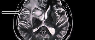
- Magnetic resonance angiography allows you to check the patient’s cerebral vessels for the following diseases: Strokes;
Heart defects;- Increased pressure inside the skull;
- Atherosclerosis of blood vessels in the neck or brain;
- Vegetovascular dystonia;
- Vasculitis;
- Stenosis;
- Aneurysm.
- Magnetic resonance angiography has several subtypes:
- Diffusion-weighted magnetic resonance imaging will be prescribed to the patient if he is suspected of ischemic brain disease;
- Diagnostics of the main cerebral arteries;
- Sinusography allows you to study the condition of the cerebral veins and the collectors of this venous system. This procedure will be performed on the patient in order to prevent the formation of blood clots in the vessels of the brain.
- It should be borne in mind that such diagnostics also have their contraindications:
- The patient is overweight (more than 150 kg);
- Metal implants;
- The presence of artificial joints;
- Electrical pacemakers.
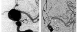
- Computed angiography. Such a study is carried out according to the following algorithm:
- The patient is given local anesthesia;
- A catheter is placed into the brain vessel through which a special substance (x-ray contrast) will flow;
- After all the preparatory work, the diagnosis itself is carried out using an angiograph;
- This X-ray machine takes pictures of the brain vessels;
- After all the necessary actions have been carried out for the patient, the place where the catheter was located should be tightly bandaged.
- The patient needs to know that such an examination should be carried out only on an empty stomach, and after diagnosis it is necessary to drink a lot in order to remove from the body the substance that was introduced through the catheter during the examination.
- Rheoencephalography is a diagnostic method that can be used to:
- Assess blood circulation in the brain;
- Determine at what speed blood moves through the vessels;
- See what condition the walls of the blood vessels are in.
Anyone who undergoes any examination of the cerebral vessels should know that a diagnosis based on these diagnostic studies can only be made by a doctor who, if necessary, will prescribe specific treatment or send the patient for additional examinations of other organs of the body.
- Vascular diseases of the head and neck: treatment with drugs and folk remedies
Angiography
Angiography is used to examine arterial dysfunction. This type of diagnosis will be called arteriography. If the goal is to determine the condition of the veins, then they talk about phlebography.
In any case, the procedure takes place in a hospital where there is an X-ray angiography room.
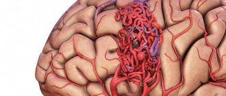
In order to determine the condition of the veins and arteries, it is necessary to inject a contrast agent and take x-rays of its progress through the body. Atherosclerosis, thrombosis, dilation of the vessel, aneurysm, thrombophlebitis will be clearly visible.
Interpretation of coronary angiography
How coronary angiography of the heart vessels is performed and what it is is clear. The main thing is not just to take a picture, but to interpret it correctly, that is, decipher it. For the most informative and accurate result, several specialists are involved in decoding: a radiologist, a cardiologist, a vascular surgeon and others.
The thickness of the vessels, their length and width are assessed from the images. Compactions and darkening are revealed. An alarming signal is a violation of the boundaries of blood vessels - blurriness and indistinctness. The pathology of the vessels will also be indicated by their incorrect location.
Prognosis and prevention
With early treatment and prevention of complications, the prognosis is favorable. Vascular pathologies today are successfully treated with conservative and surgical methods, while maintaining the patient’s high quality of life.
Vascular diseases are best prevented by using medications that lower cholesterol levels, as well as products that contain polyunsaturated fatty acids that can prevent the development of atherosclerosis. It is recommended to adhere to the principles of a healthy diet, do not consume fatty and fried foods, and alcohol. It is advisable to quit smoking and start exercising to strengthen the heart muscle and prevent the development of many diseases, including obesity and hypertension. Moderate physical activity has a positive effect on the entire cardiovascular system.
After 30-35 years, check your cholesterol levels and monitor your blood pressure numbers. If symptoms of vascular disorders appear, begin treatment promptly before complete damage to the arteries and veins occurs.
Consultations with a doctor online Taking care of your health is a life priority for everyone.
Communicate with doctors online and receive qualified assistance without leaving your home. Try it Please note! The information on this page is provided for informational purposes only. To prescribe treatment, you must consult a doctor.
How to choose an examination technique?
Ultrasound does not require special conditions for placement of equipment. This is the easiest way to diagnose. The purchase and installation of devices for CT, MRI or PET require considerable costs. In this regard, not all medical institutions can afford to carry out such procedures. For this reason, prices for these types of diagnostics will be high.
However, the popularity of the equipment and the price of diagnostics should not be the determining factors. First of all, you need to follow the recommendations of your doctor. The scope of application of the examination method should also be taken into account:
- PET detects a tumor, including a malignant one, with absolute accuracy, long before its manifestation.
- MRI is most effective in neurosurgery and neurology.
- CT is useful in detecting vascular damage and head injuries.
- From the point of view of the absence of ionization and X-ray radiation, MRI is the safest procedure. However, modern equipment for radiography and ultrasound significantly reduces the risk of gene mutations.
It is important not to forget about contraindications. Thus, PET and CT scans are strictly prohibited for pregnant women. MRI is used for expectant mothers if the potential benefit to the woman is higher than the possible risk to the baby.
Children require special preparation for the procedure. Parents should allegorically explain to their children the need to lie still. The youngest require anesthesia.
Only the attending physician can determine the need for a particular diagnosis. In some cases, different types of scanning may be required at the same time.