Diagnosis of diseases of the central nervous system (CNS) using magnetic resonance imaging makes it possible to identify and differentiate pathological processes in the early stages of development.
A series of MRI images for suspected stroke
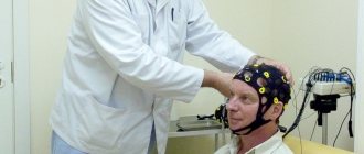
An induction field is used to visualize the state of the cerebral substance and cerebral vessels. Under the influence of a directed electromagnetic pulse, the hydrogen atoms in water molecules change position, then returning to their place. The resonance of charged particles is detected using sensitive sensors and processed using complex software algorithms.
Diagnosis of strokes using MRI is an effective way to determine pathological changes in the condition of intracranial vessels and assess the consequences of cerebrovascular accidents. Magnetic resonance imaging of the head is prescribed to clarify the diagnosis if the examination is insufficiently informative and in the case of an initial visit to a specialist. A referral for the procedure can be given by a neurologist, neurosurgeon, oncologist, or traumatologist.
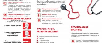
What is a stroke?
Acute cerebrovascular accident (ACVA) was previously called apoplexy, which translated from Greek means “paralysis.” The disease is classified as a severe cerebrovascular pathology, accompanied by high mortality. 70-80% of patients who have had a stroke become disabled.
The clinical picture is represented by focal and cerebral neurological symptoms. The first signs of the disease are:
- weakness, possible fainting;
- sudden agitation or drowsiness at the initial stage;
- Strong headache;
- nausea, vomiting;
- speech disorder;
- facial paresis;
- dizziness, loss of orientation in space and time;
- sweating, increased heart rate, dry mouth and other autonomic disorders.
Against the background of the development of cerebral symptoms, focal neurological phenomena occur, the nature of which depends on the location of the damaged vessel. If the blood supply to the occipital zone and the cerebellum is impaired, increasing muscle weakness of the upper and lower extremities, unsteadiness of gait, and paresthesia of the arms and legs are observed.
The focus of ischemia and necrosis of the brain (shown by arrows) on MRI
Damage to the vertebrobasilar circle leads to dizziness, loss of balance, and possible vomiting. Vision deteriorates, the eye muscles on the affected side suffer, and the sensitivity of half the body decreases.
The patient cannot repeat a simple phrase and does not answer questions. When trying to smile, one corner of the mouth remains lowered, and the tip of the tongue moves towards the affected side.
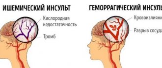
Depending on the pathogenesis of the disease, there are four main types of stroke:
- ischemic;
- hemorrhagic;
- subarachnoid hemorrhage;
- acute hypertensive encephalopathy.
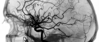
Pathologies of the great vessels play a significant role in the development of ischemic stroke. Cerebral infarction occurs when there is stenosis or thrombosis of the brachiocephalic and intracranial (intracranial) arteries. Lack of oxygen and nutrients causes necrosis of cerebral cells, which leads to the development of neurological disorders. The causes of severe hypoxia are atherosclerosis, embolism, and a sharp decrease in blood pressure (BP). In the diagnosis of ischemic stroke, MRI, CT of the brain, and other instrumental research methods are used. The prognosis largely depends on the ability to differentiate the disease in the early stages.
Hemorrhagic stroke occurs when intracerebral hemorrhage leads to the death of nerve cells. The disease develops against the background of hypertension, causing spasms and paralysis of the intracranial arteries. The walls of blood vessels become permeable to plasma and formed elements, lose their tone and passively expand with increasing blood pressure.
Picture of the development of ischemic stroke on MRI (T1- and T2-weighted images)
Subarachnoid hemorrhage occurs when an aneurysm ruptures, arteriovenous malformations, malignant diseases, or due to traumatic brain injury. Neurological symptoms appear when the cerebral substance is compressed due to the formation of hematomas in the area of the soft arachnoid membrane.
Acute hypertensive encephalopathy develops against the background of persistent arterial hypertension. A prolonged increase in blood pressure leads to progressive damage to brain tissue. In case of ineffective treatment, hypertensive encephalopathy ends in ischemic or hemorrhagic stroke, swelling of cerebral structures, and death is possible. The danger of the condition lies in the absence of a clear clinical picture.
MRI diagnostics allows for timely detection of stroke and prescribing effective treatment. The volume and nature of medical care depend on the stage of the disease. The following periods of development of the pathological process are distinguished:
- elementary;
- spicy;
- early recovery;
- late rehabilitation;
- period of long-term consequences.
Recovery time depends on the location and size of the necrosis focus, the patient’s age, the effectiveness of treatment procedures and other factors.
Meningeal symptoms
Signs of irritation of the meninges can occur with parenchymal hemorrhage due to cerebral edema, but they are more characteristic of subarachnoid hemorrhage.
The most typical sign is a stiff neck, when the patient is unable to press the chin to the chest when lying down.
If the patient bends his knees while checking for rigidity, this is another sign of irritation of the meninges, which is called Brudzinski's sign.
Is it possible to do an MRI after a stroke?
Magnetic resonance imaging does not have a negative impact on the patient's health. If you suspect the development of necrosis of brain tissue, a scan will help determine the nature and intensity of the pathological process.
Signs of acute cerebrovascular accident on MRI
The disease must be differentiated from lesions of cerebral structures caused by:
- poisoning;
- hypoglycemia;
- growth of tumors;
- epilepsy attack, etc.
MRI for cerebral stroke is highly effective during the recovery period. In the acute period, scanning is used if an ischemic lesion is suspected, characterized by the patient's disorientation without loss of consciousness and the absence of severe headache. MRI allows you to clarify the localization of the pathological focus and study in detail the damaged areas of the brain.
Rehabilitation in the acute period
At this stage (from 24 hours to 3 weeks), maintaining the achieved effect and improving exercise tolerance plays a significant role. For this purpose, methods such as:
- treatment by position;
- massage, which is equivalent to passive gymnastics;
- breathing exercises;
- active verticalization in the absence of contraindications (if necessary, using additional support);
- mechanotherapy;
- training on cyclic simulators.
During this recovery period, active correction of speech disorders is carried out with the involvement of speech therapists.
What does an MRI show after a stroke?
Tomograms visualize the state of intracranial vessels, white and gray cerebral matter, and cerebral cortex. The pathogenesis of the disease determines what an MRI shows for a stroke of one type or another.
As a result of scanning, the doctor receives photographs of thin sections of the scanned area; the layer thickness is from 1 mm. Layer-by-layer images show the area under study in three mutually perpendicular projections (lateral, central, transverse). If it is necessary to clarify the location of cerebral structures and determine the interaction of the pathological focus and healthy tissue, a 3D model of the brain is built.
Intracerebral hemorrhage on MRI
In the acute period, magnetic resonance imaging helps to identify the causes of stroke. Based on tomograms, the doctor evaluates the nature of the blood supply to the brain, the condition of the walls, tone, and lumen of the intracranial arteries.
With the development of a focus of necrosis, the location, size, and degree of damage to the cerebral substance are determined.
MRI after a stroke is prescribed during the rehabilitation period. The examination helps to monitor the processes of restoration of damaged areas and timely diagnose the development of long-term consequences.
Rehabilitation in the early recovery period
This stage begins 21 days from the onset of the disease and lasts up to six months.
Early recovery can take place in rehabilitation centers, clinics or at home. Self-execution of exercises is possible only if they are performed correctly and there is no need for constant monitoring of the patient. Therefore, all exercises at home are selected strictly individually.
The period of early rehabilitation should be a continuation of the inpatient recovery phase. Nowadays, more attention is paid to everyday skills: renewing them or teaching possible alternatives. Equally important is assistance in a person’s social adaptation.
Signs of stroke on MRI
Using magnetic resonance imaging, acute cerebrovascular accident can be detected after the first clinical symptoms appear.
A stroke on MRI of the brain has the following characteristic signs:
- thrombosis, embolism, blood vessel aneurysm;
- cerebral edema;
- necrosis of the cerebral substance (hyperintense lesion on T2-weighted images);
- displacement of the affected brain structures towards healthy tissue;
- formation of liquefaction cysts.
In the acute period (first 6 hours), diffusion-weighted (DW) MRI is performed. The resulting images reflect pathological changes in the brain matter 5 minutes after the onset of signs of stroke. DWI images help determine the area of the lesion, show swelling of the tissue around the lesion, and displacement of cerebral structures.
The acute period (1-7 days) is characterized by the appearance of clearly limited bright areas on MRI in T2-weighted mode. On T1WI, foci of necrosis have a hypointense signal.
Brain on MRI with different scanning modes
Over the next two weeks, the area of edema in the photographs decreases, and the affected area is clearly visible. The early rehabilitation period is characterized by the appearance of a cyst formed in place of necrotic tissue. Later, the formation of alternative neural connections is observed, which allows the restoration of lost brain functions.
Differential signs of stroke on MRI make it possible to clarify the nature of the disease in the first hours, when the use of thrombolysis and regeneration of areas affected by ischemia are possible.
Rehabilitation in the late recovery period
This stage lasts from six months to a year after the stroke, and sometimes longer (with persistent changes). Rehabilitation in the late recovery period involves preparing the patient for the maximum independence that he can achieve.
In addition to physical rehabilitation, work continues on social adaptation and restoration of professional skills or reorientation.
Most experts point to the positive effects of occupational therapy, especially work related to the earth.
During this period, a person may need psychological support, so psychotherapeutic consultations are carried out.
Diagnosis of strokes using MRI
The MR scan picture for stroke depends on the type of process. Various pathogenesis leads to the appearance of additional signs of stroke on MRI images. Magnetic resonance imaging is more informative in relation to cerebral infarction, the diagnosis of which can be difficult due to the absence of a clear clinical picture at an early stage.
Ischemic stroke
The disease is characterized by the appearance of foci of necrosis caused by impaired blood supply to a certain area of the brain. In the acute stage, thickening of the convolutions of the cerebral cortex is observed, the boundary between gray and white matter is erased. During the recovery period, the infarction zone is defined as an area of cystic or glial degeneration.
Lacunar stroke, which is a type of ischemia, is characterized by the appearance of small (up to 20 mm) foci and hemorrhages in the area of deep structures of the brain. The cause is thrombosis of a small intracranial artery, which caused the formation of a cyst (lacuna).
Intracranial hemorrhage on MRI (arrow indicates hematoma)
Hemorrhagic stroke
Hemorrhages are poorly detected on magnetic resonance imaging. In hemorrhagic stroke, MRI shows hematomas as outlined lesions with signal enhancement (T1-weighted).
Parenchymal hemorrhage is characterized by the accumulation of blood directly in the brain tissue. Perifocal edema appears as an area of reduced density with a rounded shape.
Subarachnoid hemorrhages
Foci of necrosis occur under the arachnoid membrane of the brain. On MRI, the picture of subarachnoid hemorrhage is similar to the changes characteristic of hemorrhagic stroke. The images visualize the affected area and allow you to determine the cause of the disease.
Intracerebral hemorrhage requires follow-up scans. As the hematoma increases, compression of the surrounding structures may increase and perifocal edema may spread.
Acute hypertensive encephalopathy
MRI of the brain shows an increase in the signal on T2 VI, characteristic of a stroke, localized mainly in the parieto-occipital regions, pons, and cerebellum. There are no signs of diffusion on DW images.
Depending on the MRI picture taken after a stroke, it is determined what nature, ischemic or hemorrhagic, the pathological process has. Information about the causes of the disease is necessary to choose the most effective treatment method.
General principles of treatment
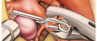
After admission to the hospital, the neurologist assesses the severity of the condition, and then determines whether the patient needs to be operated on or whether it is better to treat him conservatively.
Many doctors agree that the most dangerous are the 3rd day after a vascular accident, and stabilization of the condition occurs on days 5-7-14, depending on the severity of the pathology.
Regardless of the type of stroke, treatment begins with:
- Bed rest.
- Elimination of any physical stress. For this purpose, laxatives and other symptomatic drugs may be prescribed, for example, antitussives for severe dry cough.
- Organization of patient care for the prevention of infectious complications and bedsores.
- Adequate nutrition depending on the level of consciousness: if the patient can swallow on his own, he eats gentle food through the mouth, if he cannot, through a tube.
- Prescription of drugs that regulate blood clotting. This is necessary to stop bleeding or prevent its recurrence.
- The use of neuroprotectors - medications that reduce the “suffering” of brain cells from oxygen starvation.
Medicines are prescribed only when they are necessary and not contraindicated.
What does a stroke look like on an MRI of the brain?
Monochrome images allow you to identify altered areas that differ in color from healthy tissue. Foci of ischemia and hemorrhage have clear boundaries, making it possible to clarify the extent of damage to brain structures.
Cerebral infarction on MRI
Tomograms visualize pathologies of intracranial blood vessels, helping to determine the causes of stroke development. To increase the information content, contrast magnetic resonance scanning is used.
Whether an MRI is necessary for a stroke is decided by the doctor, taking into account the clinical picture of the disease. The method is highly effective. But since the session lasts from 15 to 30 minutes, patients in serious condition undergo an alternative CT study.
The Magnit Clinic offers an MRI scan of the brain if a stroke is suspected and during the recovery period. A modern closed tomograph from the German company Siemens creates a field of 1.5 Tesla, which allows you to obtain high-resolution photos. You can sign up for the procedure by phone or on the clinic’s website.
Management of patients with intracerebral hemorrhage
Hemorrhagic stroke is a condition that requires emergency diagnosis, adequate treatment and observation.
To do this you need:
- Conduct constant monitoring of the patient’s condition, especially in the first days after the onset of the disease: monitoring breathing and indicators of the state of the cardiovascular system.
- Monitor blood pressure numbers. They are often not reduced to normal levels, so as not to impair blood supply to the brain and provoke vasospasm.
- Prescription of hypoglycemic drugs - in almost half of patients, blood glucose levels increase.
- Use of antipyretic drugs. An increase in body temperature entails worsening cerebral edema, increased intracranial pressure and can lead to a significant deterioration in the patient’s general condition.
- Carrying out symptomatic therapy and treatment of complications.