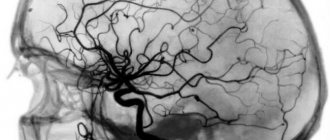
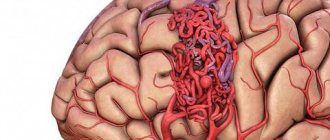
Rheoencephalography (REG) is one of the methods for diagnosing cerebral vessels using weak electrical impulses. Blood has the highest electrical conductivity compared to other media in the body. And since blood circulation is most intense in the brain, this method allows one to evaluate not only the blood vessels, but also the state of the internal structures of the brain. REG is a non-traumatic, absolutely painless and quite informative method of research; it allows the doctor to assess the speed of blood flow, tone, elasticity and blood supply of the vessels of the head. The advantages of this research method are known to every neurologist. Rheoencephalography provides a detailed picture of intracerebral vessels, regardless of their size, can be used in any conditions and allows one to obtain differentiated information about the condition of veins and arteries. At the same time, rheoencephalography is a fairly simple and absolutely comfortable procedure for the patient.
When is the study scheduled?
REG and its interpretation may be required in the following conditions:
- Changes in cerebral vessels.
- Assessing the compensatory capabilities of bypass blood flow.
- Assessing the sufficiency of venous outflow, determining the degree of hypertensive syndrome.
- Dizziness.
- Diagnosis of headaches.
- Presence of noise in the head.
- For injuries of the cervical spine and head.
- Deterioration of memory, vision, hearing.
- Hypertension.
- Vascular crises.
- Meteosensitivity and vegetative-vascular dystonia.
Referral for research
Many specialists use this study and recommend it to their patients as it is more accessible than, for example, magnetic resonance imaging. Although rheoencephalography cannot be compared with MRI in terms of information content, sometimes this study is enough to make a diagnosis.
The results of rheoencephalography of cerebral vessels may be required by neurologists, vascular and neurosurgeons, as well as general practitioners.
Indications for use
It is necessary to have a child have an REG of the head if he/she has:
- regular headaches of unknown origin;
- hearing or vision impairment;
- hypertension or hypotension;
- tinnitus;
- fainting conditions;
- swelling of the face;
- coordination problems.
The procedure is also prescribed to control blood circulation after operations, neck and head injuries. In some cases, the examination is part of an analysis of the effectiveness of drug therapy and non-drug treatment.
Methodology of the procedure
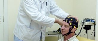
To perform rheoencephalography, you need a special device ─ rheograph. The patient is placed on his back and asked to close his eyes. The doctor fixes the electrodes on the head of the patient using rubber bands. Next, a weak high-frequency current is passed through them, and this way the condition of the vessels is recorded. During the examination, the doctor may ask you to hold your breath or, conversely, to breathe frequently.
The main condition when undergoing REG is calmness
The main difficulty of the procedure is that if the patient worries too much during the examination, this affects vascular tone and the results may be unreliable.
The result is visualized in the form of a rheoencephalogram, which is deciphered by a neurologist. REG analysis can be carried out both from the monitor screen and in paper form.
Diagnostics
Retinal angiodystonia is diagnosed using a standard set of procedures
designed to detect vascular anomalies:
- rheoencephalography - study of cerebral vessels to determine the intensity of blood supply;
- Dopplerography of blood vessels - ultrasound examination of individual basins of the bloodstream, which reveals arterial and venous insufficiency;
- electrocardiography is a classic method for recognizing signs of systemic cardiovascular disorders;
- retinal angiography.
The most informative research method is ophthalmoscopy - examination of the fundus of the eye using a special lens. This method allows the doctor to visualize vascular changes in the retina, establish their location and determine the degree of complexity. To find out the causes of the disease, laboratory blood tests are carried out (for sugar, hormones, etc.).
What the study shows
The value of the survey is that:
- Based on rheoencephalography of the vessels of the head, specialists receive significant information about the condition of the object of examination. Among them is the opportunity to study vascular tone, their elasticity, blood circulation speed and blood inflow/outflow.
- The use of rheoencephalography makes it possible not only to identify abnormalities in the vessels of the brain, but also to monitor blood flow after complex operations or severe injuries.
- With the help of REG, various pathologies are detected, and the severity of the pathological process is established.
In this case, the high speed of obtaining results is of no small importance.
What problems are being identified?
During the examination, the following are diagnosed:
- Cerebral ischemia: what is it and how to treat it?
- presence of traumatic brain injuries;
- localization of hematomas formed as a result of head trauma;
- pre-stroke condition;
- damage to blood vessels by atherosclerotic plaques (atherosclerosis);
- thrombus formation in the vessels of the brain;
- predisposition to increased blood pressure;
- diseases associated with circulatory disorders.
The procedure facilitates the task of making an accurate diagnosis, on the basis of which the doctor prescribes an adequate course of treatment. With the help of it, he subsequently monitors the effectiveness of therapy.
Due to the complete safety of such an examination for the patient’s health, it can be performed repeatedly.
One of the most significant advantages of encephalography is the ability to distinguish between pre-stroke indicators, which have certain differences for men and women.
Other features of the method
Specialists obtain even more information by conducting functional tests.
The simplest and most accessible of them is with nitroglycerin. This substance helps reduce vascular tone. This test is used to differentiate organic and functional disorders.
Symptoms
Angiodystonia of the visual organs rarely manifests itself as a disturbance in the perception of visual images.
, especially at the initial stage of development. This is why the disease is almost never detected in the early stages. As it progresses, the patient may notice:
- sensation of painless pulsation in one eye;
- the appearance of sudden flashes of light in the field of vision;
- gradual deterioration of vision - narrowing of its fields (darkening or clouding of the image at the edges), general blurriness of the picture;
- development of myopia.
Important! One of the visually recognizable signs of retinal angiodystonia is a change in the color of the sclera: yellowish spots form on it, pinpoint hemorrhages or a clear pattern of capillaries periodically appear.
The disease may also be accompanied by additional symptoms indicating the root cause of the pathology:
- in the traumatic form - headaches, paresthesia, dizziness and fainting, deterioration of cognitive functions and memory, chronic fatigue;
- with hypertension - acute headaches, sudden jumps in blood pressure to a maximum, accompanied by nausea;
- with hypotension - apathy, weakness, dizziness and nausea.
The higher the degree of pathological changes in the retina, the more intense the symptoms of the disease.
What is the technique?
REG of the brain is one of the methods for diagnosing the condition of blood vessels, which can be used to assess the functioning of the veins and arteries of the head and cervical region.
This study is carried out by recording changes in the electrical resistance of tissues after a weak electric current is passed through them. Since blood is an electrolyte (a substance that conducts electric current), when the vessels are filled with blood, the electrical resistance in them decreases, and this is detected using REG. Taking into account the speed and time of change in resistance, the doctor draws conclusions regarding the patient’s health.
Rheoencephalography is a painless method for studying the condition of cerebral vessels
Using this method, you can evaluate the pulsation of blood flow in the head arteries, determine the degree of venous outflow from the skull, and study the tone and elasticity of the walls of blood vessels. REG is a non-invasive study.
Magnetic resonance imaging (MRI), unlike rheoencephalography, is more informative and can indicate the exact location of a damaged vessel, blood clot or any other abnormality in the vascular system.
REG and USDG: what is the difference?
REG allows you to assess the tone, elasticity and blood supply of blood vessels, and reveals many pathologies of the brain. In addition, REG allows you to determine whether pathologies are functional or organic in nature. Doppler ultrasound makes it possible to judge pathologies by indirect signs: vascular patency, trajectory and speed of movement of red blood cells. REG is based on the electrical resistance of blood vessels and tissues, which is why it differs from Doppler ultrasound and other methods of ultrasound examination of the brain
.
Decoding the results
To correctly assess the data obtained, the doctor takes into account the patient’s age, since the indicators will be very different for young and elderly people.
- Treatment of cerebral atherosclerosis
The resulting rheoencephalogram is studied, which has a wave-like appearance and consists of an anacrota (growing part), catacrota (falling part), incisura (bend between them) and a dicrotic tooth that appears immediately behind it.
Based on the waves of the resulting graph, the doctor evaluates the work of blood vessels
The doctor evaluates the regularity of the waves, the nature of the construction of their peaks, the appearance of anacrota and catacrota, the location of the incisura and the depth of the dicrotic tooth. The presence of additional waves is also being studied.
After evaluating the data obtained, we can talk about the following results based on the appearance of the rheogram:
- dystonic type of REG indicates possible hypotonic deviations, decreased pulse filling and problems with blood outflow through the veins;
- angiodystonic type indicates a decrease in the tone of the vascular walls and a decrease in blood flow speed;
- The hypertensive type indicates increased pressure and tone of the vessels through which blood flows to the head and their obstructed outflow.
The amplitude indicator of the rheogram (APR) indicates volumetric pulse filling:
- APR less than normal by no more than 40% indicates a moderate decrease in pulse blood supply;
- at 40–60% - a significant decrease;
- at 60–90% - pronounced;
- at 90–100% - critical.
The coefficient of asymmetry (CA), which indicates differences in blood supply to different parts of the brain, is very important for studying. Depending on the severity of CA, several degrees of asymmetry are distinguished:
- less than 7% - no pronounced asymmetry;
- 8–14% - weak asymmetry;
- 15–25% - moderate asymmetry;
- more than 26% - severe asymmetry.
What deviations do the external characteristics of waves show - table
| Possible diagnosis | Type of rheoencephalography |
| Cerebral atherosclerosis | |
| The waves take on a strongly pronounced dome shape. | |
| Arterial hypotonicity | Increased amplitude, sharp rise with a sharp apex, shortened anacrosis. |
| Arterial hypertonicity | Reduced amplitude, extended anacrotic with additional waves, displaced apex. |
| Vascular dystonia | Floating prongs, extra waves on catacrota. |
| Obstruction of blood outflow | Catacrota increased, many small waves before the next cycle. |
| Spasm of vascular walls | Rounding the top of the wave. |
Prices
| Name of service (price list incomplete) | Price |
| Appointment (examination, consultation) with a neurologist, primary, therapeutic and diagnostic, outpatient | 1750 rub. |
| Consultation (interpretation) with analyzes from third parties | 2250 rub. |
| Prescription of treatment regimen (for up to 1 month) | 1800 rub. |
| Prescription of treatment regimen (for a period of 1 month) | 2700 rub. |
| Consultation with a candidate of medical sciences | 2500 rub. |
| Transcranial duplex scanning (TCDS) of cerebral vessels | 3600 rub. |