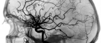
Ultrasound of the vessels of the head and neck is a modern examination technique that allows you to safely obtain data on the state of the arterial network of the brain in a few days, safely for human health. Thanks to ultrasound diagnostics, it is possible to identify pathologies that could cause the patient to complain of headaches and other ailments.
Ultrasound diagnostics includes examination of the veins located inside and outside the skull, as well as the vessels from which the arteries arise (extracranial examination).
Indications for cerebral vascular ultrasound
Ultrasound examination is prescribed for patients with angina pectoris, coronary heart disease, osteochondrosis and many other diseases. Diagnostics is indicated if the patient complains of constant headaches and if symptoms indicate the development of pathologies of the blood vessels of the head and neck.
List of indications for ultrasound of blood vessels of the head and neck:
- If you feel dizzy when changing body position.
- Systematic headache.
- Frequent fainting.
- Feeling of lack of air.
- Poor health and weakness.
- Head injuries.
- Vascular dystonia.
- Noise in the head or ears.
- Suspicion of anomalies in the development of blood vessels in the neck or brain.
- Speech impairment.
- Feeling of numbness in the limbs.
- Strong weather dependence.
- Increased cholesterol levels.
- Diabetes.
- Obesity or underweight.
- Diseases of the cervical spine.
For preventive purposes, it is recommended to undergo an ultrasound examination for people over 40 years of age, especially if there is a predisposition to cardiovascular diseases. The study is completely safe, so ultrasound of the head has no contraindications and can be prescribed even to newborns.
Doctors in this field
Where can a vascular ultrasound be performed?
“Latum Clinic” offers its services to everyone who needs a professional and comprehensive ultrasound examination of their entire body. The prices for the procedure are quite affordable, there are always free appointment slots, so you don’t have to wait months for a visit to the doctor. Sometimes the days count, so it is better to make an appointment with a doctor as early as possible to accurately determine the cause of the deterioration in your health.
The medical center is located at the crossroads between the Seligerskaya metro station and the st. 800th anniversary. Moscow's Korovinskoye Highway is also nearby. If someone knows only Dmitrovskoye Highway, then you can get to “Latum” along this route, simply turning along the signs to another road.
At any convenient working time, you can call the reception to find out all the details regarding how the procedure will take place, how long it will take and how much it will cost.
What is examined during ultrasound of cerebral vessels
Ultrasound scanning of the vessels of the brain of the head is carried out in segments. For the doctor, the reference points are the cervical cartilage, vertebrae, and skull bones.
What can be seen during diagnosis:
- Condition of the vertebral arteries.
- Causes of development of vascular diseases.
- Structure of the carotid arteries.
- The nature of the venous outflow: through the sinuses of the brain and veins.
- Blood flow through the main vessel of the brain (basilar artery).
- The condition and structure of the entire arterial chain of the head and neck.
- Pathologies of arterial trunks, etc.
Each section of the arterial system has its own designation, for example, the vertebral artery is marked with the letter V and has 5 segments to be studied.
When interpreting ultrasound and making a diagnosis, the linear velocity of blood flow must be taken into account. If the condition of the veins is assessed, then indicators of the phasing of blood flow are taken into account (depending on the diameters of the vessels, lumen and other indicators). The gaps are also checked for the presence of neoplasms: plaques in the arteries, blood clots in the veins.
The diagnostic results obtained are always compared with generally accepted standards. Only after a thorough examination of the entire venous-arterial network of the brain can the doctor find the cause of the development of the ailment and select an effective treatment.
How is ultrasound of blood vessels performed?
The operating principle of the device is based on the effect of reflecting sound waves, thanks to which the doctor can obtain a visual image of the internal cavities of the body. To study blood vessels, ultrasound machines with the Doppler effect are used, thanks to which you can see the movement of blood in the vessel. The doctor can determine the speed and direction of blood flow, vessel fullness, level of blood supply to tissues and other indicators of the circulatory system.
Special preparation is needed only before the abdominal vascular ultrasound procedure. For 3 days you must follow a special diet that reduces gas formation in the intestines and take sorbents. Before the examination, you need to take a shower; you cannot use medicated creams and ointments on the examination sites. The day before you should not drink alcohol, energy drinks, strong tea and coffee, or smoke. In consultation with the attending physician, it is advisable to discontinue vascular medications.
Types of vascular studies:
- Doppler ultrasound (USDG). The study allows you to obtain a two-dimensional image of the vessels being examined.
- Ultrasound duplex scanning (USDS). During the study, the condition of the vessels and the nature of blood flow are assessed.
How is ultrasound of the vessels of the brain and neck performed?
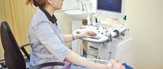
Diagnostics together with decoding takes about 40-50 minutes. The patient is asked to lie on the couch and tilt his head back slightly so that the doctor can comfortably reach the vessels being examined. A conductor gel must be applied to the skin. Next, a sensor transmitting ultrasonic waves is applied to certain areas on the head and neck. First, the condition of the vessels of the brain is assessed, and then the cervical spine. The received data is projected onto the monitor screen.
During an ultrasound of the cervical vessels, the patient should generally lie still and not move, although the doctor may ask you to turn your head or hold your breath. After the procedure, the uzist will issue a sheet with the examination results, based on which the attending physician will be able to make the correct diagnosis.
How is an ultrasound of the veins of the lower extremities performed?
An ultrasound examination is important if you suspect the presence of serious diseases, such as thrombosis or varicose veins. A specialist will determine with 100% accuracy the area where pathological processes occur (formation of blood clots, slowing of blood flow, expansion and inflammation, damage to the walls).
To perform an ultrasound, you do not need to prepare and shave hair from the area, limit physical activity, follow a diet, etc. The procedure does not take much time and does not require complex and uncomfortable preparation.
Since ultrasound of the veins of the lower extremities is performed to determine the disease, excellent quality equipment is required. The beam must be accurately reflected from the red blood cells. Thanks to this, the doctor sees what is happening in the veins, vessels and arteries. Therefore, in a few minutes you can assess the condition of valves, deep and superficial veins, blood flow speed, etc.
What is duplex scanning?
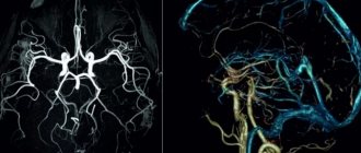
The best modern diagnostic method is ultrasound scanning, since the quality of the results obtained is maximum. In the West, preference is given to color mapping. It increases the accuracy of the information if you need to track the speed of blood flow towards and away from the sensor (marking is done in red and blue). The method allows you to perform the following tasks:
- Monitor damage and deformation of the venous walls.
- Find out the speed of blood flow in the deep and superficial veins.
- Check the functionality of the valves.
- Assess the dimensions and density of the thrombus (determine the degree of thrombosis).
Classical and minimally invasive operations are always performed with 100% accuracy, as ultrasound of the veins of the lower extremities is performed in combination with duplex scanning.
Before visiting a specialist, be sure to take a shower. There is no need to shave your leg hair or deep clean your skin. It is advisable not to drink alcohol on the eve of an ultrasound examination. The procedure is performed as follows:
- In the office you undress from the waist down (down to your underwear).
- Ultrasound gel is then applied to the legs.
- The specialist checks the condition of the veins, valves, vessels and arteries using a device with a sensor.
- If necessary, the intensity of pressure and the power of the beam are increased to more clearly see the condition of the deep veins.
- Your body position changes—your doctor may ask you to stand up or lie down to see how the blood flows. For more information, you will need to hold your breath (take a deep breath).
Now you know exactly how an ultrasound scan of the veins of the lower extremities is performed. The session lasts a few minutes and you will not experience any discomfort. There is no pain. During the change in ultrasound intensity, you will not feel anything (many people fear pain or discomfort).
Do not forget that only a competent phlebologist with extensive experience can decipher the result. Therefore, immediately after a duplex scan or ultrasound, go to an appointment with a specialist so that he can make a final conclusion.
The First Phlebology Center will determine the condition of the veins of the lower extremities and prescribe an effective course of treatment for you. You can sign up for an examination right now by phone.
Service cost*
| NAME | PRICE (Kolomenskaya) | PRICE (Vidnoe) |
| Duplex scanning of brachiocephalic arteries with color Doppler mapping of blood flow (on a Voluson, Mindray, Logiq device) | 2900 rubles | 2900 rubles |
| Duplex scanning of the intracranial sections of the brachiocephalic arteries (on a Voluson, Mindray, Logiq device) | 5700 rubles | 5700 rubles |
| Duplex scanning of extracranial sections of the brachiocephalic arteries (on a Voluson, Mindray, Logiq device) | 3100 rubles | 3100 rubles |
We accept payment:
*Attention! The indicated prices are provided as reference information and do not constitute a public offer. Check current prices by phone and directly in clinics.
What veins and arteries are examined using ultrasound?
- Doppler ultrasound of the vessels of the neck, brain, eyes.
- Ultrasound diagnostics of veins and arteries of the lower and upper extremities.
- Ultrasound scanning of the vessels of the pelvis, abdominal cavity and kidneys.
- Ultrasound examination of heart vessels.
- Duplex scanning of the embryo.
- Ultrasound scanning/ultrasound scanning of the penis, scrotum.
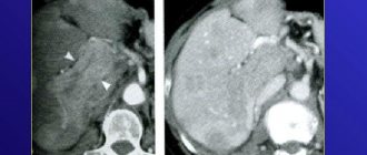
During an ultrasound examination, the doctor can identify a wide range of vascular pathologies - aneurysm of the aorta or other vessels, varicose veins, phlebitis and thrombophlebitis, dissection and occlusion of vessels, atherosclerosis, etc.
In a medical clinic you can do ultrasound of blood vessels at the best price in St. Petersburg. The study is carried out using modern expert-level ultrasound equipment, and the clinic’s specialists have extensive experience in performing ultrasound diagnostics. Based on the results of the examination, you can get advice from a specialized specialist at the clinic. You can make an appointment by phone: +7.
Advantages and disadvantages of the procedure
Today, almost every medical institution has the necessary equipment to conduct ultrasound of the neck vessels. Let's consider the advantages of this technique:
- This method is absolutely safe;
- The procedure can be performed at any age;
- She has no direct contraindications;
- The process is absolutely painless and does not cause discomfort.
- The only disadvantage we can note is the lack of information content - this is not the main method of diagnosing diseases, but only an auxiliary one.
Rules for preparing for research
On the day of the procedure, you must refrain from:
- Taking antispasmodics such as “Riabal”, “No-shpa”, “Drotaverine”, “Papaverine”, “Baralgin”, “Cinnarizine”.
- Smoking.
- Drinking black tea and drinks containing caffeine.
- Staying in unventilated, stuffy rooms with large crowds of people, which can have a detrimental effect on vascular tone.
If the patient is undergoing treatment with cardiovascular medications, then the advisability of discontinuing them before the procedure must be agreed with a neurologist. In any case, the sonologist must be informed about the treatment being performed before the ultrasound.
Stages of extracranial Dopplerography:
1. In the office, remove all jewelry from your neck and head.
2. Remove clothing from those parts of the body through which the sensor will be passed: neck, collarbones, shoulder blades.
3. Lie on the couch with your head facing the monitor. The doctor will apply acoustic gel to open areas of the body and begin to move the device over them, placing it in areas where medium and large arteries and veins are located. To obtain more accurate data on the state of vascular tone, the patient will need to hold his breath at the doctor’s request, take special medications and change body position.
Doppler ultrasound of vessels located in the cranial area has a different specificity. Acoustic gel is applied to the temples, the back of the head and the area above the eyes. The device is installed there because in these places the bones of the skull are the thinnest and are able to transmit ultrasonic waves.