Cyanosis is a symptom characterized by a blue tint of the skin and mucous membranes, which occurs due to an increased level of carbhemoglobin in the blood. Cyanosis, which is associated with the entry of chemical compounds into the body or the deposition of any substances in the skin, is considered to be false cyanosis.
Cyanosis resulting from hypoxemia is considered true. Capillary blood in the presence of true cyanosis contains more than fifty g/l of reduced hemoglobin.
Cyanosis is a fairly common symptom of heart disease. The further a part of the body is from the heart muscle, the more intense its cyanosis.
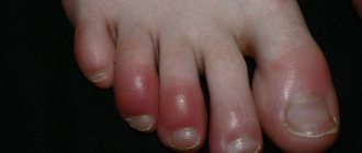
Cyanosis of the fingers and toes, nasolabial triangle, and ears is called acrocyanosis. Acrocyanosis occurs when the level of reduced hemoglobin increases in the venous blood. Acrocyanosis occurs due to excessive absorption of oxygen by tissues when blood flow slows down. Central cyanosis is a widespread cyanosis. The cause of diffuse cyanosis is a lack of oxygen in the pulmonary circulation.
Patients with both peripheral and central cyanosis often come to the Yusupov Hospital. Cyanosis of the face brings aesthetic discomfort to patients, while cyanosis of the extremities may remain unnoticeable for a long time. By signing up for a consultation by phone or online, during an individual conversation you can find out the differences between cyanosis and acrocyanosis, learn about the possible causes of cyanosis, view photos of patients with cyanosis of the skin, photos of patients with cyanosis of the nasolabial triangle and calculate the financial side of the issue.
The Yusupov Hospital is engaged in scientific activities, diagnostics, treatment, rehabilitation and prevention. This is the only medical institution in Moscow of such a high level.
Causes of cyanosis
Whether constant cyanosis is characteristic of a particular disease or is transient in nature depends on the cause of its occurrence.
If after examination the cause of cyanosis of the skin cannot be determined, then it is called primary, or idiopathic.
Cyanosis may not have a pathological basis, but in order to protect the patient, it is necessary to undergo an examination and exclude organic pathology. Secondary cyanosis occurs as a result of some disease.
Central cyanosis most often occurs in respiratory failure, when there is high pressure in the pulmonary arteries, pathology of the bronchi, lungs, or if the patient has a congenital defect of the interventricular or interatrial septum. The most intense cyanosis will be in those areas where the skin is the thinnest; for example, cyanosis of the lips will be characterized by an almost purple color. A characteristic sign is also cyanosis of the nails.
Peripheral cyanosis is more often observed in patients with heart failure, as the speed of blood flow is significantly reduced. Areas of the body that are exposed to low temperatures have a bluish color.
Diagnosis of the disease in a newborn
At the neurorehabilitation center, diagnostic measures are used that are aimed at identifying the cause of the disease - its main cause of occurrence.
First of all, a small patient is listened to to identify heart sounds and breathing stability.
More detailed information is obtained using special instruments and laboratory tests:
- checking vital parameters such as temperature, breathing, pulse and blood pressure;
- objective examination with medical history;
- X-ray of the chest or respiratory organs;
- blood analysis;
- response to oxygen therapy.
The specialist also takes into account the presence of provoking factors in the patient’s body and the time of manifestation of cyanosis (in infants the disease is noticeable immediately).
Cyanosis in children
Cyanosis of the nasolabial triangle in an infant is not uncommon and can be observed both in healthy children and in children with diseases of organs and systems (nervous, cardiovascular, respiratory, etc.). In children, the level of oxygen in the blood decreases during strong screaming or crying, but normally it becomes no lower than ninety-two percent. Cyanosis in an infant occurs when the oxygen level drops below this level.
During the newborn period, cyanosis during screaming or crying is the norm, and occurs due to imperfect systems, but as the child grows, the cyanosis disappears. Cyanosis that persists several weeks after birth should be alerted and examined so as not to miss the pathology that causes hypoxia. If cyanosis is associated with extremely thin tissue, this is considered as a variant of the norm.
Sometimes cyanotic nasolabial triangle is observed during infectious diseases such as pneumonia, true croup, etc. In addition to cyanosis, they are accompanied by shortness of breath, difficulty breathing, intoxication, and increased body temperature. If cyanosis and shortness of breath occur instantly - this may be due to foreign objects entering the respiratory tract, parents should urgently call an ambulance.
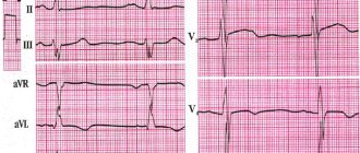
Pathological conditions that often cause cyanosis in children are congenital heart defects, abnormal development of blood vessels, etc. Therefore, cyanosis in a child should be examined in any case. Carrying out an ultrasound examination of the heart, radiographic methods and an electrocardiogram will help the doctor confirm or exclude the diagnosis and plan further studies. The child should also be examined by a neurologist, because sometimes the cause of cyanosis is underdevelopment of the respiratory system.
Treatment of bluish skin
Cyanosis is a clinical sign of a number of pathologies in which the skin of patients acquires a blue color. The reason for such changes is the accumulation of deoxyhemoglobin in the blood - hemoglobin that gives oxygen to the tissues. Blood depleted of oxygen becomes dark, visible through the skin and makes it bluish. This is most clearly noticeable in places with thin skin - on the face and ears.
Cyanosis occurs in individuals with circulatory disorders leading to generalized or local hypoxemia.
With insufficient blood supply to the capillaries, acrocyanosis develops, which is manifested by cyanosis of the skin of the fingers and toes, and the tip of the nose. This term translated from ancient Greek means “dark blue limb.”
The severity of cyanosis varies from barely noticeable cyanosis to purple skin color. Temporary cyanosis occurs with excessive physical exertion, persistent cyanosis occurs with long-term cardiac or pulmonary diseases.
Classification
Central cyanosis is diffuse and of maximum severity. It develops with weak arterialization of the blood, leading to hypoxia. Gas exchange is disrupted in the lungs, excess carbon dioxide accumulates in the arterial blood, which is clinically manifested by blueness of the conjunctiva of the eyes, palate, tongue, mucous membranes of the lips and cheeks, and facial skin. Qualitative and quantitative changes in hemoglobin in the blood lead to disruption of its transport function and hypoxia. Acrocyanosis is localized on the feet, hands, tip of the nose, ears, lips. Peripheral cyanosis is considered a normal variant in the first days of a newborn’s life. Its origin can be easily explained by the incompletely eliminated germinal type of blood circulation, especially in premature infants. Blueness of the skin increases with swaddling, feeding, crying, and anxiety. When the infant fully adapts to the outside world, cyanosis will disappear.
Acrocyanosis in adults is a sign of varicose veins, heart failure, thrombophlebitis, vegetative-vascular dystonia, atherosclerosis, arteritis.
There are several types of cyanosis based on origin:
- The respiratory type is caused by an insufficient volume of oxygen in the lungs and a disruption in the transport chain of its supply to cells and tissues. It develops when there is a complete or partial disruption to the movement of air along the respiratory tract.
- Cardiac type - insufficient blood supply to organs and tissues leads to oxygen deficiency and bluish skin.
- The cerebral type develops when the blood loses its ability to attach oxygen to hemoglobin and deliver it to brain cells.
- The metabolic type develops when the absorption of oxygen by tissues is impaired.
- Respiratory cyanosis disappears 10 minutes after oxygen therapy; all other types persist for a long time. Massaging the earlobe helps get rid of acrocyanosis.
Symptoms
Cyanosis is a symptom of life-threatening diseases. With central cyanosis, the skin of the periorbital and perioral areas first turns blue, then it spreads to areas of the body with the thinnest skin. Peripheral cyanosis is most pronounced in areas distant from the heart. It is often combined with swelling and swelling of the veins of the neck.
Depending on the time of occurrence, cyanosis can be acute, subacute and chronic.
Cyanosis does not have a negative impact on the general well-being of patients, but in combination with other signs of the underlying pathology it becomes a reason to consult a doctor. If cyanosis occurs suddenly, increases rapidly and has a significant degree of severity, then it requires emergency care.
Cyanosis, depending on the etiology of the disease, is accompanied by various symptoms: severe cough, shortness of breath, tachycardia, weakness, fever and other signs of intoxication.
Cyanosis in bronchopulmonary diseases is manifested by a purple tint of the skin and mucous membranes and is combined with shortness of breath, wet cough, fever, sweating, and moist rales. These symptoms are characteristic of an attack of bronchial asthma, acute bronchitis and bronchiolitis, pneumonia. With pulmonary embolism, intense cyanosis develops against the background of chest pain and shortness of breath, and with pulmonary infarction it is combined with hemoptysis. Patients with similar symptoms require urgent hospitalization and respiratory resuscitation.
In heart disease, cyanosis is one of the main symptoms. It is combined with shortness of breath, characteristic auscultatory findings, moist rales, and hemoptysis. Cyanosis with heart defects is accompanied by secondary erythrocytosis, increased hematocrit, and the development of capillary stasis. Patients experience deformation of fingers like drumsticks and nails like watch glasses.
Cyanosis is not subject to special treatment. When it appears, oxygen therapy is carried out and the main treatment is intensified. Therapy is considered effective when the severity of cyanosis decreases and disappears.
In the absence of timely and effective treatment for diseases manifested by cyanosis, patients develop a disorder of the nervous system, the general resistance of the body decreases, sleep and appetite are disturbed, and in severe cases the person may fall into a coma. This condition requires emergency medical care in the intensive care unit.
Diagnostics
Diagnosis of diseases manifested by cyanosis begins with listening to complaints and collecting anamnesis. The patient is asked when the cyanosis of the skin appeared, under what circumstances cyanosis occurred, whether it is permanent or paroxysmal. Then the localization of cyanosis is determined and it is clarified how its shade changes during the day.
After a conversation with the patient, a general examination begins, the severity of his condition and the presence of concomitant diseases are established. The doctor performs auscultation of the heart and lungs.
Then they move on to laboratory and instrumental research methods
Therapy
During neurorehabilitation
Doctors are trying to normalize hemoglobin and increase oxygen levels; after using oxygen therapy, cyanosis should completely disappear in infants. If cyanosis becomes chronic, then it will not be possible to quickly eliminate the blue color of the skin; it may reappear even after complete disappearance.
During the treatment of facial cyanosis in a newborn, medications belonging to the following groups are often used:
- cardiac glycosides;
- bronchodilators;
- antihypoxants;
- analeptics.
Only a specialist can determine the reasons for the appearance of a bluish color on the skin of newborns, and he will also prescribe the most effective treatment in a particular case. Most often, the disease is stopped in the early stages, but if a child was born with a heart defect, cyanosis can only be removed surgically.
Kinds
Before prescribing treatment, the specialist must accurately determine the type of pathology. Based on their origin, the following types of cyanosis are distinguished:
- Cordial. Weakened blood flow and inadequate nutrition of tissues - not enough blood and oxygen reach them.
- Respiratory. Oxygen starvation of the lungs and failure of oxygen supply to almost all tissues and organs.
- Cerebral. Oxygen does not attach to hemoglobin, causing ischemia of brain cells.
- Metabolic. Tissue cells cannot absorb oxygen properly.
- Hematological. Caused by blood diseases.
Ten minutes of oxygen therapy is generally sufficient to eliminate respiratory cyanosis. Other types do not go away so easily and require a competent approach to treatment.
The nature of the spread of pathology varies, so experts distinguish:
- How to raise hemoglobin using folk remedies - 7 proven recipes
- Central cyanosis or diffuse cyanosis. Localized throughout the body, it is provoked by a failure of breathing or the entire blood flow.
- Peripheral. For problems with the performance of the heart, arteries, or the development of oxygen starvation of the limbs or face.
- Acrocyanosis. Only the skin on the fingers, on the edge of the nose, around the lips becomes bluish, which occurs due to stagnation of blood in the veins.
- Local. This type is determined when the mucous membranes and genitals are examined. Caused by stagnation of blood.
Cyanosis manifests itself in different ways, so it can be in acute, subacute and chronic forms.