- home
- Functional diagnostics
- Ultrasound of the abdominal organs
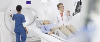
Ultrasound diagnostics (ultrasound examination, echography) – scanning the human body to obtain images of internal structures and organs. High frequency sound waves penetrate tissue. Their reflected echoes come back, being converted into images on the computer monitor. Later you can get a video or photo of the area being examined. Abdominal ultrasound is a non-invasive method used in the diagnosis of a wide range of diseases and conditions.
Types of research
Classic ultrasound of the abdomen gives a detailed idea of the size, structure of parenchymal and hollow organs. An ultrasound is performed:
- Liver;
- Pancreas;
- Spleen;
- Ultrasound of the bladder;
- Gallbladder with definition of functions;
- Small intestine;
- Large intestine;
- Gallbladder;
- Abdominal aorta;
- Intestines;
- Stomach.
The method is completely safe and does not cause discomfort or pain. You can undergo an ultrasound as prescribed by a doctor or at your own request if there are disturbing symptoms. Compared to other diagnostic procedures such as CT and MRI, ultrasound diagnostics are cheaper, faster, simpler and do not use ionizing radiation.
Ultrasound of the liver
The doctor prescribes a liver examination if there are certain symptoms that may indicate that the patient is not well. Among them is discomfort in the right hypochondrium, which manifests itself more strongly after drinking alcohol, fatty foods, smoked and fried foods.
A signal to perform an ultrasound of the liver may be a lump detected during palpation - this may be a tumor, the exact location of which and its inherent signs will be identified by the doctor using ultrasound. It is important to undergo an examination after a course of therapy if hepatitis and other diseases are detected.
Ultrasound of the pancreas
Sonography makes it possible to identify the size of an organ, its inherent shape, and the presence of changes in structure and structure. Attention is paid to the location of the pancreas relative to other structures and vascular formations. If pathological changes are identified, the study expands the boundaries, and the question arises of using additional diagnostic methods.
Inflammation of the pancreas can manifest itself in reduced size of the organ if it becomes chronic. This may also be indicated by excessive tissue scarring and calcifications. Cysts formed in the structure of the organ are reflected during examination as compacted areas with clearly defined boundaries.
Ultrasound of the spleen
Prescribed when liver pathologies are detected. And also if there is a suspicion of injuries resulting from falls, mechanical shocks and bruises. Indications for the procedure include leukemia, the formation of compactions and tumors, diseases of an infectious nature (syphilis, tuberculosis, sepsis).
For the examination, the patient is placed on his back. A special gel is applied to his stomach, on which the ultrasonic signal transducer glides more easily. The session takes 15 minutes, and the results are immediately available. For better visualization, the doctor may ask the patient to turn on their side or assume another position that makes it easier to view.
Ultrasound of the bladder
The reason for performing an ultrasound of the bladder may be a long-term change in the color of urine, which is not associated with eating foods and medications that can affect its tone. The doctor should be alerted to the patient's complaints about pain and discomfort when urinating, the release of a small amount of fluid with frequent urge to go to the toilet.
It is recommended to undergo an examination if pain appears in the suprapubic region, bloody discharge in the urine and flakes visible from the side in the liquid. As a result of viewing the internal structures, stones, foreign bodies in the organ, neoplasms and other anomalies may be detected.
Ultrasound of the gallbladder with determination of functions
The procedure is preceded by a trial choleretic breakfast, with the help of which it is easier for the doctor to examine the organ. The study is carried out in several stages, when after 10-15 minutes a specialist examines the organ with ultrasound at rest and after exercise. The patient is asked to lie on his back, and when necessary, move to his side, on all fours, or stand up straight.
Ultrasound of the bile duct shows whether cholecystitis or cholelithiasis exists, and reveals biliary dyskinesia. A quick and painless examination reveals five degrees of abnormality. Such accuracy allows you to subsequently draw up a competent therapeutic protocol and achieve good results in the treatment of the disease.
Diagnostic measures
The main diagnostic technique for detecting changes in the portal vein remains ultrasound. The study can be prescribed to pregnant women, children and elderly patients. Doppler ultrasound, used in conjunction with ultrasound, helps assess the speed and direction of blood flow. Normally, it should be directed towards the organ.
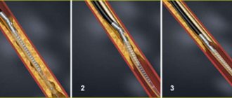
With the development of thrombosis, a hyperechoic (dense) heterogeneous formation is detected in the lumen of the vessel. It can fill the entire lumen of the vessel, or cover it only partially. In the first case, blood movement stops completely.
One of the most common liver vascular pathologies
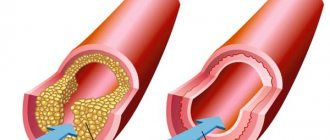
With the development of portal hypertension syndrome, dilation of the vascular lumen is detected. In addition, the doctor detects an enlarged liver and fluid accumulation. Doppler ultrasound will show a decrease in blood flow velocity.
A possible sign of portal hypertension is a cavernoma. The patient is required to undergo an FGDS to assess the condition of the esophageal anastomoses. Additionally, esophagoscopy and radiology of the esophagus and stomach may be recommended.
In addition to ultrasound examination, computed tomography with a contrast agent can be used. The advantage of using CT is the visualization of the liver parenchyma, lymph nodes and other formations located in close proximity.
Angiography is the most accurate method for diagnosing portal vein thrombosis. Instrumental studies are complemented by blood testing. Of clinical interest are indicators of leukocytes, liver enzymes, and bilirubin.
Ultrasound of the abdominal organs for children (child)
Parents are concerned about the question of whether it is possible to perform an ultrasound of the abdominal cavity on a child, and whether the procedure will cause harm. The answer is obvious - it is possible, and if necessary, with repeated monitoring. Due to the simplicity and safety of the examination, the test is prescribed for a child under 1 year of age in order to:
- Conduct an inspection of internal structures;
- Identify deviations if they exist and take measures to eliminate anomalies.
Thanks to the scan, the size of the abdominal organs, tissue composition, and structure density become visible.
Normal kidney sizes:
- length 9-12 cm [1,5,7] (length less than 9 cm can be considered as hypoplasia), sometimes in tall people the kidneys can be longer than normal, also when lengthening the kidney, it is necessary to exclude doubling of the kidney, which is often asymptomatic (t i.e. often not dangerous to health) and is detected by chance (i.e. for another reason) on urography;
— transverse size 4-6cm[1,5,7];
— parenchyma thickness 1.3-25 mm[1].
The adrenal glands are normally visible only in infants, usually up to 1-2 months of life. On ultrasound they appear as hypoechoic curved linear structures.
https://www.uzgraph.ru/forum/ugadayka/7/479/nadpoc...
Sometimes you can find a description of the size of the adrenal glands in some literature sources[6] and in reports on adults; most often this is simply a measurement of a piece of fatty tissue in the area of the upper pole of the kidney, because according to classical ideas about anatomy, this is where the adrenal gland should be located (as the name suggests). In fact, in adults, the adrenal glands are not visible on ultrasound (i.e., they are indistinguishable on ultrasound from the fatty tissue around the kidney), and they can be located not only in the area of the upper pole of the kidney. And the main task of ultrasound is to search for formations of the adrenal gland, which are often visible on ultrasound!
Normally, the ureters are also not visible on ultrasound; they become visible when they expand due to diseases. The exception is when the bladder is full. In such cases, the patient should be asked to go to the toilet and look again.
The bladder normally has an oval shape and an anechoic lumen, the thickness of the wall when filled is up to 4 mm [1,7], if the bladder is empty or poorly filled, the wall may be thicker.
Indications for the study
Immediate ultrasound is required for abdominal pain, prolonged nausea, vomiting, bloating, unexplained weight loss, injury, fever of unknown origin. Ultrasound diagnostics is prescribed as part of a set of measures when identifying a tumor to clarify its nature.
Ultrasound is an imaging method. It is often resorted to when a biopsy is necessary, but the organ is located deep. In this case, it is necessary to view the structures of the abdomen and the needle placed in it, aimed at taking biological material, which is later sent for histological and cytological analysis.
Preparation for ultrasound of the abdominal organs
Ideally, an ultrasound examination when scanning the hollow organs of the digestive system is carried out after special preparation for an ultrasound of the abdominal organs, when gas-forming products are excluded from the diet for three days. Otherwise, the air interferes with obtaining a detailed image of the structures.
What foods are prohibited to eat before the study?
Prohibited foods include cabbage, apples, milk, and other fruits and vegetables containing cellulose. Legumes and grains should also be avoided. The day before the study, experts indicate low-fat soup, cottage cheese, and yogurt among what you can eat. For 8-12 hours it is necessary to exclude food altogether.
Is it possible to drink water before the test?
To view the bladder and prostate in men, you need to fill your bladder (drink about 3-5 cups of liquid 1.5 hours before the exam and do not urinate). It is strictly forbidden to drink soda, energy drinks, or coffee.
For women, when asked whether they can drink water before an ultrasound of the abdominal organs, the answer is similar. It is possible and necessary, but only if the water is simple, not saturated with sugar and gases. If a transvaginal ultrasound is performed, it is not necessary to drink water.
It is important that the patient cooperates with the doctor during the examination and assumes the required position or holds his breath when asked to do so. In the event of an emergency, an abdominal ultrasound is performed without special preparation in emergency mode.
Normal dimensions of the gallbladder:
- length up to 8-12cm[1,5,7];
— transverse size up to 3.5-5 cm [1,5,7];
— wall up to 3-4mm*[1,5,7].
*All measurements are given on an empty stomach, i.e. when the gallbladder is full. The wall of the gallbladder in a contracted state can be thicker!
When conducting a food test to determine the function of the gallbladder using ultrasound, it should shrink by 2/3 of its original volume by about 30 minutes after eating.
The pancreas normally has an even contour, a fine-grained structure and an echogenicity slightly lower than the hepatic (but may be higher than the hepatic, which, with normal sizes, can also be regarded as a variant of the norm - slight fatty involution, against the background of an “active” lifestyle, in particular abuse fatty foods, smoked meats, alcohol...), measured by transverse scanning, usually three dimensions are measured (anterior-posterior dimensions): head, body and tail; although some authors [5] also suggest measuring the isthmus of the pancreas (the border of the head and body), but in practice this is rarely used.
How is it done and what does an abdominal ultrasound show?
An abdominal ultrasound is performed with the patient lying on his back while the doctor passes a transducer across his skin in the area of the organs being examined. To facilitate the sliding of the transducer over the body, a water-based profile gel is used, which can be easily removed with a damp cloth after the session.
In order to get a more complete picture of some organs, the doctor himself tells the patient how they do it and what an ultrasound of the abdominal cavity shows - the patient may be asked to turn over on the couch to the right or left side. The test lasts about 15 minutes. During the examination, the patient does not feel any pain. During ultrasound, the size of the organ, its structure, clarity of boundaries, and the presence of changes in tissues are visualized.
Standards for organs
- Normally, the right lobe of the liver does not exceed 125 mm, the left 70 mm. The portal vein is up to 14 mm, the thickness of the left lobe is 50-60 mm, the right one is 120-125 mm.
- The width of the gallbladder is 30-50 mm, length 60-100 mm, volume from 30 to 70 cubic meters. cm.
- Includes an interpretation of the ultrasound of the abdominal cavity, the length of the spleen, which normally does not exceed 110 mm, a thickness of 50 mm, and an index of up to 20 square meters. cm.
- The head of the pancreas is 32 mm, the body is 21 mm, and the tail is 35 mm.
- The dimensions of the urinary tract are wall thickness no more than 2 mm, the urine norm for men is 350-750 ml, for women 250-550 mm.
Portal vein system
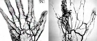
The anatomical structure of the portal vein is complex. The trunk has many branches into venules and other blood vessels of different diameters. The portal system is another circle of blood flow, the purpose of which is to cleanse the blood plasma of decay products and toxic components.
A number of diseases affect changes in blood flow through the portal vein system
The changed dimensions of the portal vein make it possible to diagnose certain pathologies. Its normal length is 6–8 cm, and its diameter is no more than 1.5 cm.
Prices for ultrasound of the abdominal organs
We offer attractive prices for abdominal ultrasound in Khabarovsk, and we carry out diagnostics using the latest equipment - Sonoscape s35 and LOGIQ-C5 scanners. Highly qualified doctors, mid-level staff attentive to the needs of patients, no queues and professional assistance in identifying and treating diseases - we offer all this to medical patients. The cost of our services is affordable and affordable:
| Code | Name of procedure | price |
| 7.01 | Comprehensive ultrasound of the abdominal organs (liver, gall bladder, pancreas, spleen) | 1800 |
| 7.02 | Ultrasound of the liver, gallbladder | 1000 |
| 7.03 | Ultrasound of the liver, gallbladder with Dopplerography of blood vessels | 1200 |
| 7.04 | Ultrasound of the pancreas | 600 |
| 7.05 | Ultrasound of the gallbladder | 600 |
| 7.06 | Ultrasound of the gallbladder with functional tests | 1000 |
| 7.07 | Ultrasound of the spleen | 600 |
| 7.08 | Ultrasound of the kidneys and adrenal glands | 900 |
| 7.09 | Kidney ultrasound | 800 |
| 7.10 | Ultrasound of the bladder (examination with transvaginal/transrectal and abdominal sensors) | 1200 |
| 7.11 | Ultrasound of the bladder with determination of residual urine (abdominal sensor) | 800 |
| 7.12 | Ultrasound of the uterus, appendages (examination with transvaginal and abdominal sensors) | 1300 |
| 7.13 | Ultrasound of the uterus, appendages (examination with a transvaginal sensor) | 1300 |
| 7.14 | Ultrasound of the uterus, appendages (examination with an abdominal sensor) | 1000 |
| 7.15 | Folliculometry (primary) | 1200 |
| 7.16 | Folliculometry (repeated) | 1000 |
| 7.17 | Ultrasound of the scrotum | 800 |
| 7.18 | Ultrasound of the prostate gland (examination with an abdominal probe) | 1000 |
| 7.19 | Ultrasound of the prostate gland (examination with a transrectal probe) | 1000 |
| 7.20 | Ultrasound of the prostate gland (examination with transrectal and abdominal sensors) | 1400 |
| 7.23 | Doppler ultrasound of vessels of various locations (fetus; scrotum; kidneys; abdominal aorta and its branches, etc.) | 2200 |
| 7.40 | Ultrasound examination of the hip joints (children up to one year old) | 900 |
| 7.41 | Screening “Children under one year old” - neurosonography, examination of the thymus, heart, liver, gallbladder, pancreas, spleen, kidneys, hip joints | 4300 |
| 7.42 | Ultrasound of the bladder | 600 |
| 7.44 | Control of the uterine cavity after m/a | 500 |
Check out the prices by studying the price list for our services. In addition to functional diagnostic services, we offer to attend an appointment with specialized specialists, whose work schedule is also presented on our website.
Treatment of pathology
Treatment of the disease involves an integrated approach and includes taking medications and surgery. Drug therapy includes the following medications:
- drugs from the group of anticoagulants - prevents the formation of blood clots and improves vascular patency;
- thrombolytics - dissolve existing blood clots, freeing the lumen of the portal vein.
Medicines are prescribed by the attending physician based on current symptoms
If there is no therapeutic result from the selected drug therapy, the person is prescribed surgical treatment. Transhepatic angioplasty or thrombolysis may be performed.
The main complication of surgical treatment is bleeding of the esophageal veins and the development of intestinal ischemia. Any pathology of the portal vein of the liver is a serious condition that requires the appointment of therapy adequate to the condition.
Where can I get a paid ultrasound of organs?
If you are interested in where you can get a paid ultrasound of your organs, pay attention to the medical one. We work every day without lunch breaks. You can always see on the map how to find us, as well as check our opening hours, on our website.
If it’s difficult to make a decision, read the reviews of our patients, who speak about the quality of services better than any advertising. Come visit us and see for yourself. We will be glad to see you at the address on the street. Panfilovtsev, 38.