Author Marina Lebedeva
10/28/2004 13:33 (Updated: 07/05/2021 13:58)
Health
Hemoglobin level in children is one of the most important indicators of health. How dangerous are deviations from the norm, what could be their cause and are there ways to correct the situation?
What is the difference between “children’s” hemoglobin and “adult” hemoglobin?
Hemoglobin begins to be produced in a child in the womb at the earliest stages of fetal formation. This hemoglobin is fundamentally different from the hemoglobin in the blood of an adult and is called fetal hemoglobin. Shortly before birth, fetal hemoglobin begins to be replaced by the “adult” form of this protein - glycosylated hemoglobin. The complete replacement process is completed at approximately one year of age.
In the first years of life, the norm of hemoglobin in a child’s blood varies significantly depending on age. This indicator must be carefully monitored in order to notice critical deviations from the norm in time.
It is hemoglobin, which contains iron, that gives blood its red color. But not all creatures on Earth have red blood. For example, the blood of octopuses, lobsters and crabs has a blue-green tint because another protein, hemocyanin, which contains copper rather than iron, performs the function of hemoglobin in it.
Iron deficiency anemia in infants and young children
Anemia and anemic syndrome, caused by many causes, can be mentioned among the most common pathological conditions that general pediatricians have to deal with every day. This group includes various diseases and pathological conditions characterized by a decrease in the content of hemoglobin and/or red blood cells per unit volume of blood, leading to disruption of the oxygen supply to tissues. The following laboratory criteria for anemia are applied (N.P. Shabalov, 2003). Depending on the age of the children, the hemoglobin level is:
- 0–1 day of life - < 145 g/l;
- 1–14 days of life - < 130 g/l;
- 14–28 days of life - < 120 g/l;
- 1 month - 6 years - < 110 g/l.
Of all anemias, the most common is iron deficiency (IDA), which accounts for approximately 80% of all anemias. According to the World Health Organization (WHO), more than 500 thousand people worldwide suffer from IDA. The prevalence of IDA in children in Russia and developed European countries is: about 50% in young children; more than 20% - in older children.
IDA is a clinical and hematological syndrome characterized by impaired hemoglobin synthesis as a result of iron deficiency, developing against the background of various pathological (physiological) processes, and manifested by signs of anemia and sideropenia.
Iron is one of the main microelements in the human body. Normally, the adult body contains 3–5 g of iron in bound form. 70% of the total amount of iron is part of hemoproteins. The iron in these compounds is bound to porphyrin. The main representative of this group is hemoglobin (58% iron); Iron is also contained in myoglobin (8%), cytochromes, peroxidases, catalases - up to 4%. Iron is also part of non-heme enzymes (xanthine oxidase, nicotinamide adenine dinucleotide (NADH) dehydrogenase, aconitase, localized in mitochondria); transport form of iron (transferrin, lactoferrin). Iron reserves in the body exist in two forms: in the form of ferritin (up to 70%) and hemosiderin (up to 30%). The peculiarity of iron distribution in young children is that they have a higher iron content in erythroid cells and less iron in muscle tissue.
Iron absorption occurs predominantly in the duodenum and proximal jejunum. The daily diet usually contains about 5–20 mg of iron, and only about 1–2 mg per day is absorbed. The degree of iron absorption depends both on its amount in food consumed and bioavailability, and on the state of the gastrointestinal tract (GIT).
Iron is more easily absorbed in heme (meat products) - 9–22%. Absorption of non-heme iron is determined by diet and gastrointestinal secretion patterns.
Iron absorption is especially active from breast milk, although its content is low - only 1.5 mg per liter; The bioavailability of iron in breast milk is up to 60%. This is facilitated by the special form in which it is presented - in the form of the iron-containing protein lactoferrin. In the lactoferrin molecule, two active binding sites for Fe3+ ions are identified. Lactoferrin is found in breast milk in saturated and unsaturated forms. The ratio of lactoferrin forms varies depending on the lactation period. During the first 1–3 months of life, the saturated iron transport form of lactoferrin predominates. The presence of specific receptors for lactoferrin on the epithelial cells of the intestinal mucosa promotes the adhesion of lactoferrin to them and its more complete utilization. In addition, lactoferrin, by binding excess iron that is not absorbed in the intestine, deprives the opportunistic microflora of the microelement necessary for its life and triggers nonspecific bactericidal mechanisms. It has been established that the bactericidal function of immunoglobulin A is realized only in the presence of lactoferrin.
Physiological losses of iron in urine, sweat, feces, through the skin, hair and nails do not depend on gender and amount to 1-2 mg per day, in women during menstruation - 2-3 mg per day. In children, iron loss is 0.1–0.3 mg per day, increasing to 0.5–1.0 mg per day in adolescents.
The daily requirement of a child's body for iron is 0.5–1.2 mg per day. In young children, due to rapid growth and development, there is an increased need for iron. During this period of life, iron reserves are quickly depleted due to increased consumption from the depot: in premature infants by the 3rd month, in full-term infants by the 5th–6th month of life. To ensure the normal development of a child, the daily diet of a newborn should contain 1.5 mg of iron, and for a child 1–3 years old - at least 10 mg.
Iron deficiency in children leads to an increase in infectious diseases of the respiratory system and gastrointestinal tract. Iron is necessary for the normal functioning of brain structures; if it is insufficient, the child’s neuropsychic development is disrupted. It has been established that in children who had iron deficiency anemia in infancy, at the age of 3–4 years, disturbances in the transmission of nerve impulses from the centers of the brain to the organs of hearing and vision are determined due to impaired myelination and, as a consequence, impaired nerve conduction.
The causes of iron deficiency in children are very diverse. The main cause of IDA in newborns is considered to be the presence of IDA or hidden iron deficiency in the mother during pregnancy. Antenatal causes also include complicated pregnancy, impaired uteroplacental circulation, fetomaternal and fetoplacental bleeding, fetal transfusion syndrome in multiple pregnancies. Intrapartum causes of iron deficiency are: fetoplacental transfusion, premature or late ligation of the umbilical cord, intrapartum bleeding due to traumatic obstetric care or abnormal development of the placenta or umbilical cord. Among the postnatal causes of sideropenic conditions, the first place is taken by insufficient intake of iron from food. In this case, newborns who are bottle-fed with unadapted milk formulas, cow's and goat's milk suffer the most. Other postnatal causes of IDA are: increased body need for iron; iron losses exceeding physiological ones; gastrointestinal diseases, malabsorption syndrome; deficiency of iron stores at birth; anatomical congenital anomalies (Meckel's diverticulum, intestinal polyposis); consumption of foods that inhibit iron absorption.
Premature children and children born with a very large weight, children with a lymphatic-hypoplastic type of constitution are always at risk.
In children of the first year of life, iron deficiency is most often caused by an unbalanced diet, in particular, feeding exclusively with milk, vegetarianism, and insufficient consumption of meat products.
Bleeding of various etiologies can lead to sideropenia. The source of this may be: hiatal hernia, esophageal varices, gastrointestinal ulcers, tumors, diverticula, ulcerative colitis, hemorrhoids, as well as bleeding from the genitourinary tract and respiratory tract. Taking certain medications, such as nonsteroidal anti-inflammatory drugs, salicylates, coumarins, and glucocorticosteroids, can also lead to iron loss. Iron deficiency always accompanies diseases accompanied by impaired intestinal absorption (enteritis, Crohn's disease, parasitic infestations, etc.). Intestinal dysbiosis also interferes with normal digestion of food and thereby reduces the body's ability to absorb iron. In addition, there may be a disruption in iron transport due to insufficient activity and decreased transferrin levels in the body.
It is extremely important to recognize the cause of the development of IDA in each specific case. Focus on nosological diagnosis is necessary, since in most cases, when treating anemia, it is possible to influence the underlying pathological process.
IDA manifests itself with general symptoms. One of the main and visible signs is pallor of the skin, mucous membranes, and conjunctiva of the eyes. Noteworthy are general lethargy, moodiness, tearfulness, easy excitability of children, decreased overall body tone, sweating, lack or decreased appetite, shallow sleep, regurgitation, vomiting after feeding, decreased visual acuity. Changes in the muscular system are detected: the child has difficulty overcoming physical activity, weakness and fatigue are noted. In children of the first year of life, regression of motor skills may be observed.
In the second half of life and in children older than one year, signs of damage to epithelial tissue are observed - roughness, dry skin, angular stomatitis, painful cracks in the corners of the mouth, glossitis or atrophy of the oral mucosa, fragility and dullness of hair, hair loss, dullness and brittleness of nails, tooth decay (caries), retardation in physical and psychomotor development.
Depending on the severity of the disease, symptoms of damage to organs and systems are identified: cardiovascular - in the form of a functional heart murmur, tachycardia; nervous system - in the form of headaches, dizziness, fainting, orthostatic collapse. Possible increase in the size of the liver and spleen. From the gastrointestinal tract, there is difficulty swallowing, bloating, diarrhea, constipation, perversion of taste - the desire to eat clay, earth.
The diagnosis of IDA is made based on the clinical picture, laboratory signs of anemia and iron deficiency in the body: hypochromic (color index < 0.85) anemia of varying severity, hypochromia of erythrocytes, decrease in the average hemoglobin concentration in erythrocytes (less than 24 pg), microcytosis and poikilocytosis of erythrocytes (in peripheral blood smear); decrease in the number of sideroblasts in bone marrow aspirate; decrease in iron content in blood serum (<12.5 µmol/l); an increase in the total iron-binding capacity of serum (TIBC) of more than 85 µmol/l (an indicator of “starvation”); an increase in the level of transferrin in the blood serum, with a decrease in its saturation with iron (less than 15%); decreased serum ferritin levels (<15 µg/L).
Treatment of IDA
Treatment of IDA in young children should be comprehensive and based on four principles: normalization of the child’s regimen and nutrition; possible correction of the cause of iron deficiency; prescription of iron supplements; concomitant therapy.
The most important factor in correcting iron deficiency is a balanced diet, and primarily breastfeeding. Breast milk not only contains iron in a highly bioavailable form, but also increases the absorption of iron from other foods consumed at the same time. However, intense metabolic processes in infants lead to the fact that by the 5th–6th month of life, antenatal iron reserves are depleted even in children with a good perinatal history and babies fed with breast milk.
Among other foods, the greatest amount of iron is found in pork liver, beef tongue, veal kidneys, egg yolk, oysters, beans, sesame seeds, seaweed, wheat bran, buckwheat, pistachios, chick peas, peaches, oatmeal, spinach, hazelnuts and etc. (table).
Iron absorption is inhibited by tannins contained in tea, carbonates, oxalates, phosphates, ethylenediaminetetraacetic acid used as a preservative, antacids, and tetracyclines. Ascorbic, citric, succinic and malic acids, fructose, cysteine, sorbitol, nicotinamide enhance iron absorption.
Long walks in the fresh air, normalization of sleep, a favorable psychological climate, prevention of acute respiratory viral infections (ARVI), and limitation of physical activity are necessary. The child's diet should be balanced and include foods rich in iron and substances that enhance its absorption in the intestines. Children suffering from IDA need to be introduced to complementary foods 2–4 weeks earlier than healthy ones. It is advisable to start introducing meat complementary foods at 6 months. You should avoid introducing cereals such as semolina, rice, and bearberry into your child’s diet, giving preference to buckwheat, barley, and millet.
However, these measures are insufficient and do not lead to the cure of IDA, so the basis of therapy is iron supplements. The main ones used orally include: ferric iron compounds - hydroxide-polymaltose complex (iron polymaltose), maltofer, maltofer foul, ferrum lek and iron-protein complex (iron protein succinylate) - ferlatum; divalent iron compounds - actiferrin, ferroplex, tardiferon, hemofer, totema, ferrous fumarate, ferronate.
Therapy should be started with drugs for oral administration and only if they are poorly tolerated (nausea, vomiting, diarrhea), malabsorption syndrome, resection of the small intestine, etc. - iron supplements are prescribed parenterally. When prescribing oral forms, preference should be given to nonionic iron compounds - protein (ferlatum) and hydroxide-polymaltose Fe3+ complexes (maltofer, maltofer foul, ferrum lek). These compounds have a large molecular weight, which makes it difficult for them to diffuse across the intestinal mucosal membrane. They enter the blood from the intestines as a result of active absorption. This explains the impossibility of overdosing on drugs, unlike iron salt compounds, the absorption of which occurs along a concentration gradient. There is no interaction between them and food components and medications, which allows the use of non-ionic iron compounds without disturbing the diet and treatment of concomitant pathologies. Their use significantly reduces the incidence of side effects usually observed when prescribing oral iron supplements (nausea, vomiting, diarrhea, constipation, etc.). In addition, in young children, the dosage form of the drug is of great importance. At this age, it is convenient to use drops and syrups, which also provides the possibility of precise dosing of drugs and does not cause a negative attitude from the child.
When prescribing any iron supplements, it is necessary to calculate the individual need for it for each patient, based on the fact that the optimal daily dose of elemental iron is 4–6 mg/kg. The average daily dose of iron in the treatment of IDA is 5 mg/kg. The use of higher doses does not make sense, since the amount of iron absorption does not increase.
The use of parenteral iron supplements is indicated to quickly achieve an effect in severe anemia; gastrointestinal pathology combined with malabsorption; nonspecific ulcerative colitis; chronic enterocolitis; with severe intolerance to oral forms of drugs. Today in the Russian Federation, only one drug is approved for intravenous administration - venofer (iron sucrose), while ferrum lek can be used for intramuscular administration.
It must be remembered that in young children, iron deficiency is never isolated and is often combined with a deficiency of vitamins C, B12, B6, PP, A, E, folic acid, zinc, copper, etc. This is due to the fact that nutritional deficiency and impaired intestinal absorption, leading to iron deficiency, also affects saturation with these micronutrients. Therefore, it is necessary to include multivitamin preparations in complex therapy for IDA.
The effectiveness of IDA therapy can be judged after 7–10 days by an increase in reticulocytes by 2 times compared to the initial number (the so-called reticulocyte crisis). The increase in hemoglobin is also assessed, which should be 10 g/l or more per week. Accordingly, achievement of the target hemoglobin level is observed on average 3–5 weeks from the start of therapy, depending on the severity of anemia. However, treatment with iron supplements should be carried out in sufficient doses and for a long time (at least 3 months) even after normalization of hemoglobin levels in order to replenish iron reserves in the depot.
If within 3-4 weeks there is no significant improvement in hemoglobin levels, then it is necessary to find out why the treatment was ineffective. Most often we are talking about: an inadequate dose of iron supplement; ongoing or unknown blood loss; the presence of chronic inflammatory diseases or neoplasms; concomitant vitamin B12 deficiency; incorrect diagnosis; helminthic infestation and other parasitic infections.
Contraindications to the use of iron supplements are:
- lack of laboratory confirmation of iron deficiency;
- sideroachrestic anemia;
- hemolytic anemia;
- hemosiderosis and hemochromatosis;
- infection caused by gram-negative flora (enterobacteria, Pseudomonas aeruginosa, Klebsiella).
With the development of severe anemia, accompanied by inhibition of erythropoiesis and a decrease in erythropoietin production, the administration of recombinant human erythropoietin (rhEPO) preparations is indicated. The use of rhEPO is of particular importance in the development of early anemia of prematurity, which develops in the second month of life and occurs, according to various authors, in 20–90% of cases. The administration of rhEPO drugs (Recormon, Eprex, Epocrine) leads to a sharp activation of erythropoiesis and, as a consequence, to a significant increase in iron requirements.
Therefore, the use of rhEPO is an indication for the administration of iron supplements, usually parenteral. Currently, a- and b-epoetins are approved for use in the Russian Federation and are included in the list of additional medicinal products. Prescribing rhEPO allows, in most cases, to avoid blood transfusions, in which there is a high probability of complications (transfusion reactions, sensitization, etc.). The preferred route of administration of rhEPO preparations, especially in early childhood, is subcutaneous. The subcutaneous route of administration is safer and more economical, since smaller doses are required to achieve an effect than with intravenous administration. Until recently, in the countries of the European Union and in the Russian Federation, mainly β-erythropoietins were used for the treatment of hyporegenerative anemia in children, which, when administered subcutaneously, did not cause significant adverse reactions, unlike a-erythropoietins, when administered subcutaneously, there was a high risk of developing red cell aplasia. The most widely used drug among β-erythropoietins is Recormon (F. Hoffmann-La Roche), which is easy to use and leads to a rapid increase in the level of erythrocytes and reticulocytes without affecting leukopoiesis, increases the level of hemoglobin, as well as the rate of incorporation of iron into cells.
Since 2004, European countries have allowed subcutaneous administration of a-erythropoietins, among which in our country the most commonly used are Eprex (Jansen-Silag) and Epocrine (Sotex-GosNII OCHB).
The goal of rhEPO treatment is to achieve hematocrit levels of 30–35% and eliminate the need for blood transfusions. The target hemoglobin concentration values may vary depending on the days and months of the child’s life, but cannot be lower than 100–110 g/l. Depending on the dose, target hemoglobin concentrations and hematocrit are achieved after approximately 8–16 weeks of rhEPO treatment.
To prevent iron deficiency anemia, rhEPO is prescribed to premature newborns born weighing 750–1500 g before the 34th week of pregnancy.
Treatment with erythropoietin should begin as early as possible and continue for 6 weeks. The drug Recormon is administered subcutaneously at a dose of 250 IU/kg 3 times a week. However, it must be taken into account that the younger the child is, the higher doses of erythropoietin he requires, so the dose can be increased.
As mentioned above, rhEPO therapy leads to a sharp increase in iron intake, therefore, in most cases, especially in premature infants, along with an increase in hematocrit, serum ferritin levels decrease. Rapid depletion of iron reserves in the body can lead to IDA. Therefore, all patients receiving rhEPO therapy are prescribed iron supplements. Therapy with iron supplements should continue until serum ferritin levels are normalized (at least 100 mcg/ml) and transferrin saturation (at least 20%). If serum ferritin concentration remains persistently below 100 mcg/ml or there are other signs of iron deficiency, the dose of iron should be increased, including the use of parenteral drugs.
Prevention of IDA in young children includes: antenatal (correct regimen and nutrition of the pregnant woman, timely detection and treatment of anemia in the pregnant woman, preventive administration of iron supplements to women at risk for developing IDA); postnatal (observance of hygienic living conditions for the child, long-term breastfeeding and timely introduction of complementary foods, adequate choice of formula for children on mixed and artificial feeding, prevention of the development of rickets, malnutrition and ARVI in the child). The following people need prophylactic administration of iron supplements:
- women of reproductive age suffering from heavy and prolonged menstrual bleeding;
- regular donors;
- pregnant women, especially repeat pregnancies following a short interval;
- women with iron deficiency during lactation.
Preventive administration of iron supplements is indicated for children at risk for developing IDA:
- premature babies (from 2 months of age);
- children from multiple pregnancies, complicated pregnancies and childbirths;
- large children with high rates of weight gain and height;
- children with constitutional anomalies;
- suffering from atopic diseases;
- those who are artificially fed with unadapted formulas;
- with chronic diseases;
- after blood loss and surgical interventions;
- with malabsorption syndrome.
The dose of iron prescribed for preventive purposes depends on the degree of prematurity of the child:
- for children with birth weight less than 1000 g - 4 mg Fe / kg / day;
- for children with birth weight from 1000 to 1500 g - 3 mg Fe/kg/day;
- for children with birth weight from 1500 to 3000 g - 2 mg Fe/kg/day.
The significance of the problem of IDA in young children is due to its high prevalence in the population and its frequent development in various diseases, which requires constant vigilance among doctors of all specialties. Nevertheless, at the present stage, the doctor’s arsenal has enough diagnostic and therapeutic capabilities for early detection and timely correction of sideropenic conditions.
Literature
- Anemia in children / ed. V. I. Kalinicheva. L.: Medicine, 1983. 360 p.
- Anemia in children: diagnosis and treatment / ed. A. G. Rumyantseva, Yu. N. Tokareva. M., 2000. 128 p.
- Arkadyeva G.V. Diagnosis and treatment of iron deficiency anemia. M., 1999. 59 p.
- Beloshevsky V. A. Iron deficiency in adults, children and pregnant women. Voronezh, 2000. 121 p.
- Borisova I.P., Skobin V.B., Pavlov A.D. Early administration of recombinant erythropoietin in premature infants/7th National Congress “Man and Medicine”. M., 2000. P. 125.
- Vakhrameeva S.N., Denisova S.N. Latent form of iron deficiency anemia in pregnant women and the health status of their children // Russian Bulletin of Perinatology and Pediatrics. 1996. No. 3. pp. 26–29.
- Dvoretsky L. I., Vorobyov P. A. Differential diagnosis and treatment for anemic syndrome. M.: Newdiamed, 1994. 24 p.
- Dvoretsky L.I. Iron deficiency anemia//Russian Medical Journal. 1997. No. 19. pp. 1234–1242.
- Idelson L.I. Hypochromic anemia. M.: Medicine, 1981. 190 p.
- Kazakova L. M., Makrushin I. M. Immunity in iron deficiency // Pediatrics. 1992. No. 10–12. pp. 54–59.
- Kazyukova T.V., Samsygina G.A., Levina A.A. Iron deficiency in children: problems and solutions//Consilium medicum. 2002. pp. 17–19.
- Malakhovsky Yu. E., Manerov F. K., Sarycheva E. G. Mild form of iron deficiency anemia and latent iron deficiency are borderline conditions in children of the first two years of life // Pediatrics. 1988. No. 3. pp. 27–34.
- Papayan A.V., Zhukova L.Yu. Anemia in children: a guide for doctors. St. Petersburg: Peter, 2001. 382 p.
- Prigozhina T. A. Efficacy of recombinant erythropoietin in the complex prevention and treatment of early anemia of prematurity: abstract. dis. ...cand. honey. Sci. M., 2001. 19 p.
- Rumyantsev A. G., Morshchakova E. F. Pavlov A. D. Erythropoietin. Biological properties. Age-related regulation of erythropoiesis. Clinical application. M., 2002. S. 137–144; 266–270.
- Rumyantsev A. G., Morshchakova E. F., Pavlov A. D. Erythropoietin in the diagnosis, prevention and treatment of anemia. M., 2003. 568 p.
- Sergeeva A. I., Sultanova K. F., Levina A. A. et al. Indicators of iron metabolism in pregnant women and young children // Hematology and Transfusiology. 1993. No. 9–10. pp. 30–33.
- Tetyukhina L.N., Kazakova L.M. Prevention of iron deficiency as a measure to reduce morbidity in children // Pediatrics. 1987. No. 4. pp. 72–73.
- Dallman PR, Looker AC, Johnson CL et al. Iron Nutrition in Health and Disease. Eds. Hallberg L., Asp N. G. Libbey; London. 1996; 65–74.
- Messer Y., Escande B. Erytropoietin and iron in the anemia of prematurity. TATM 1999; 15–17.
- Ohls RK The use of erythropoetin in neonatoles//Clin Perinatol. 2000; 20(3):681–696.
- Ulman J. The role of erythropoietin in erythropoiesis regulation in fetuses and newborn infants//Ginekol. Pol. 1996; 67:205–209.
L. A. Anastasevich , Candidate of Medical Sciences A. V. Malkoch , Candidate of Medical Sciences, Russian State Medical University, Moscow
How to find out your hemoglobin level?
It is quite easy to detect the hemoglobin level - you just need to donate blood for a general analysis. This test involves determining the hemoglobin concentration, which is measured in grams per liter. To obtain accurate information, blood should be donated before feeding, preferably in the morning. Within one and a half to two hours after feeding, hemoglobin decreases.
There are also indirect signs indicating low hemoglobin in children, in particular,
- pale skin
- and peeling.
The hemoglobin content in capillary blood is 10–20% higher than in venous blood. This must be taken into account when interpreting test results.
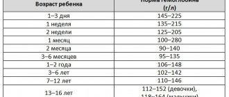
What is the normal level of hemoglobin in a child’s blood?
The hemoglobin level in newborns in the first days of life is very high - 180–240 g/l. This is explained by the fact that the baby has a reserve of iron accumulated while still in the womb. In premature babies, hemoglobin is often below normal - 160–220 g/l. Such children have a high risk of anemia - a critical decrease in hemoglobin in the blood.
By the end of the first week, the hemoglobin level in children drops to 160–200 g/l. In the following weeks and months, hemoglobin gradually increases - by the age of one month its norm is already 120–160 g/l, and by the age of one year it is 110–130 g/l.
At 5 years of age, the norm is 110–140 g/l, at 10 years – 120–140 g/l[5].
During puberty, around the age of 15, the level of hemoglobin in children reaches “adult” levels - 125–165 g/l.
Grodno Regional Children's Clinical Hospital
Details Published March 25, 2015
Pediatrician of the 5th pediatric department, regional freelance pediatric hematologist Guzarevich V.B.
Iron deficiency anemia in adolescents
Puberty is a period of puberty, maturation, growth and development of the adolescent’s body. It has its own unique characteristics, due to powerful vegetative, endocrine and immune restructuring of the body against the background of a physiological growth spurt, as well as disorders of psychosocial adaptation.
It is at this time that numerous transient functional disorders form and previously hidden organic defects appear: hypothalamic dysfunction, with a wide range of clinical manifestations (vegetative-vascular dystonia, metabolic syndrome, etc.), congenital inferiority of connective tissue (visceroptosis, articular hypermobility, osteochondrosis, etc. .), which contributes to a decrease in adaptation to the influence of the external environment. It should be noted that nosological forms during this age period have significant differences. Thus, diseases of the endocrine, nervous and musculoskeletal systems dominate in adolescents, while infectious and colds are more often registered in young children, and in adults the frequency of neoplasms and diseases of the circulatory system increases. This age is characterized by polysystemic (polymorbidity) diseases. For example, in a girl who considers herself practically healthy, a thorough examination is diagnosed with dysmenorrhea, iron deficiency, poor posture, flat feet, nephroptosis, instability of the cervical spine, chronic tonsillitis, myopia, irritable bowel syndrome with constipation, biliary tract dysfunction, crystalluria. Iron deficiency is one of the pressing problems of adolescence. A decrease in the amount of iron in the body leads to impaired hemoglobin formation, the development of hypochromic anemia and trophic disorders in organs and tissues.
At present, there is no doubt that the main factor contributing to the development of iron deficiency anemia in adolescents is the discrepancy between iron reserves in the body and its needs. Iron deficiency in adolescence is much more common in girls, which may be due to heavy menstruation. However, in cases where the need for iron far exceeds its supply, iron deficiency anemia can also develop in young men. The most common reasons in this case are the intensive growth of a teenager, participation in strenuous sports, initial low iron levels against the background of an unbalanced diet, and vegetarianism. It should be remembered that iron deficiency anemia is often observed in conditions such as anorexia nervosa, diseases of the stomach and duodenum, including those associated with H. pylori, repeated intestinal infections, giardiasis, helminthic infestations, and inflammatory bowel diseases.
Clinical manifestations of iron deficiency anemia are pallor of the skin and mucous membranes, loss of appetite, increased physical and mental fatigue, decreased performance, irritability, emotional instability, increased sweating, dizziness, headaches, tinnitus, flashing “flies” before the eyes, increased heart rate, pain in heart. Characterized by dryness and flaking of the skin, dystrophic changes in the nails, dullness and increased fragility of the hair, “jams” in the corners of the mouth; burning of the tongue, difficulty swallowing solid food, perversion of taste and smell, muscle pain, decreased muscle tone, night and day urinary incontinence due to muscle weakness, constipation. Girls may experience menstrual irregularities.
Diagnosis of iron deficiency anemia is based on a decrease in hemoglobin levels below 120 g/l in a general blood test, as well as a decrease in ferritin (iron reserves in the body) in the blood serum.
In all cases of iron deficiency, it is necessary to establish the immediate cause of its occurrence and, if possible, eliminate it. Treatment should be comprehensive and aimed not only at eliminating anemia as a symptom, but also at eliminating iron deficiency and replenishing its reserves in the body.
It is necessary to enrich a teenager’s diet with foods that are the main sources of iron, and not only the amount of iron in a particular product matters, but also the degree of its absorption and the possibility of assimilation by the body.
Meat, especially red meat (veal, beef, lamb, beef tongue), is the best source of heme iron; 20–25% of iron is absorbed from it. Iron from chicken and pork is absorbed to a lesser extent. And in liver and fish, iron is contained in the form of ferritin and hemosiderin, so it is absorbed even worse.
Non-heme iron (vegetables, fruits, nuts) is poorly absorbed (1–5%), and its absorption is influenced by many factors that can either improve or worsen its absorption. Thus, they enhance the absorption of iron: vitamin C (especially abundant in citrus fruits), hydrochloric acid (found in gastric juice), fructose (fruits, honey). Impair iron absorption: drugs that reduce the acidity of gastric juice (antisecretory drugs, antacids), calcium (a lot in dairy products), oxalates (spinach, cabbage, beets, nuts, chocolate, tea, etc.), polyphenols (cocoa, coffee, black and some types of herbal tea), phytic acid (walnuts, almonds, legumes, bran)
However, once anemia has developed, it cannot be cured with diet alone: iron deficiency anemia is treated with iron supplements. Nutrition helps maintain a normal iron balance in the body after treatment. Preference is given to oral forms of iron supplements, since compared to parenteral administration, the rate of restoration of hemoglobin levels is almost the same, and the number of side effects is sharply reduced. Taking iron supplements is recommended 1 hour before meals, preferably in the evening, since the absorption of iron increases in the second half of the day. The therapeutic effect of oral iron intake appears gradually. The first positive clinical sign is the disappearance or reduction of muscle weakness. On days 8–12 from the start of treatment, the content of reticulocytes in the peripheral blood increases. Normalization of hemoglobin occurs by 4–5 weeks from the start of therapy.
In recent years, in the treatment of iron deficiency conditions in adolescents, a new generation of iron-containing drugs based on the polymaltase complex of ferric hydroxide - Ferrum Lek, Maltofer and Maltofer Fol - has been actively used, which in addition to iron contains folic acid, which takes part in the production of nucleic acids, purines, amino acids, stimulating formation of red blood cells. One of the attractive characteristics of these drugs is their low toxicity. The drugs are well tolerated, have a pleasant taste and are in the form of chewable tablets, which undoubtedly increases adherence to therapy. For iron deficiency anemia, children over 12 years of age and adolescents - 100-200 mg per day, and for latent iron deficiency and for preventive purposes - 50-100 mg per day. The duration of therapy, as a rule, is at least 2 months, which is determined by the degree of iron deficiency in the body and depletion of its reserves, the rate of hematopoiesis, and absorption. In the case of clinically pronounced iron deficiency, normalization of hemoglobin is achieved only 2–3 months after the start of treatment. After normalization of hemeglobin levels, it is necessary to continue taking iron supplements in prophylactic doses to restore internal iron reserves for several months.
The prognosis of the disease is favorable: cure occurs in the vast majority of cases. The problem of iron deficiency is, first of all, a nutritional problem, so the primary prevention of iron deficiency anemia is an adequate, balanced diet for a person at any age.
Causes and signs of increased hemoglobin levels
Various reasons can lead to an increase in the level of hemoglobin in a baby. The most common are:
- dehydration - both as a result of insufficient fluid intake and overheating, and as a result of diarrhea;
- congenital heart diseases, including pulmonary heart failure;
- oncological diseases;
- disturbances in the synthesis of the hormone erythropoietin;
- intestinal obstruction;
- blood diseases.
Sometimes a slightly elevated hemoglobin level can be considered normal. In people living at high altitudes, hemoglobin is slightly increased - this is a reaction to thin air. Also, hemoglobin may be slightly higher than normal in children who are very active in sports.
Children with elevated hemoglobin are usually lethargic and irritable, they suffer from drowsiness and fatigue, and the skin sometimes takes on a reddish tint.
Iron deficiency conditions in adolescents: principles of correction
One of the important areas in pediatrics is adolescent medicine, or hebiatics, a branch of medicine that studies the processes of puberty, maturation, growth and development of the adolescent’s body, as well as specific diseases characteristic of the teenage period of life.
The puberty period has its own unique characteristics, due to powerful vegetative, endocrine and immune restructuring of the body against the background of a physiological growth spurt, as well as disorders of psychosocial adaptation. It is at this time that numerous transient functional disorders form and previously hidden organic defects appear: hypothalamic dysfunction, with a wide range of clinical manifestations (vegetative-vascular dystonia, metabolic syndrome, etc.), congenital inferiority of connective tissue (visceroptosis, articular hypermobility, osteochondrosis, etc.) , which helps reduce adaptation to the influence of the external environment. It should be noted that nosological forms during this age period have significant differences. Thus, diseases of the endocrine, nervous and musculoskeletal systems dominate in adolescents, while infectious and colds are more often registered in young children, and in adults the frequency of neoplasms and diseases of the circulatory system increases. This age is characterized by polysystemic (polymorbidity) diseases. For example, in a girl who considers herself practically healthy, a thorough examination is diagnosed with dysmenorrhea, iron deficiency, poor posture, flat feet, nephroptosis, instability of the cervical spine, chronic tonsillitis, myopia, irritable bowel syndrome with constipation, biliary tract dysfunction, crystalluria.
Iron deficiency is one of the pressing problems of adolescence. A decrease in the amount of iron in the body leads to a disruption in the formation of hemoglobin and a decrease in the rate of its synthesis, the accumulation of free protoporphyrin in erythrocytes, the development of hypochromic anemia and trophic disorders in organs and tissues.
At present, there is no doubt that the main factor contributing to the development of iron deficiency anemia in adolescents is the discrepancy between iron reserves in the body and its needs.
Iron deficiency in adolescence is much more common in girls, which may be due to heavy menstruation. However, in cases where the need for iron far exceeds its supply, iron deficiency anemia can also develop in young men. The most common reasons in this case are the intensive growth of a teenager, participation in strenuous sports, initial low iron levels against the background of an unbalanced diet, and vegetarianism. It should be remembered that iron deficiency anemia is often observed in conditions such as anorexia nervosa, idiopathic pulmonary hemosiderosis, Goodpasture's syndrome, atransferrinemia, collagenosis, diseases of the stomach and duodenum, including those associated with H. pylori, repeated intestinal infections, giardiasis, helminthic infestations, inflammatory bowel diseases, infectious diseases (tuberculosis, brucellosis, mycosis, etc.), as well as in chronic renal failure and during dialysis treatment [1, 2].
There are prelatent iron deficiency (iron reserves are depleted only from the depot while maintaining the transport and hemoglobin funds of iron), latent iron deficiency (accounts for 70% of all iron deficiency anemias) and iron deficiency anemia (accounts for 30% of all iron deficiency conditions) (Table).
The clinical picture of iron deficiency anemia consists of two leading syndromes: anemic and sideropenic. The latter is characterized by dystrophic changes in the skin and its appendages; perversion of taste and smell, muscle pain (due to myoglobin deficiency), muscle hypotension, changes in nervous regulation.
It should be noted that the clinical manifestations of the disease in adolescence differ from those in young children. Thus, koilonychia, blue sclera (the choroid is visible through the thinned sclera), taste and olfactory disturbances, dysphagia, and dyspepsia are more typical for them.
Diagnosis of iron deficiency conditions is based on the determination of the laboratory parameters presented above. The main criterion is the ferritin content in the blood serum [3]. However, if it is impossible to determine this indicator, you can focus on the level of hemoglobin, the determination of which is available in any medical institution.
It is advisable to annually determine the hemoglobin level in girls who have heavy blood loss during menstruation or another nature, low dietary iron intake (dietary restrictions for weight loss, vegetarianism, etc.), and a history of iron deficiency anemia of various nature. Boys need to monitor their hemoglobin levels if they are intensely involved in strenuous sports (athlete's anemia).
In all cases of iron deficiency, it is necessary to establish the immediate cause of its occurrence and, if possible, eliminate it (eliminate the source of blood loss, treat the underlying disease, complicated by sideropenia). Treatment should be comprehensive and aimed not only at eliminating anemia as a symptom, but also at eliminating iron deficiency and replenishing its reserves in the body.
It is necessary to enrich a teenager’s diet with foods that are the main sources of iron, and not only the amount of iron in a particular product matters, but also the degree of its absorption and the possibility of assimilation by the body.
Meat, especially red meat (veal, beef, lamb), is the best source of heme iron; 20–25% of iron is absorbed from it. Iron from chicken and pork is absorbed to a lesser extent. And in liver and fish, iron is contained in the form of ferritin and hemosiderin, so it is absorbed even worse.
Non-heme iron (vegetables, fruits, nuts) is poorly absorbed (1–5%), and its absorption is influenced by many factors that can either improve or worsen its absorption. Thus, they enhance the absorption of iron: vitamin C (especially abundant in citrus fruits), hydrochloric acid (found in gastric juice), fructose (fruits, honey). Impair iron absorption: drugs that reduce the acidity of gastric juice (antisecretory drugs, antacids), calcium (a lot in dairy products), oxalates (spinach, cabbage, beets, nuts, chocolate, tea, etc.), polyphenols (cocoa, coffee, black and some types of herbal tea), phytic acid (walnuts, almonds, legumes, bran) [4].
However, once anemia has developed, it cannot be cured with diet alone: iron deficiency anemia is treated with iron supplements. Nutrition helps maintain a normal iron balance in the body after treatment.
The most commonly used iron compounds in pediatric practice include ferric iron preparations, polymaltose hydroxide and ferrous iron salts - sulfate, fumarate, chloride and gluconate. To improve absorption, ferrous salts are combined with organic acids, amino acids and other compounds. Preference is given to oral forms, since compared to parenteral administration, the rate of recovery of hemoglobin levels is almost the same, and the number of side effects is sharply reduced. Taking iron supplements is recommended 1 hour before meals, preferably in the evening, since the process of iron absorption increases in the second half of the day.
The therapeutic effect of oral iron intake appears gradually. The first positive clinical sign is the disappearance or reduction of muscle weakness. On days 8–12 from the start of treatment, the content of reticulocytes in the peripheral blood increases. Normalization of hemoglobin occurs by 4–5 weeks from the start of therapy.
In recent years, in the treatment of iron deficiency conditions in adolescents, a new generation of iron-containing drugs based on the polymaltase complex of ferric hydroxide - Maltofer and Maltofer Fol - has been actively used, which in addition to iron contains folic acid, which takes part in the production of nucleic acids, purines, amino acids that stimulate erythropoiesis.
The results of using Maltofer are assessed positively by various authors [5]. One of the attractive characteristics of this drug is its low toxicity. The drug is well tolerated, has a pleasant taste and is in the form of chewable tablets, which undoubtedly increases adherence to therapy. For iron deficiency anemia, children and adolescents aged from one to 12 years are prescribed Maltofer at a dose of 50–100 mg per day, for latent deficiency and for the prevention of iron deficiency - 25–50 mg per day. Children and adolescents over 12 years old - 100-300 mg per day, and for latent iron deficiency and for preventive purposes - 50-100 mg per day. The duration of therapy, as a rule, is at least 2 months, which is determined by the degree of iron deficiency in the body and depletion of its reserves, the rate of hematopoiesis, and absorption. In the case of clinically pronounced iron deficiency, normalization of hemoglobin is achieved only 2–3 months after the start of treatment. To restore internal iron reserves, administration in prophylactic doses should be continued for several months. If blood loss continues, it is necessary to carry out preventive courses of treatment with an iron preparation (1 month 2–4 times a year).
Literature
- Korovina N. A., Zaplatnikov A. L., Zakharova I. N. Iron deficiency anemia in children (a guide for doctors). M., 1999. P. 64.
- Finogenova N. A., Chernov V. M., Morshchakova E. F. et al. Anemia in children: diagnosis and treatment. M., 2000. pp. 9–17.
- Finogenova N. A., Chernov V. M., Morshchakova E. F. et al. Anemia in children. Diagnosis, differential diagnosis, treatment. M., 2004. pp. 20–64.
- Gorodetsky V.V., Godulyan O.V. Iron deficiency conditions and iron deficiency anemia: treatment and diagnosis (methodological recommendations). M., 2006. P. 25.
- Soboleva M.K. Experience in using the drugs Maltofer and Maltofer Fol in the treatment of iron deficiency anemia in children and adolescents // Siberian Consilium. 2003, No. 1, p. 79–82.
T. A. Bokova1, Candidate of Medical Sciences G. V. Maslikova, Candidate of Medical Sciences
GBUZ MO MONIKI named after. M. F. Vladimirsky, Moscow
1 Contact information
What is dangerous about low hemoglobin in a child?
Low hemoglobin in children is more common than high hemoglobin. According to WHO, a lack of hemoglobin - anemia - occurs in 47.4% of preschool children and 25.4% of schoolchildren.
Low hemoglobin in a child is a significant risk factor. It can lead to developmental delays, the development of heart and kidney diseases (they, along with the brain, are the first to suffer from oxygen starvation), disruption of the functioning of the immune system, skin diseases and edema. Without treatment, anemia can result in cardiomyopathy and heart failure, and these pathologies are deadly. With a critical decrease in hemoglobin levels, even a hypoxic coma is possible.
Signs of low hemoglobin in children are numerous, but nonspecific, in other words, they accompany not only anemia, but also many other diseases. In any case, parents should be alert to symptoms such as weakness, lethargy, tearfulness and drowsiness of the baby, decreased appetite, and pale skin. Older children may complain of dizziness and headaches. With low hemoglobin, cognitive functions - memory, ability to concentrate, and learning - are noticeably affected. Many cases of poor academic performance are not due to laziness, but to a lack of iron and low hemoglobin.
You need to take a hemoglobin test regularly, at least once every three months. It is not easy to determine the development of anemia by indirect signs; very often its symptoms are ignored or attributed to other causes.
A blood test for hemoglobin is necessary after any infectious diseases, sudden changes in diet (for example, after prescribing a diet when allergies or gastrointestinal diseases are detected) and any unexpected changes in the baby’s behavior. If your child has suddenly turned from an active and cheerful baby into a sleepyhead and a crybaby, you need to immediately take a referral for analysis.
Low hemoglobin often affects physical development - such children grow slowly and lag behind healthy peers in weight and height.
Sometimes tests show that the child’s hemoglobin is low, and this causes reasonable concern for parents. What can cause low hemoglobin in a child?
Causes of low hemoglobin in children
- Infectious diseases and parasitic lesions.
- Lack of iron in the mother’s diet during pregnancy and lactation or its lack in the child’s diet. Low hemoglobin is often observed in children of women following a vegan diet. Plant foods also contain iron, but it is less absorbed than what is present in animal products.
- Deficiency of vitamins necessary for iron to be absorbed into the blood - these include folic acid and vitamin C. Copper also plays an important role in the absorption of iron.
- Diseases of the digestive system in which iron absorption is impaired.
- Blood loss after injuries and operations, as well as after regular bleeding, such as nasal bleeding.
- Lack of physical activity and fresh air.
- Congenital pathologies, in particular thalassemia.
Causes of anemia
There are many reasons for a decrease in the level of red blood cells, the main ones can be divided into three groups:
- insufficient production of red blood cells;
- premature destruction of red blood cells (hemolysis);
- blood loss
Insufficient production of red blood cells
For the formation of hemoglobin and red blood cells, the body requires microelements such as iron, as well as vitamins B12 and folic acid. With a significant deficiency of these elements, the production of red blood cells decreases and iron-, B12- or folate-deficiency anemia develops. Iron deficiency is the most common cause of anemia in children, especially under 5 years of age.
Red blood cells are formed from progenitor stem cells that are found in the bone marrow. A decrease in the number of stem cells or disruption of their function can also lead to a decrease in the number of red blood cells and the occurrence of anemia. Such conditions may be congenital or acquired.
Premature destruction of red blood cells
Anemia resulting from hemolysis can be observed in children with congenital genetically determined pathologies of erythrocytes, for example, thalassemia or spherocytosis. At older ages, the causes of hemolysis can be autoimmune diseases, infections, taking certain medications, liver and kidney diseases, and a number of other diseases, such as microangiopathy.
Blood loss
Increased loss of red blood cells can be observed with obvious or hidden blood loss. In children and adolescents, among obvious blood losses, nasal and uterine bleeding (heavy menstruation) are most common. Hidden blood loss can be observed in kidney diseases, as well as inflammatory diseases of the gastrointestinal tract.
Prevention of deviations from the norm
The most important measure to prevent the development of complications associated with changes in hemoglobin levels is regular tests and examinations with a pediatrician. Sometimes low or high hemoglobin in a child indicates the presence of serious diseases that require immediate treatment. But a lot also depends on the parents, since deviations from the norm are often found in healthy children due to errors in diet and routine.
Prevention of elevated hemoglobin
- During pregnancy and breastfeeding, mothers should not take vitamin complexes with iron without consulting a doctor. In many cases, supplemental sources of iron are necessary, but it is extremely important not to exceed the recommended dosage.
- Do not buy children's vitamin complexes without consulting your doctor. If a child gets enough iron and vitamins from food, taking extra vitamins and minerals may do more harm than good.
- Review your child's diet - it should contain meat, vegetables and fruits rich in iron.