Heart murmurs
- an acoustic phenomenon associated with changes in blood flow in the heart and blood vessels. There is a point of view according to which heart murmurs are a problem exclusively for children. Indeed, about 90% of functional noise is found in children and adolescents. However, scientific experts have revealed a fairly wide prevalence of heart murmurs (about 77%) in young people aged 20 to 28 years.
Most cardiologists agree that in adults, the presence of a heart murmur clearly indicates some kind of cardiac pathology and is a reason for a complete cardiac examination.
Types and types of heart murmurs
In medicine, it is customary to distinguish between organic heart murmurs
(indicating cardiac pathology) and functional (“innocent” murmurs). Organic heart murmurs are murmurs caused by a defect (congenital or acquired) or other heart disease.
Most often the cause
organic noise is a valve or septal defect (ventricular or atrial septal defect). In terms of timbre, organic noises are hard, rough, persistent; by intensity – loud, sharp; long in duration, carried out beyond the borders of the heart, into the interscapular or axillary region; persist and intensify after physical activity; are equally audible in any body position and are associated with heart sounds.
Functional noise
in the heart were called “innocent”, because are not associated with the presence of heart disease, can be heard in other diseases not related to heart pathology.
In terms of timbre, functional noises are soft, musical, inconsistent, of low intensity, short, not carried out beyond the heart, weaken after physical activity, their character changes depending on the position of the body, and are not associated with heart sounds.
A more detailed classification of heart murmurs is represented by the following types of murmurs:
Systolic murmurs
- Holosystolic (pansystolic) murmurs.
- Midsystolic (systolic ejection) murmurs.
- Early systolic murmurs.
- Mid-late systolic murmurs.
Diastolic murmurs
- Early high-frequency diastolic murmurs.
- Mid-diastolic murmurs.
- Presystolic murmurs.
Continuous (systole-diastolic) murmurs
Why does a child's heart make noise?
There are many reasons, the most common:
- Abnormal state of venous return of the lungs - the pulmonary veins either do not communicate with the atrium on the right, or can fuse with the veins of the systemic circle.
- Pathology of the interventricular septum can develop independently or be combined with other cardiac pathologies.
- Coarctation of the aorta is a complex disease that requires consultation with a doctor: only a specialist can answer what a heart murmur at 2-3 years old means.
- Atrial septal disorder is a congenital heart defect characterized by a hole between the right and left atrium.
Causes of heart murmurs
The following causes of heart murmurs are identified:
- High blood flow speed.
- The flow of blood through a narrowed or deformed opening into the dilated chamber of the heart.
- Regurgitation of blood (return flow) through an incompetent valve.
heart murmurs in children
may occur due to minor anomalies in the development of the heart (additional trabeculae, chords, open oval window, elongated Eustachian valve, septal aneurysm, etc.), which do not affect the development of the child and do not cause harm to his health. Sometimes a functional heart murmur occurs in a child due to intense, rapid and uneven growth of the child (growth murmur).
If a child has a functional heart murmur
may be truly “innocent”, then in adults it may indicate the presence of some pathology, for example, the presence of a heart defect.
In any case, if you have a heart murmur
, it is better to immediately contact a cardiologist for an examination.
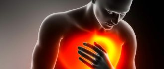
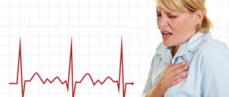
Heart murmurs in adults
Sometimes, when listening (auscultating) the heart, doctors talk about the presence of some kind of “murmur”. What are these sounds? Are they dangerous to health? Are these signs of heart disease? Let's try to figure it out.
The article was published in the new issue of the journal of the Center for High Medical Technologies.
Read the entire issue
The work of the heart is always accompanied by sounds. In the vast majority of cases there are two of them. They are called the first and second heart sounds because they follow each other and together make up the cardiac cycle. The sounds are associated with vibrations of various structures of the heart, as well as the muscles and walls of blood vessels during the movement of blood. The cardiac cycle consists of two large phases. The first phase, which begins with the first sound, is systole, when blood is expelled from the heart into large vessels (aorta and pulmonary artery) by the force of muscle contraction. Therefore, the first heart sound is also called systolic. The second phase begins with the second sound and is called diastole. At this time, the heart muscle relaxes, and the chambers of the heart are filled with a new portion of blood. Another name for the second sound is diastolic. Heart sounds have certain characteristics that characterize their duration, sonority, and time of occurrence.
All other sounds that occur while listening to the heart are usually called “murmurs”. The characteristics of noise depend on the general condition of a person, his constitution, rhythm frequency and many other factors. It is important to understand that any sound outside the normal two-tone cardiac cycle requires full evaluation and attention.
Noises are divided into two large groups: organic and functional.
The first group - organic murmurs - occur with various structural changes in the heart. This means that they are based on heart defects - congenital or acquired. The murmur may indicate, for example, damage to the heart valves - valve stenosis, valvular insufficiency, or a combination of both. Organic noise also appears in cardiomyopathy (expansion of the cavity of the heart chambers or hypertrophy (thickening) of the myocardium), endocarditis (inflammation of the inner lining of the heart (endocardium), acute pericarditis (inflammation of the pericardial layers lining the outside of the heart). All of these pathologies are very serious and life-threatening conditions requiring urgent medical intervention. Therefore, the presence of a “murmur” in the heart is an important signal for additional diagnostics and detection of the disease.
The second group of murmurs, the so-called functional murmurs, are either not associated with the heart or are based on anomalies that do not affect its functioning. The causes of functional noise are varied. They can occur as a result of vegetative-vascular dystonia, as well as during periods of rapid growth in children and adolescents. Also, sometimes they indicate some structural features of the human heart that arose during prenatal development. In this case, systolic murmur can accompany a person throughout his life. In addition, functional noises can be caused by the anatomical features of the large bronchi, located next to the aorta and pulmonary artery, which simply “squeeze” these vessels with accelerated blood flow through their valves. Long-term overstrain, both mental and physical, can contribute to the appearance of noise. One of the common causes of sound phenomena is pregnancy, during which the volume of circulating blood in the mother’s body increases for optimal blood supply to the fetus. In this regard, during pregnancy, changes in intracardiac blood flow also occur with the auscultation of a systolic murmur. In some cases, functional murmurs, although not associated with problems in the heart itself, often indicate other, sometimes serious, diseases. Functional noise can also appear against the background of metabolic disorders. For example, with anemia (decreased hemoglobin in the blood), thyrotoxicosis (excess of thyroid hormones) or fever.
Very often, the patient does not even suspect the presence of a heart murmur, much less the type and cause of its occurrence. A heart murmur can be diagnosed by a doctor by listening to it. You need to see a doctor if you have the following symptoms:
Symptoms of a heart murmur:
- Increased heart rate at rest and during light exercise.
- Labored breathing. Pay attention to your health if even walking causes you to feel short of breath.
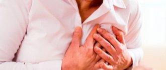
- Chest pain that gets worse after exercise.
- Blue lips and fingertips that occur after fast walking or running.
- Swelling of the limbs.
The presence of one or more of the above symptoms is a reason to consult a doctor. And remember, a heart murmur is not always caused by a serious illness. But you should still undergo a timely examination to exclude such a disease or, if it is detected, to begin treatment in a timely manner.
Diagnosis of heart murmurs
- Auscultation is a highly informative method for diagnosing diseases of the cardiovascular system, based on listening to sound phenomena associated with the activity of the cardiovascular system.
- Angiography.
- Chest X-ray.
- Echocardiography (ECG).
- Cardiac catheterization.
- Load tests.
Samples used to measure heart murmur intensity:
- Breathing (increased sounds from the right side of the heart on inspiration, increased sounds from the left side of the heart on exhalation).
- Valsalva maneuver (forced exhalation with the nose and mouth closed).
- Physical activity (isotonic, isometric, wrist dynamometry).
- Positional changes (changing the patient’s body position, squats, raising legs from a lying position).
- Extrasystole or atrial fibrillation.
- Pharmacological interventions (inhaled drugs).
- Temporary arterial occlusion (external compression of both arms with a bilateral air cuff).
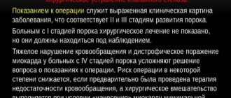
If any pathologies of the cardiovascular system
It is recommended to carry out treatment depending on the etiology of the disease.
Treatment and prevention
Therapy directly depends on the cause. For murmurs not associated with cardiac pathology, the main emphasis is on treating the underlying disease. In case of endocrine disorders, therapy and adjustment will be carried out by an endocrinologist. If anemia causes noise, the help of a therapist is necessary. Treatment of murmurs associated with cardiac abnormalities is the responsibility of a cardiologist. In this case, both conservative therapy and surgical intervention may be required.
The main prevention will be a healthy lifestyle, including a balanced diet, feasible physical exercise, 8 hours of sleep, preventive intake of vitamins in the spring and autumn, and giving up bad habits.
If you have any questions or need advice from a specialist, please use the registration form on the website. Our specialist will help you understand the problem and tell you what functional heart murmurs are. Registration is available 24 hours a day.
Related services: Cardiac Check-up Diagnosis of heart rhythm disorders by ECG monitoring
Preventive recommendations
The main preventive measure includes careful attention to your health; you should pay attention to even minor diseases in order to prevent them from developing into inflammatory processes or rheumatism.
The next point is a balanced diet and healthy food. The diet should include fresh vegetables and fruits, cottage cheese, meat, and fish. Dried fruits are a must - dried apricots, raisins.
Every day you need to take walks in the fresh air, go out into nature, and walk in the park more often. It is also important to completely give up bad habits, especially smoking.
Noise classification
Typifications that are used in clinical practice.
Based on localization:
- Cordial. They are the only ones of interest to the cardiologist. In terms of differential diagnosis, it is difficult to understand where the sound is coming from. Additional examination is required. There are options here. Noises due to regurgitation, disruption of the integrity of the closure of valves (tricuspid, aortic, mitral), too fast blood flow, will produce noises of varying intensity and tone during auscultation.
- Extracardiac. Relatively rare. As a rule, it is located in the pericardium, a special membrane enclosing a muscular organ. When the structures of the bag rub, a specific sound occurs. The second possible reason is damage to the pleura. Also identified is the pulmonary factor, a deviation of respiratory activity.
The nature:
- Organic. Accompanying heart defects, congenital and acquired. Caused by a violation of the anatomical development of tissues. Classic pathological processes are mitral valve stenosis, insufficiency, prolapse. It is difficult to say more precisely what caused the noise; verification by cardiographic methods is needed.
- Inorganic. Associated with impaired functional activity of the heart. For example, when the rheological properties of blood change, its fluidity increases, viscosity increases, blood pressure increases and current accelerates through the structures of the muscular organ.
Based on the moment of registration:
- Systolic. As the name suggests, they are fixed by a doctor during the period of myocardial contraction. The main possible causes: regurgitation, return of blood to the atria due to dysfunction of the mitral valve, and also reverse flow into the left ventricle from the aorta. It can be organic or functional in nature, which complicates the initial diagnosis.
- Diastolic. Slightly less common. They are observed when the myocardium relaxes. Always associated with heart defects.
- Continuous. They are recorded both at the moment of contraction and during pause. Usually accompanied by congenital defects associated with underdevelopment of the interatrial septum and other factors. Found mainly in young patients and children.
There are also more detailed classifications. Thus, systolic murmur is divided into pansystolic (full, noted in any position, merges with the first sound), mesosystolic, indistinguishable from the second tone, holosystolic. Does not mix with normal sound.
Diastolic is typed in the same way. These are rather medical points that are important for theorists. In complex clinical situations, detailed classifications can play a role.
Now our path lies in the functional diagnostics room
Electrocardiography (ECG) is a method of graphically recording electrical phenomena occurring in the heart. An ECG is recorded using a special device - an electrocardiograph. In this case, the electrical potentials are amplified 600-700 times and recorded in the form of a curve on a moving tape. ECG recording is carried out using electrodes placed on various parts of the body. Using this method, various disturbances of the rhythm and conduction of the heart, as well as overloads of its various parts, are determined.
The sound characteristics of noise can be assessed not only by simple auscultation, but also using a research method such as phonocardiography (PCG). It is based on graphic recording of sounds accompanying heart contractions. Mainly heart sounds and murmurs are recorded. The resulting image is called a phonocardiogram. It significantly complements auscultation and makes it possible to objectively determine the frequency, shape and duration of sounds, as well as their changes in the process of dynamic observation of the patient. This method is used to diagnose functional murmurs and cardiac malformations. It is also important for rhythm disturbances, when with the help of auscultation alone it is difficult to decide in which phase of the cardiac cycle the sound phenomena occurred. The analysis of phonocardiography and the diagnostic conclusion on it are carried out only by a specialist. In this case, auscultatory data are taken into account. For correct interpretation of PCG, synchronous recording of a phonocardiogram and an electrocardiogram is used. The method is good and absolutely safe, but is currently being replaced by
Echocardiography (EchoCG) is a simple ultrasound examination, only the point of application is the heart. Using this method, you can accurately determine the source of noise or exclude heart disease.
How to choose a clinic to visit?
The need for multiple visits to a cardiologist in order to determine the motility of the changes that are occurring requires choosing a medical center in the patient’s area of residence, which simultaneously includes all the required parameters for diagnosing the condition.
Our Help Desk for private clinics in Moscow “Your Doctor” will help you make your choice, the information base of which is systematized and convenient for viewing data, allowing you not only to choose a medical institution, but also to immediately arrange a call to a cardiologist at home or register the time for his visit within the medical center.
This article is posted for educational purposes only and does not constitute scientific material or professional medical advice.
Diagnostics
If murmurs are detected during listening by a pediatrician, neurologist or cardiologist, then the patient must undergo a comprehensive diagnosis. It includes:
- conducting an electrocardiogram (shows disturbances in the functioning of the heart, for example, disturbances in rhythmic activity or chamber hypertrophy);
- performing radiography (demonstrates the boundaries of the hematopoietic organ and the condition of both lungs);
- carrying out echocardioscopy (ultrasound examination will show the condition of the valves, vessels and cavity of the heart, growths, narrowings and pathologies).
The main task of the doctor is to accurately differentiate the type of eshum. We need to thoroughly understand what is the true reason for their appearance. This will help you not waste time and prevent a serious illness from developing.
You need to remember that even if you listen to noises, but the child does not have any complaints, there is nothing to worry about. Those noises that are accompanied by pallor of the skin, blueness of the nasolabial triangle, shortness of breath and elevated temperature should be alarming. The baby may complain of chest pain, be capricious and refuse to eat. In such a situation, urgent examination and consultation with a specialist is necessary.
Treatment methods
Treatment options are multiple. Actually, noise control is not required; the root cause must be eradicated. That is, the main pathological condition. When the process is stopped, the abnormal sound disappears by itself.
| Diagnosis | Treatment options |
| Rheumatism | Prescription of medications: glucocorticoids (Prednisolone), immunosuppressants at the discretion of the leading specialist in the acute phase. In chronic cases, cardioprotectors are indicated to normalize metabolic processes in the muscular layer of the heart. Surgery - in case of development of threatening anatomical defects: valve defects, destruction of chambers. |
| Myocarditis | Antibiotics are used for infectious diseases, and drugs to suppress the immune system otherwise. The duration of therapy is about a week. It makes sense to be observed in a hospital. Correction of acquired defects is surgical. |
| Stenosis of vascular structures | Surgery with ballooning or stenting. Forced expansion of the lumen of the arteries. In case of ineffectiveness or initial futility, prosthetic replacement of the affected area is indicated. |
| Aneurysms and other enlargements | Urgent surgical therapy within the walls of a hospital. Dissection of the wall protrusion, prevention of bleeding, suturing. In the future, antiplatelet agents and cardioprotectors are prescribed to prevent relapse of the pathological condition. |
| Mitral valve stenosis | Planned surgery for prosthetics of an anatomical structure. Plastic surgery of the patient does not make sense, since pathology usually develops against the background of underdevelopment or abnormalities in the functioning of connective tissues. An artificial substitute is the best and correct solution. |
| Defects of the tricuspid and aortic valves | The methods are identical. Conservative therapy makes no sense. In all three cases, if there are no signs of progression of the process and there is no dynamics, observation is indicated. Surgery may not be required, depending on the rate of deterioration and the fact of its presence. |
| Disturbances in the formation of heart septa | Integrity is restored promptly. Prosthetics or plastic surgery is indicated. |
| Hyperthyroidism | Iodine preparations, other medications, correction of organic thyroid lesions, diet. |
| Anemia | Administration of vitamins, iron, and other etiotropic techniques, depending on the origin of the process. |
| Pregnancy | No treatment required. Dynamic observation by a cardiologist is indicated. |
| Increased body temperature | Antipyretics (antipyretics), preferably based on Ibuprofen. Paracetamol and Aspirin are dangerous for the liver and heart. |
Symptomatic measures depend on the underlying diagnosis. Blood pressure disorders of the growth type are treated with calcium antagonists, beta blockers, centrally acting drugs, and ACE inhibitors.
To relieve an acute attack, emergency medications like Captopril are prescribed, but with precise dosing so as not to cause a sharp drop in the indicator.
Antiarrhythmics (Amiodarone) and diuretics may be prescribed to eliminate peripheral edema. Also cardioprotectors (Mildronat) and others. At the discretion of the specialist. The pain is relieved with Nitroglycerin.
OFFICE OF CARDIOLOGY
| Name of service | Price |
| Initial appointment with a CMN cardiologist, with prescription of treatment, ECG reading/with interpretation | 5000 |
| Initial appointment with a cardiologist | 2200 |
| Repeated appointment with a cardiologist | 3700 |
| Consultation with a cardiologist based on the results of studies and analyzes of third-party organizations | 3000 |
| ABPM - 24-hour blood pressure monitoring | 4600 |
| Health certificate, including consultation | 3700 |
| Holter (24-hour) ECG monitoring | 4500 |
Make an appointment