Heart transplant - indications
Diseases that are indications for transplantation:
- cardiomyopathy - replacement of myocardial fibers with scar tissue;
- inoperable heart defects of congenital and acquired nature;
- tumor processes in the area of the heart muscle;
- angina pectoris and heart rhythm disturbances that cannot be corrected with medication;
- congenital disorders of the development of the heart muscle that are not subject to plastic correction;
- end-stage heart failure.
Heart transplantation is indicated in extreme cases. When all other options for correcting the patient’s condition have already been tried and have not given the desired result.
4 Consequences of the operation
The early postoperative period is the most difficult in terms of adaptation of the new organ to the changed operating conditions. If the outcome of the operation is positive and there are no serious complications, normal heart function returns in about 3-5 days. It is during this period that complications such as:
- donor heart rejection;
- thrombosis of the heart arteries;
- dysfunction of the brain;
- disruption of the lungs, liver, kidneys and other organs.
In the next 7-10 days, complications such as:
- infectious infection;
- coronary artery disease;
- internal bleeding;
- pneumonia;
- arrhythmia;
- development of oncological tumors against the background of immunosuppressive drugs in the preoperative period.
All these consequences can appear in different orders and at different time intervals after the operation. Complications after transplantation occur in more than 90% of cases, most often arrhythmia, coronary artery disease and internal bleeding.
Any complications, to one degree or another, can cause the patient’s death.
Correction of artrioventricular valve insufficiency.
Functional insufficiency of atrioventricular valves is observed in almost all patients with ischemic or dilated cardiomyopathy. Ischemia leads to the development of direct dysfunction or rupture of the papillary muscles and the formation of mitral regurgitation. A decrease in the contractility of the LV myocardium causes dilatation of its cavity, distortion of the posterior papillary muscle and increases mitral regurgitation. Backward shunting of blood into the left atrium increases LV volume overload and further increases myocardial ischemia. A vicious circle arises, which can only be interrupted by restoring the obturator function of the mitral valve.
For this correction of mitral regurgitation, support rings with modified spatial geometry are used, which reduce the recurrence of mitral regurgitation in the long-term postoperative period.
What may interfere with the operation?
Unfortunately, not every patient will be able to receive a donor heart. There are many different reasons for this:
- Very small number of donors. This can only happen to a person who has registered brain death and whose heart is absolutely healthy.
- There is a very long queue to receive this organ (waiting list), this is most important for children. The organ must fully comply with all the stated requirements and, perhaps, several dozen more patients who are much earlier on the list will have such parameters.
- Sometimes the donor organ cannot be delivered to the right place on time, since the operation must be performed no later than six hours after removal.
- Many people do not agree to undergo a transplant due to ethical or religious reasons. For example, in Christianity a person is alive as long as his heart beats.
- The patient can be stopped by the fear of long and rather expensive rehabilitation.
- Advanced age. Typically, operations are not performed on people over 60 years of age, but there are exceptions.
In addition to the listed obstacles, transplantation will not be performed for a number of other diseases not related to cardiology. It can be:
- pulmonary hypertension in severe form;
- diabetes mellitus at a stage in which negative changes in the retina, blood vessels or kidneys have already begun;
- infectious diseases in the acute stage;
- HIV and tuberculosis;
- autoimmune diseases - rheumatism, arthritis, lupus erythematosus, etc.;
- severe liver or kidney failure;
- chronic severe forms of lung disease;
- oncology;
- addiction to alcohol or drugs;
- severe mental disorders.
What should a donor be like?
The donor is a person with confirmed brain death. These are people who are in a coma after a stroke or emergency.
With the help of medical equipment and medications, the vital activity of the cardiopulmonary system is supported. Therefore, the heart muscle functions even after brain death and is suitable for transplantation.
To remove a donor heart, written permission from relatives or lifetime and legally certified consent of the donor himself is required.
If the intended donor does not have blood relatives or his identity has not been established, organ removal can occur without completing the above documents.
Conditions for heart removal as a transplant:
- the donor must be no older than 65 years;
- the donor should not have infectious diseases that could be transmitted to the recipient;
- the heart must be absolutely healthy;
- the blood groups of the donor and recipient must match;
- the tissues of the donor and recipient must be compatible at the level of the antigenic structure of the receptors;
- the donor's brain death must be recorded and confirmed clinically;
- the difference in the size of the heart muscle of the donor and recipient should not exceed 50%.
The reason preventing transplantation may be a religious factor, because Some religions do not recognize a person as dead if the heart is still functioning.
Another obstacle to organ retrieval may be the distance that must be covered to transport the organ to the appropriate facility. Since the viability of the heart after the death of the donor does not exceed 6 hours.
How much does a heart transplant cost?
All citizens of the Russian Federation have the right to free high-tech medical care, which includes transplant operations. Based on this, if a suitable heart is available, it will not cost the patient anything. But, in addition to the heart transplant operation, the patient will face long and expensive rehabilitation. This period may drag on and require the recipient to pay at least 50 thousand dollars. For children, such a transplant will cost much more, since both the selection of a donor and the operation itself will have to be performed in a foreign clinic.
Historical reference
The first heart transplant was performed in 1964 by James Hardy. The patient received the heart of a chimpanzee. After this, it was possible to keep the patient alive for only an hour and a half.
A significant milestone in successful transplantology is considered to be the transplantation of a human donor heart, performed in South Africa in 1967 by Christian Bernard. The donor was a young woman, 25 years old, who died in an accident. And the recipient is a sick 55-year-old man who has no chance of further treatment. Despite the skill of the surgeon, the patient died of bilateral pneumonia 18 days later.
The main problem of survival is the degree of rejection of the donor organ by the human immune system. Currently, many specialized cardiology centers have access to surgical technology.
Carrying out a heart transplant operation
Heart transplantation requires modern equipment and highly qualified doctors to perform the operation.
For this reason, transplantation is carried out in special research centers equipped with specific equipment.
- Pancreas transplant for diabetes mellitus: indications, features of the operation, results
The operation is performed by several teams of surgeons who replace each other. During the procedure, the patient's condition is monitored by a team of anesthesiologists.
There are two methods used for heart transplantation:
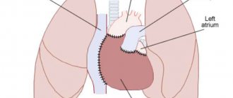
- biatrial - the patient’s heart is partially removed, preserving the atria, to which the donor organ is brought;
- bicavia - the donor heart is brought to the superior and inferior vena cava, without preserving the recipient's atria.
Progress of the operation
- The working field is disinfected. The sternum is cut along the chest using an electric knife.
- An expander is inserted into the surgical access area, fixing the tissue. The folds of the pleura shift.
- The pericardium is opened and a device is brought to the vena cava to stimulate blood circulation. The heart is restricted with clamps to exclude it from the circulatory system.
- It is removed along the lines corresponding to the chosen method of transplantation.
- The donor's organ is prepared for transplantation.
- The donor heart is introduced into the recipient's body and sutures are placed according to the previously chosen technique.
- Blood flow is restored by removing the clamps on the vessels.
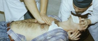
- If the heart has not begun to function on its own, it is started with a cavity defibrillator or rhythmic compressions.
- The chest cavity is cleared of blood.
- The expander is removed, and the bones are fixed with wire sutures or plates. Soft tissues are sewn together.
How is rehabilitation of a postoperative patient carried out?
Rehabilitation begins with restoration of pulmonary ventilation.
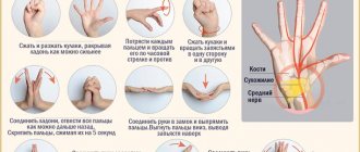
- The patient is recommended to do breathing exercises several times a day and inflate a balloon.
- To prevent thrombosis of the veins of the legs, massage and passive movements in the ankles, bending the knees alternately, are performed.
- The patient can receive the most complete range of rehabilitation measures in a special center or sanatorium. Referral should be discussed with your doctor.
- It is not recommended to quickly increase the load on the heart.
- Hot baths are excluded. You can use a warm shower for washing.
All medications prescribed by a doctor must be taken in the correct dosage.
Operative technology.
Circular sutures running through the tissue at a depth of at least 5 millimeters are applied from the endocardium side with a step calculated based on echocardiographic data. An LV cone is formed with a pre-calculated end-diastolic volume.
After cardiac recovery, surgical LV remodeling is assessed using transesophageal echocardiography.
| before surgery | after operation |
Surgical remodeling of the left ventricle.
Pathological LV remodeling in patients with cardiomyopathy leads to an increase in ventricular volume and stress on the ventricular wall. After the LV becomes spherical, the orientation of the cardiomyocytes changes. At the same shortening fraction, the ejection fraction in the spherical ventricle decreases significantly. In the natural course of the disease, the 5-year survival rate for SILV < 100 ml/m2 is 85%, and for SILV > 100 ml/m2 - 53%. Thus, any intervention aimed at optimizing these relationships is justified, and a favorable prognosis for the patient is possible when the LVSI decreases < 90 ml/m2.
Operative technology.
Correction of these disorders is carried out by implantation of a multi-chamber pacemaker with separate stimulation of the right atrium and ventricles. To do this, three electrodes are passed through the subclavian vein and fixed to the wall of the right atrium, at the apex of the RV and through the coronary sinus in the left lateral vein of the LV at the base of the heart.
During open myocardial revascularization, LV remodeling or mitral valve repair, electrodes are fixed epicardially - in the right atrium and at the base of the heart to both ventricles. A prerequisite for the effectiveness of resynchronization is optimization of the pacemaker.