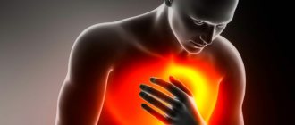
© Author: Ilya Vitalievich Soldatenkov, general practitioner, especially for SosudInfo.ru (about the authors)
Dextrocardia is a congenital pathology or abnormal development of the heart, characterized by its partial or complete displacement to the right thoracic cavity with a mirror arrangement of blood vessels. The anatomical structure of the heart does not change. Those with a “right heart” usually live full lives and live to a ripe old age.
The human heart is formed and begins to actively develop in the second week of embryo formation in the form of two cardiac primordia. In the first phase of embryonic development, a congenital defect is formed, characterized by curvature of the heart tube in the direction opposite to normal. Very often, dextrocardia in the fetus is complicated by other heart pathologies.
Uncomplicated cardiac dextrocardia has no clinical manifestations or negative consequences for its owner, does not cause complaints and does not require treatment.
Main types of dextrocardia:
- Simple - the heart is located on the right, there are no other pathologies, the body functions normally.
- Dextrocardia with transposition of internal organs.
- Complicated - the heart is on the right, there are various associated pathologies.
Dextrocardia happens:
- isolated - altered location of the heart and normal localization of internal organs,
- partially combined - reverse arrangement of the organs of the chest cavity,
- complete - reverse arrangement of the organs of the thoracic and abdominal cavities.
dextrocardia with organ transposition
Married couples who have had cases of dextrocardia in their family should be attentive to their health, pregnancy planning and follow the basic principles of a healthy lifestyle.
Recommendations for treatment and prevention at home
At the bottom of the article is a selection of drugs to prevent the disease
General description of the disease
A very rare anomaly of natural origin, in which all internal organs or one individual organ are located in a mirror order.
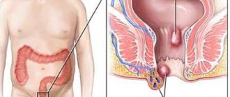
That is, the organs are located the other way around: the heart is on the right side, and not, as we are used to, on the left, the gallbladder and liver are located on the left side, and the stomach and spleen are located on the right. This reverse arrangement can also affect the lungs. With lung transposition, there will be a trilobed lung on the left, and a bilobed lung on the right. This also applies to all blood and lymphatic vessels, nerves and intestines.
Prevalence and types of transposition of internal organs
If the apex of the heart is directed to the right, and all other organs are located in a mirror image, this anomaly is called transposition of organs with dextrocardia.
If the heart is located on the left side of the chest, and all other internal organs are inverted, then such cases are called transposition of organs with levocardia.
The first type of anomaly is more common; dextrocardia occurs in 1 person in 10 thousand. With the second type of transposition, out of 22 thousand people there is only one person with levocardia.
Organs located in a mirror image compared to the normal position of organs with levocardia and dextrocardia without transposition of internal organs are very dangerous for human life.
Reasons for the reverse arrangement of organs
Medical workers have not yet established any reasons for the development of such a serious natural anomaly.
The location of the organs is not affected by the age of the parents, nor by nationality, nor by genetics. All such special people give birth to children with a normal arrangement of internal organs. This means that transposition is not a hereditary disease.
Scientists have noticed that relatively many cases of dextrocardia occur in people with trisomy on the thirteenth chromosome (with the so-called Patau syndrome). In this case, only the heart is reversed, and all unpaired internal organs are located in the normal order.
Symptoms and diagnosis of organ transposition
If a person does not have a congenital heart defect, then the specific location of the organs cannot be detected by external signs.
Many people find out about their peculiarity after many years of life, when they are faced with some serious health problems that do not at all relate to the placement of organs.
With a congenital heart defect, the baby is immediately diagnosed with transposition during a cardiogram and ultrasound.
In people with dextrocardia, congenital heart defects occur in 5-10 percent. As for transposition with normal placement of the heart (with levocardia), cardiac defects are detected in almost 95% of people.
Nowadays, in order for a person to know his anatomical features, even at the age of several months, doctors prescribe medical examinations for babies in order to early diagnose this anomaly.
Complications during transposition of internal organs
The arrangement of organs in a mirror image, if a person is not aware of it, often makes it difficult to make a correct diagnosis. After all, all the signs and symptoms (pain in the side, stomach) will occur on the “wrong” side. Let’s say a person with transposition develops appendicitis and complains of pain in the lower left corner of the abdomen; there will be problems with the spleen, the doctor may attribute it to problems with the liver or gall bladder.
Therefore, it is extremely important to know about your anatomical features. In the West, people with such features wear special keychains, bracelets or get tattoos with the exact diagnosis and type of transposition.
The field of transplantology in people with transposition causes great difficulties. After all, donors are mainly people with the correct arrangement of internal organs and blood vessels. Replacing one organ with another in the presence of a reverse arrangement is a very complex process and requires a highly qualified transplantologist, because correctly located vessels and nerves must be turned in a mirror image so that the new organ takes root and is not rejected.
Ultrasound and medicine
Ultrasound scanner HS60
Professional diagnostic tools.
Assessment of tissue elasticity, advanced 3D/4D/5D scanning capabilities, BI-RADS classifier, options for expert cardiological studies.
Basic principles of the method and physical characteristics
Ultrasound
- high-frequency vibrations lying in the range above the frequency band perceived by the human ear (more than 20,000 Hz).
Radiated into the patient's body, ultrasonic vibrations are reflected from tissues, blood, and surfaces such as boundaries between organs and, returning to the ultrasound scanner
, are processed and measured after a preliminary delay to produce a focused image.
The resulting data appears on the monitor screen, allowing you to assess the condition of the internal organs. Even though ultrasound cannot effectively penetrate media such as air or other gases, as well as bone, it is widely used in the study of soft tissue. The use of ultrasound gels
and other liquids, along with improvements in transducer performance, is increasing the applications of ultrasound scanners for various medical examinations.
The speed of ultrasonic waves in the soft tissues of the human body is on average 1.540 m/sec and is practically independent of frequency. Sensor
is one of the main components of diagnostic systems that converts electrical signals into ultrasonic vibrations and produces electrical signals by receiving reflected echoes from the patient's internal tissues.
An ideal sensor should be effective as an emitter and sensitive as a receiver, have good characteristics of the pulses it emits with strictly defined indicators, and also accept a wide range of frequencies
reflected from the tissues being studied.
In electronic sensors, ultrasonic vibrations are excited by applying high-voltage pulses to the piezo crystals that make up the sensor (the piezoelectric effect was discovered by Pierre and Marie Curie in 1880). The number of times the crystal vibrates per second determines the frequency of the sensor. As the frequency increases, the wavelength of the generated vibrations decreases, which is reflected in improved resolution; however, the absorption of ultrasonic vibrations by body tissues is proportional to the increase in frequency, which entails a decrease in penetration depth. Therefore, high-frequency transducers provide better image resolution when examining shallow tissues, just as low-frequency transducers allow examination of deeper organs, while yielding high-frequency image quality
. This disagreement is a major determining factor in the use of sensors.
In daily clinical practice
Various sensor designs are used, which are discs with one element, as well as combining several elements located around the circumference or along the length of the sensor, producing different image formats that are necessary or preferable when
diagnosing
various organs.
Five types of sensors are traditionally and mainly used
- Mechanical sector sensors.
- Annular sensors.
- Linear sensors.
- Convex sensors.
- Phased scanning sensors.
These five main types of sensors differ according to
- method of generating ultrasonic vibrations;
- radiation method;
- the image format they create on the monitor screen.
Image formats obtained using various sensors
Phased sensors
Linear sensors
Convex sensors
Mechanical sector sensors
Annular sensors
* Areas with the best resolution are highlighted with a dark background.
For diagnostic purposes it is usually used
sensors with frequencies: 3.0 MHz, 3.5 MHz, 5.0 MHz, 6.5 MHz, 7.5 MHz.
In addition, in recent years, devices equipped with high-frequency
sensors of 10-20 MHz have appeared on the ultrasonic technology market.
Sensor Applications
- 3.0 MHz (convex and sector) are used in cardiology;
- 3.5 MHz (convex and sector) - in abdominal diagnostics and studies of the pelvic organs;
- 5.0 MHz (convex and sector) - in pediatrics;
- 5.0 MHz short focus can be used for breast examination;
- 6.0-6.5 MHz (convex, linear, sector, annular) - in cavity sensors;
- 7.5 MHz (linear, sensors with a water attachment) - for the study of superficially located organs - the thyroid gland, mammary glands, lymphatic system.
Basic Picture Settings
- Gain
- “amplification” of the detected signal by changing the ratio of the amplitudes of the input and output signals. (Excessively high gain levels cause the image to blur and appear “white”). - Dynamic range
- the range between the recorded signals with maximum and minimum intensity. (The wider it is, the better signals that differ little in intensity are perceived). - Contrast
- characterizes the system's ability to distinguish echoes with small differences in amplitude or brightness. - Focusing
- used to improve resolution in a specific area of interest. (Increasing the number of focal areas improves image quality, but reduces frame rate.) - TGC
- depth compensated gain. - Frame average
- allows you to smooth the image by superimposing a certain number of frames on top of each other per unit of time or make it hard, bringing it closer to real time. - Direction
- changes the orientation of the image on the screen (left to right or top to bottom).
image artifacts often appear
, and some acoustic phenomena are also observed.
Image Artifacts
- Reverberation.
Occurs when an ultrasonic wave hits between two or more reflective surfaces, partially experiencing multiple reflections. In this case, non-existent surfaces will appear on the screen, which will be located behind the second reflector at a distance equal to the distance between the first and second. This most often occurs when the beam passes through liquid-containing structures. - Mirror artifacts.
This is the appearance in the image of an object located on one side of a strong reflector on its other side. This phenomenon often occurs near the diaphragm. - "The Tail of a Comet"
This is the name given to small echo-positive signals that appear behind gas bubbles and are caused by their own vibrations. - Refraction artifact.
Occurs when the ultrasound path from the transducer to the reflecting structure and back is not the same. In this case, an incorrect position of the object appears in the image. - Effective reflective surface artifact.
The problem is that the actual reflective surface is larger than that shown in the image, since the reflected signal does not always return entirely to the sensor. - Beam thickness artifacts.
This is the appearance, mainly in liquid-containing structures, of wall reflections, due to the fact that the ultrasound beam has a specific thickness and part of this beam can simultaneously form an image of an organ and an image of adjacent structures. - Ultrasound velocity artifacts.
The average speed of ultrasound in soft tissues, 1.54 m/s, for which the device is programmed, is slightly higher or lower than the speed in one or another tissue. Therefore, slight image distortion is inevitable. - Acoustic shadow artifact.
Occurs behind structures that strongly reflect or strongly absorb ultrasound. - Artifact of distal pseudoenhancement.
Occurs behind structures that weakly absorb ultrasound. - Side shadow artifact.
Occurs when a beam falls tangentially onto a convex surface of a structure, the speed of ultrasound transmission in which is significantly different from the surrounding tissue. Refraction and, sometimes, interference of ultrasonic waves occurs.
Basic terms used to describe the acoustic characteristics of formations and pathological processes
- anechoic;
- hypoechoic;
- isoechoic;
- hyperechoic;
- cystic formation;
- solid education;
- cystic-solid formation;
- echo-dense formation with acoustic shadow;
- diffuse damage;
- nodal (focal) lesion;
- diffuse nodular lesion.
Echogenicity
- a characteristic of tissues that reflects their ability to form an echo.
Homogeneous structure
is an area that produces a homogeneous echo.
Some ultrasound symptoms of pathological processes and formations
- "Hallo."
It is a rim of reduced echogenicity around a formation, such as a liver metastasis. - Bull's eye sign.
A volumetric formation of uneven acoustic density with a hypoechoic rim and a hypoechoic area in the center looks similar, observed with metastases in the liver. - Symptom of “pseudotumor”.
Against the background of pronounced fatty infiltration of the liver, a hypoechoic area of unchanged parenchyma, usually located near the gallbladder, may appear as an additional formation. - "Rail" symptom.
Occurs with pronounced dilatation of the intrahepatic bile ducts, when the liver vein and duct are presented in the form of parallel tubular structures. - Symptom of "double-barreled shotgun".
This is what a significantly dilated common bile duct and portal vein look like in the projection of the portal of the liver. - "Snow flake" symptom.
Multiple small formations of increased echogenicity in the lumen of the gallbladder, appearing immediately after a change in the patient’s body position, observed in chronic cholecystitis. - Symptom of a "blizzard".
Areas of increased echogenicity in the liver with fuzzy contours of uncertain shape and varying sizes, observed in cirrhosis. Also, multiple heterogeneous formations of oval shape, increased echogenicity, located in the uterine cavity with hydatidiform mole or in the ovaries with luteal cysts. - Symptom of “pseudo-kidney”.
Manifests itself with tumor damage to the gastrointestinal tract. When scanning transversely, the image of the affected area of the intestine resembles a kidney - the peripheral zone is low echogenic, and the central zone has increased echogenicity.
Terms to describe the location of anatomical structures
- cranial (upper);
- caudal (lower);
- ventral (anterior);
- dorsal (lower);
- medial (middle);
- lateral (side);
- proximal (description of structures located close to their place of origin or attachment);
- distal (description of structures located far from their place of origin or attachment).
The study evaluates
- location and relative position of organs and their parts;
- their shape and size;
- contours;
- structure (with sound conductivity assessment);
- presence or absence of additional education;
- condition of intra- and periorgan vessels.
Main scanning planes
- sagittal
(longitudinal) - scanning plane, when the long axis of the sensor is oriented in the direction of the patient's head - legs; - frontal
- scanning plane, when the sensor is located on the side surface of the patient’s body with its long axis head-legs oriented; - transverse
—scanning plane when the long axis of the sensor is oriented perpendicular to the long axis of the patient’s body.
Longitudinal scanning
Scanning plane and sensor position during longitudinal scanning.
Transverse scanning
Scanning plane and sensor position during transverse scanning.
Ultrasound scanner HS60
Professional diagnostic tools.
Assessment of tissue elasticity, advanced 3D/4D/5D scanning capabilities, BI-RADS classifier, options for expert cardiological studies.
Useful products for organ transposition
In the absence of heart defects or other congenital diseases, a person can lead absolutely normal life activities.
Food must be high in calories, healthy, contain all macro- and microelements, vitamins, enzymes required for normal human life. If you have any diseases, you need to adjust your diet depending on the identified problem. Any form of nutrition or diet should be discussed with qualified healthcare professionals who will provide any recommendations.
Traditional medicine for organ transposition
With organ transposition, folk remedies can only act as an addition to solving the problem that has befallen such a “special” person.
Any serious disturbances in the functioning of the organ require qualified medical assistance. Under no circumstances should you make your own diagnosis or prescribe treatment. If you do not know about your peculiarity, then you can “heal” a healthy organ, but the suffering organ will continue to hurt and the disease will only progress. Diagnosis must be carried out using medical examinations and modern equipment.
Dangerous and harmful products for organ transposition
A person with a mirror arrangement of organs is strongly recommended to lead a healthy lifestyle and include only healthy foods in their diet.
Alcohol, tobacco, trans fats, spreads, vegetable mixtures, sweet sodas, fast food and all other non-living foods should be excluded from the diet. If you have allergic reactions, you should exclude products containing allergens. The list of harmful products may be expanded due to other congenital or acquired diseases. What is important here is a personal approach to each person individually, taking into account all the characteristics of his body.
TRANSPOSITION OF INTERNAL ORGANS, HUMAN
Leave a comment at the bottom of the page »Go«
Causes
The pathology is based on a gene mutation that leads to disruption of intrauterine development of the fetus. Dextrocardia is an autosomal recessive hereditary disease with pathological localization of internal organs. For unknown reasons, the heart tube becomes bent and shifted to the right during embryogenesis.
Dextrocardia is often confused with an acquired disease - dextroposition of the heart (see figure below), caused by various dysfunctions. Dystopia of the heart in the chest with mechanical displacement of organs to the right is caused by the following pathologies: pulmonary atelectasis, accumulation of fluid in the chest cavity, tumors. Long-term or short-term displacement of the heart occurs when the stomach and intestines are full of food and gases, in the presence of ascites, hepatosplenomegaly, after removal of the right lung. As a result of treatment of the underlying disease during dextroposition, the patients' condition quickly normalizes. With dextrocardia, it is impossible to change the location of the heart.
dextrocardia and other cardiac abnormalities
Diagnostics
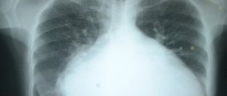
Diagnostic examination of patients includes examination, percussion, auscultation, additional instrumental techniques: radiography, electrocardiography, ultrasound examination of the heart and blood vessels, tomography, angiocardiography.
- By percussion and auscultation, specialists determine the apical impulse and cardiac dullness on the right, an unusual arrangement of heart sounds.
- X-ray diagnostics can detect abnormal location of the heart.
- Electrocardiographic signs are actually reversed, and it appears that the electrodes were incorrectly installed . An ECG for dextrocardia is of great diagnostic importance. ECG signs can confirm or refute a preliminary diagnosis and differentiate dextrocardia from other heart pathologies.
ECG leads for dextrocardia
Modern diagnostic methods make it possible to detect dextrocardia during intrauterine development of the fetus. Newborns with a similar defect are examined in more depth: cardiac echocardiography is performed, which allows one to see the main structures of the heart and assess blood flow in the vessels. Using ultrasound of internal organs, their location is determined.