All about the tricuspid valve: structure, mechanism of operation, main tasks
The heart is the central organ of the cardiovascular system, providing blood circulation through rhythmic contractions and pushing blood from the cavities into the great vessels. The correct direction of blood flow is carried out by the valve apparatus. Between the right atrium and the ventricle is the tricuspid valve, also called the atrioventricular orifice (AV). The main pathologies are congenital or acquired defects, complications after infectious diseases. If pathology is detected, treatment by a cardiologist is indicated.
How does it all work? A few words about the structure of the heart and its parts
The structure of the heart in mammals with 2 circles of blood circulation is approximately the same. The heart consists of two atria (the first chambers along the path of incoming blood), two ventricles, valves between these chambers, and vessels entering and leaving the heart with valves at their beginnings.
Between the right atrium and the right ventricle is the right atrioventricular
, or a
trioventricular
, which consists of 3 valves.
Therefore, it is called tricuspid
, or
tricuspid
.
Between the left atrium and the left ventricle is the left atrioventricular valve
, which consists of two valves and is called
mitral
.
Valves
, located at the mouths of the vessels extending from the heart, or the main vessels, namely
the aorta
and
pulmonary artery
, are called
“aortic”
and
“pulmonary”
.
Atrioventricular valves
– casement, i.e. their structure resembles doors on the wings: opened and closed, down - up.
Aortic valves
and
the pulmonary artery are different in structure
. Each of them consists of 3 semilunar valves that close in the center. When they open, they press against the wall of their vessel (aorta or pulmonary artery), and close, completely closing the lumen of the vessel. Moreover, their appearance resembles a brand name.
The tissue of the valves themselves, both atrioventricular and semilunar, is thin, even transparent in children, but amazingly elastic and durable, designed by nature for continuous rhythmic work, amounting to billions of monotonous actions.
Between the cavities of the heart, or its chambers, there are partitions that separate the flow of venous and arterial blood. This is the interatrial septum, i.e. between the right and left atria, and the interventricular septum - between the right and left ventricles. In a normal, formed heart, they are completely closed, there are no holes or defects in them
and thus blood never flows from one half of the heart to the other.
Let us dwell in more detail on the anatomical structure of the heart and its chambers. After all, even those that have the same name (atria or ventricles) are structured completely differently and perform different functions.
The shape of the heart resembles a pear lying somewhat on its side, with the top located to the left and below, and the base to the right and above. The apex of the heart is the part of the heart whose movements can be felt if you place your palm on the chest in the fifth intercostal space to the left of the sternum. You can easily feel its push both in yourself and in your child. These are the movements of the apex of the heart with each contraction. The contractions are almost synchronous with the pulse, which can also be easily felt on the arm (where the forearm meets the hand) or on the cervical vessels. The pulse is the filling of the vessels with a wave of blood coming from the heart with each contraction. Pulse frequency and its rhythm are an indirect and easily accessible reflection of the activity of the heart itself.
The apex is the most mobile part of the heart, although the whole thing, all its departments, are in constant motion.
The work of the heart, its movement, consists of two alternating phases - contraction (systole) and relaxation (diastole).
The rhythmic, constant alternation of these phases, necessary for normal functioning, is ensured by the occurrence and conduction of an electrical impulse through a system of special cells - through the nodes and fibers of the conduction system of the heart
.
Impulses arise first in the uppermost, so-called sinus node
, then pass to the second,
atrioventricular node
, and from it - along thinner fibers - to the muscles of the right and left ventricles, causing contraction of all their muscles.
Right atrium
accepts venous blood from the vena cava, i.e. from the whole body and in addition the venous blood of the heart itself. This is the largest chamber in volume and, perhaps, the most stretchable chamber of the heart. If necessary, it can accommodate several times more blood than under normal conditions, i.e. has a gigantic “reserve” of volume. The wall of the right atrium consists of a layer of thin muscle fibers. In addition to the function of “receiving” venous blood, the right atrium performs the function of a cardiac pacemaker. Both main nodes of the conduction system of the heart lie in its walls.
How does the tricuspid valve work?
The human heart consists of four sections. The aortic and pulmonary valves communicate with the atrial cavity and the arteries of the same name. The left and right atrioventricular openings are located between the ventricle and the atrium of the heart on the corresponding side.
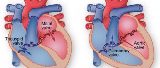
The tricuspid valve (right AV orifice) is a window with three flexible plates - leaflets on a ring of fibrous tissue, communicating with the right parts of the heart. Normally it has three valves: anterior, posterior and septal, due to which it is also called tricuspid. There may be four or six leaves.
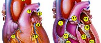
The AV orifice contains papillary muscles and chordae tendineae extending from them, which are attached to the base of each leaflet and ensure proper operation of the valve, tension or relaxation during heart contraction cycles. The right atrioventricular valve is shown in the figure below:
Mechanism of operation
During diastole (relaxation of the heart muscle), the tricuspid valve opens and allows venous blood to flow from the right atrium to the right ventricle. During systole (contraction of the heart), the valves close tightly and do not wrap due to fixation by the chordae and muscles. From the ventricle the blood is ejected into the pulmonary trunk. Next, along the right and left main arteries it is sent to the lungs for gas exchange. In this case, the muscles and functional apparatus of the valve prevent the backflow of blood into the heart cavity from large vessels.
- Holding the valve leaflets open by the papillary muscles and chordae during the filling phase.
- Tight closure to prevent regurgitation (backflow into the atrium) during the phase of expulsion of blood into the pulmonary trunk.
- The difference between the larger size of the valve leaflets and the smaller diameter of the AV orifice, due to which the valve closes tightly during the contraction phase as the volume of the ventricle changes.
- Anatomically, the valve is shaped like a funnel, which ensures passive blood flow when pressure changes in the cavities of the heart.
What phases does the cardiac cycle consist of?
The cardiac cycle consists of three phases sequentially replacing each other:
- Atrial systole (from the Greek systole - compression, contraction)
Lasts 0.1 sec. During this phase, the atria contract, their volume decreases, and blood flows from them into the ventricles. The leaf valves are open during this phase.
Lasts 0.3 seconds. The leaflet (atrioventricular) valves close to prevent blood from flowing back into the atria. The muscle tissue of the ventricles begins to contract, their volume decreases: the semilunar valves open. Blood is expelled from the ventricles into the aorta (from the left ventricle) and the pulmonary trunk (from the right ventricle).
Total diastole (from the Greek diastole - expansion)
Lasts 0.4 seconds. In diastole, the cavities of the heart expand - the muscles relax, the semilunar valves close. Flap valves are open. During this phase, the atria fill with blood, which passively flows into the ventricles. Then the cycle repeats.
We have already discussed the cardiac cycle, but I want to focus your attention on some details. In total, one cycle lasts 0.8 seconds. The atria rest for 0.7 seconds during ventricular systole and total diastole, and the ventricles rest for 0.5 seconds during atrial systole and total diastole. Thanks to this energetically beneficial cycle, the heart muscle gets less tired during work.
PS We found an article that relates to this topic, study it - Heart and blood vessels